Section 1 of 8Two kinds of microscope
| Feature | Light microscope | Electron microscope |
|---|---|---|
| Radiation used | Visible light | Beam of electrons |
| Magnification | Up to about ×1500 | Up to about ×2 000 000 |
| Resolution | About 200 nm | As fine as 0.2 nm or better |
| Specimen | Can be living | Must be in a vacuum, so dead |
| Cost and size | Cheap, portable | Expensive, large and needs training |
Section 2 of 8Why resolution matters
Resolving power
- Resolving power Resolution is the smallest distance between two points that can still be seen as separate.
Wavelength
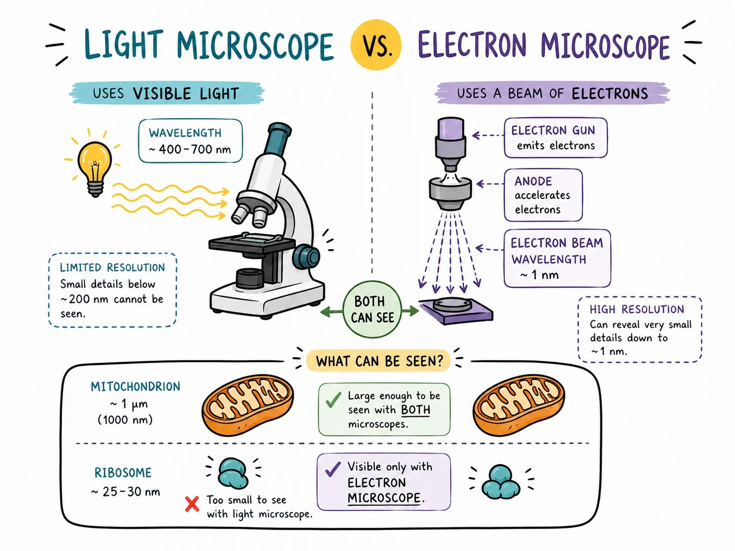
- Wavelength Electrons have a much shorter wavelength than light, so they can show finer detail.
Magnification alone
- Magnification alone Enlarging a blurred image more does not add detail; only better resolution does.
Section 3 of 8Types of electron microscope


Electron microscopes
Transmission (TEM)
- Transmission (TEM) Electrons pass through a very thin specimen to give a flat, high-detail image of internal structure.
Scanning (SEM)
- Scanning (SEM) Electrons bounce off the surface to give a three-dimensional view of the exterior.
Discoveries
- Discoveries They revealed ribosomes, the fine structure of mitochondria and chloroplasts, and much more.
Section 4 of 8Magnification calculations
1
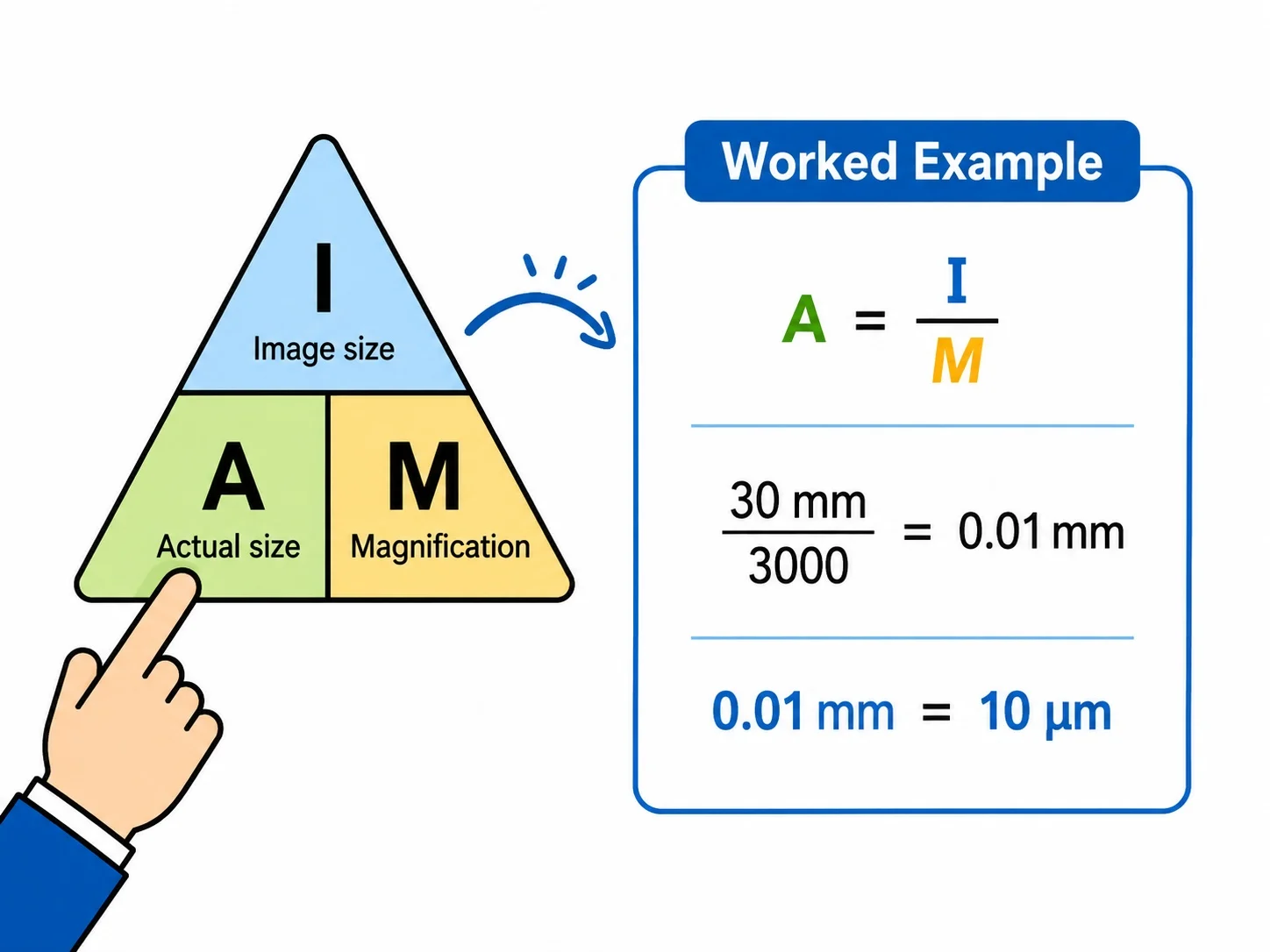
Use the equation magnification = image size ÷ real size.
2
Measure the image in millimetres with a ruler and convert it to the same unit as the real size, usually μm (1 mm = 1000 μm).
3
Rearrange to find the real size = image size ÷ magnification.
4
Example: an image of 30 mm with magnification ×1500 gives a real size of 30 000 μm ÷ 1500 = 20 μm.
Section 5 of 8Worked example: finding real size
1
Question: a mitochondrion appears 45 mm long in an electron micrograph with magnification ×30 000. Find its real length.
2
Convert the image size to micrometres: 45 mm × 1000 = 45 000 μm.
3
Rearrange the equation: real size = image size ÷ magnification.
4
Calculate: 45 000 ÷ 30 000 = 1.5 μm.
5
State the answer with its unit, 1.5 μm, which is 1.5 × 10⁻⁶ m.
Section 6 of 8Choosing a microscope
Living cells
- Living cells A light microscope is used to watch living cells, for example moving protists or dividing cells,
- because the specimen is not placed in a vacuum.
Cell organelles
- Cell organelles Structures such as ribosomes and the inner membranes of mitochondria are too small for a light microscope and need an electron microscope.
Surface detail
- Surface detail A scanning electron microscope is chosen when the 3D shape of the outside of a specimen, such as a pollen grain, is needed.
Section 7 of 8Key terms
| Term | Meaning |
|---|---|
| Magnification | How many times larger an image is than the real object. |
| Resolution | The ability to distinguish between two points that are close together. |
| Micrometre (μm) | One millionth of a metre; 1000 μm = 1 mm. |
| Wavelength | The distance between two matching points on a wave. |
| Transmission electron microscope | A microscope in which electrons pass through a thin specimen to show internal structure. |
| Scanning electron microscope | A microscope in which electrons bounce off the surface of a specimen to show its 3D shape. |
Section 8 of 8Exam technique
1
Mixing units, such as dividing millimetres by micrometres, in magnification calculations.
2
Describing resolution as the same thing as magnification.
3
Stating that electron microscopes can look at living cells; specimens are in a vacuum.
4
Quoting the wrong direction when rearranging the magnification triangle.
5
Leaving the image size in millimetres when the real size is in micrometres.