Skip to content
Dabour HubLEARN · PRACTICE · EXPLORESign inStudent

2.2.1Organisation

The Lungs

5 min read 9 parts Lesson 8 of 19 2 quizzes

0%read

Section 1 of 9The gas exchange system

The Human Gas-Exchange System
The Human Gas Exchange System
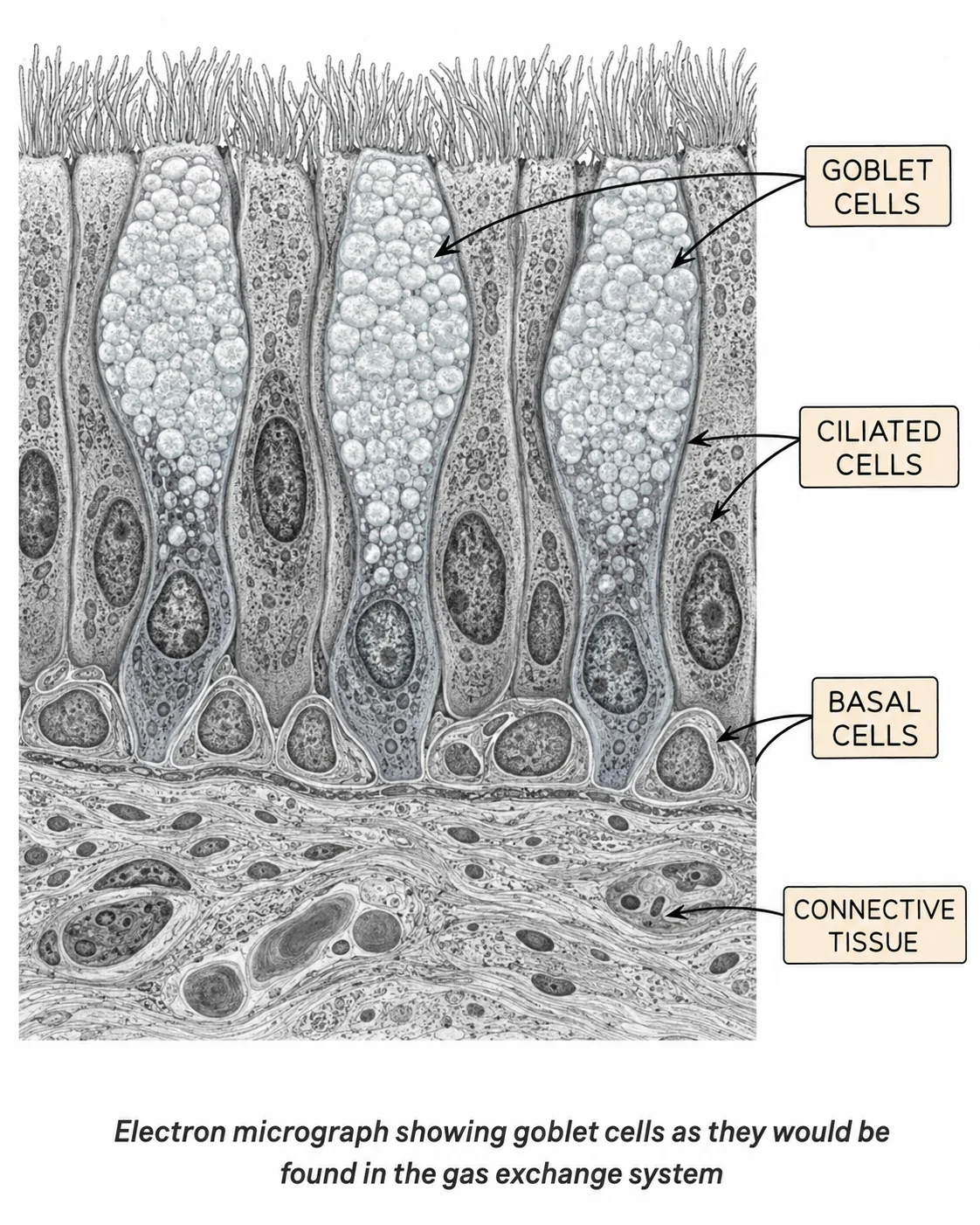
Goblet Cells — Gas Exchange System
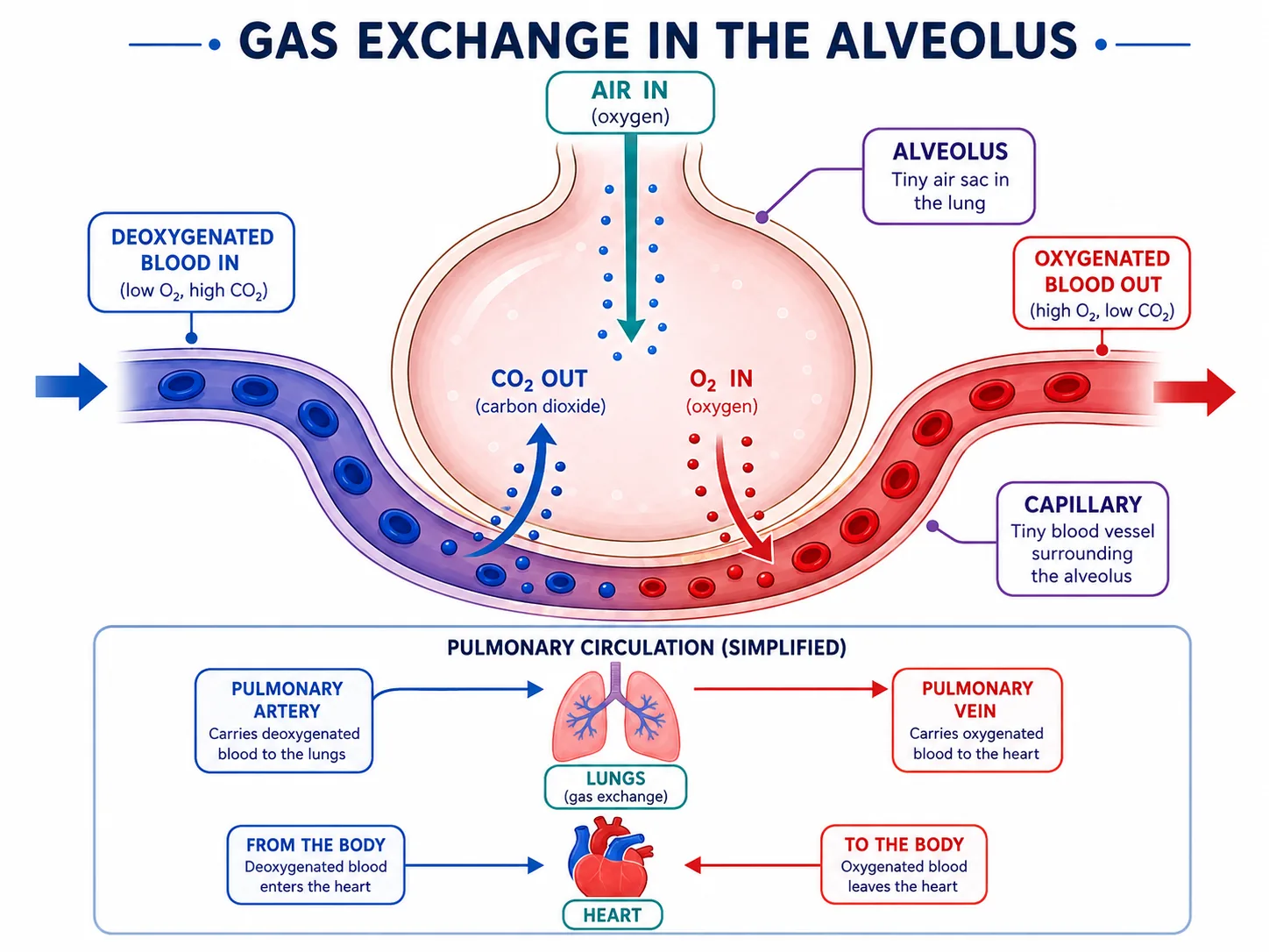
Gas Exchange at an Alveolus
Gas Exchange in the Alveolus
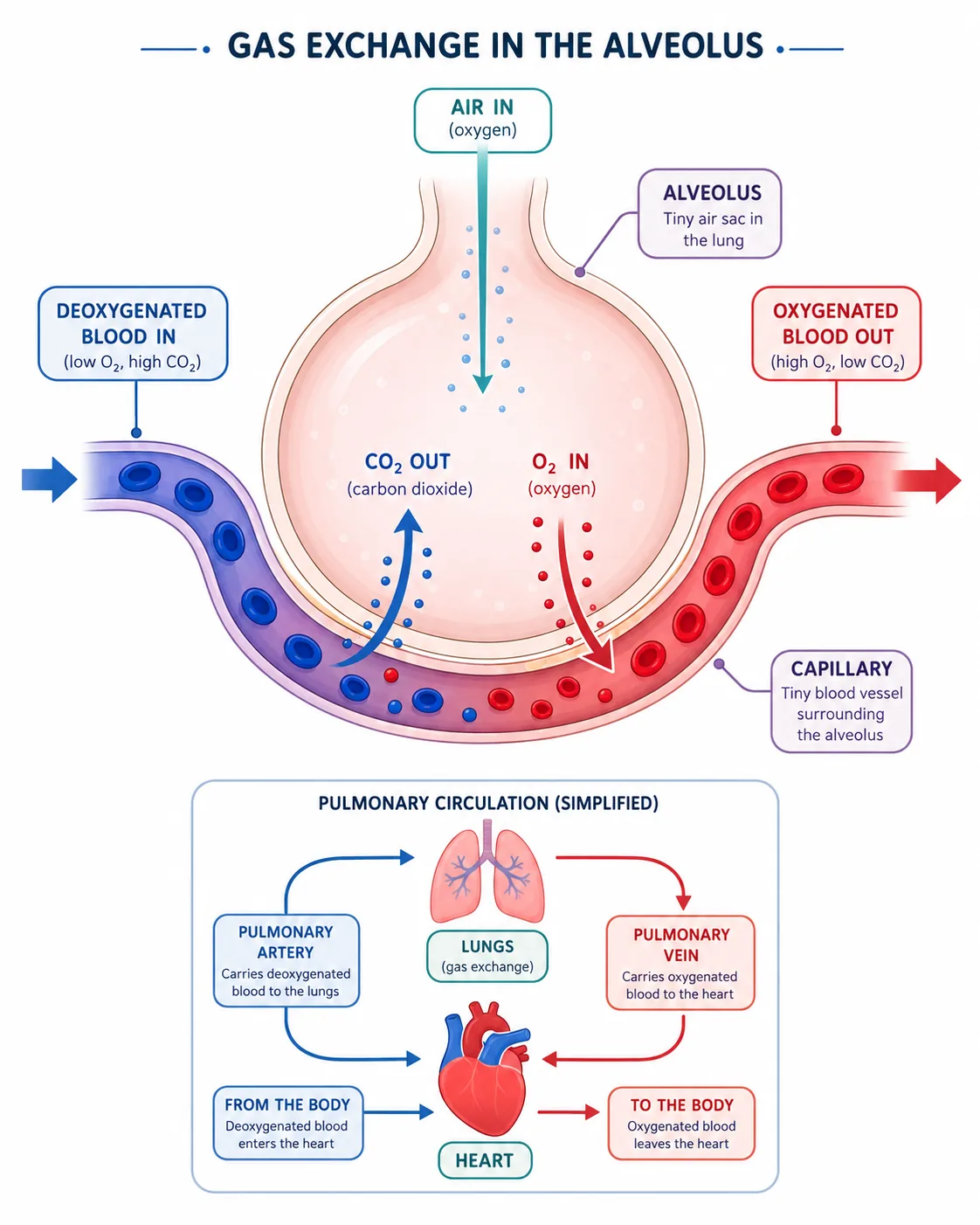
Gas Exchange at an Alveolus
alveolus gas exchange landscape
pasted 1782296383125 0
Alveolus — Gas Exchange (Continuous Simulation)
Cross Section of a Bronchus
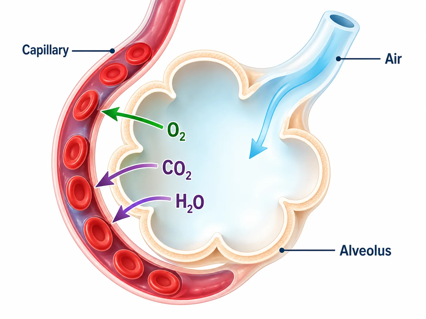
Gas exchange at an alveolus
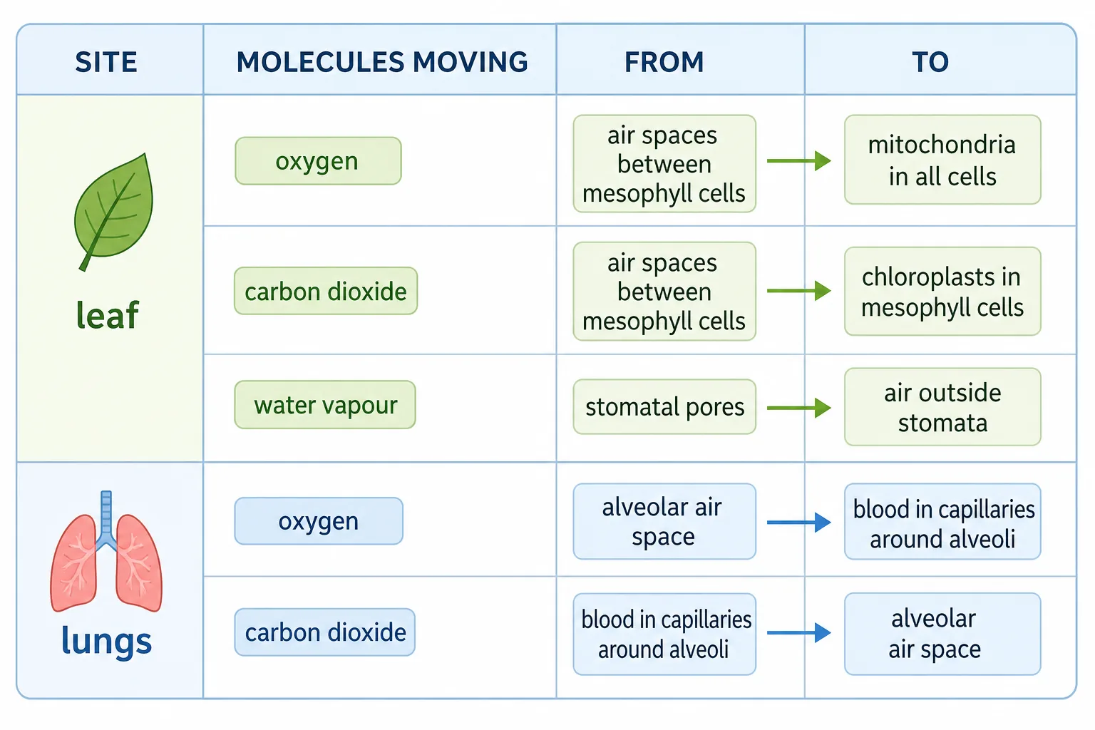
Gas exchange table
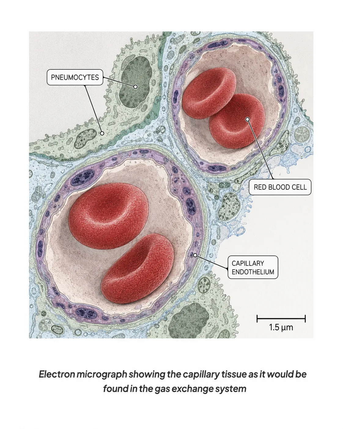
Capillary tissue in the gas exchange system
pasted 1782303915205 0
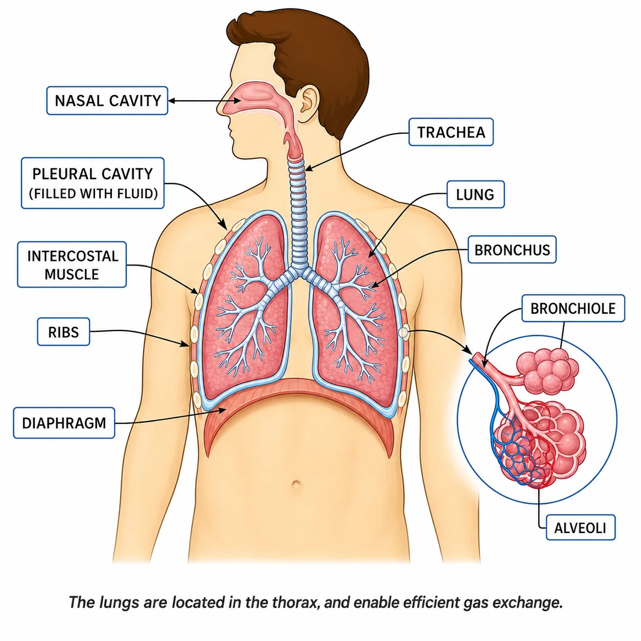
The Human Thorax
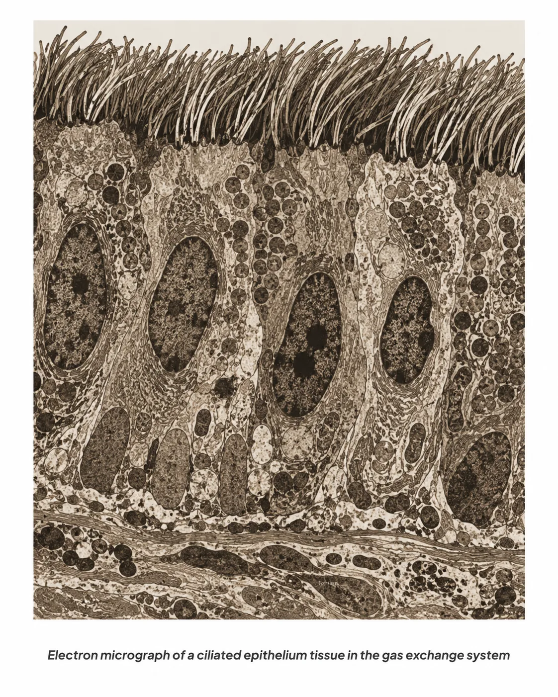
Ciliated Epithelium
The Human Gas Exchange System
Respiratory system in the thorax (lungs, trachea, alveoli)
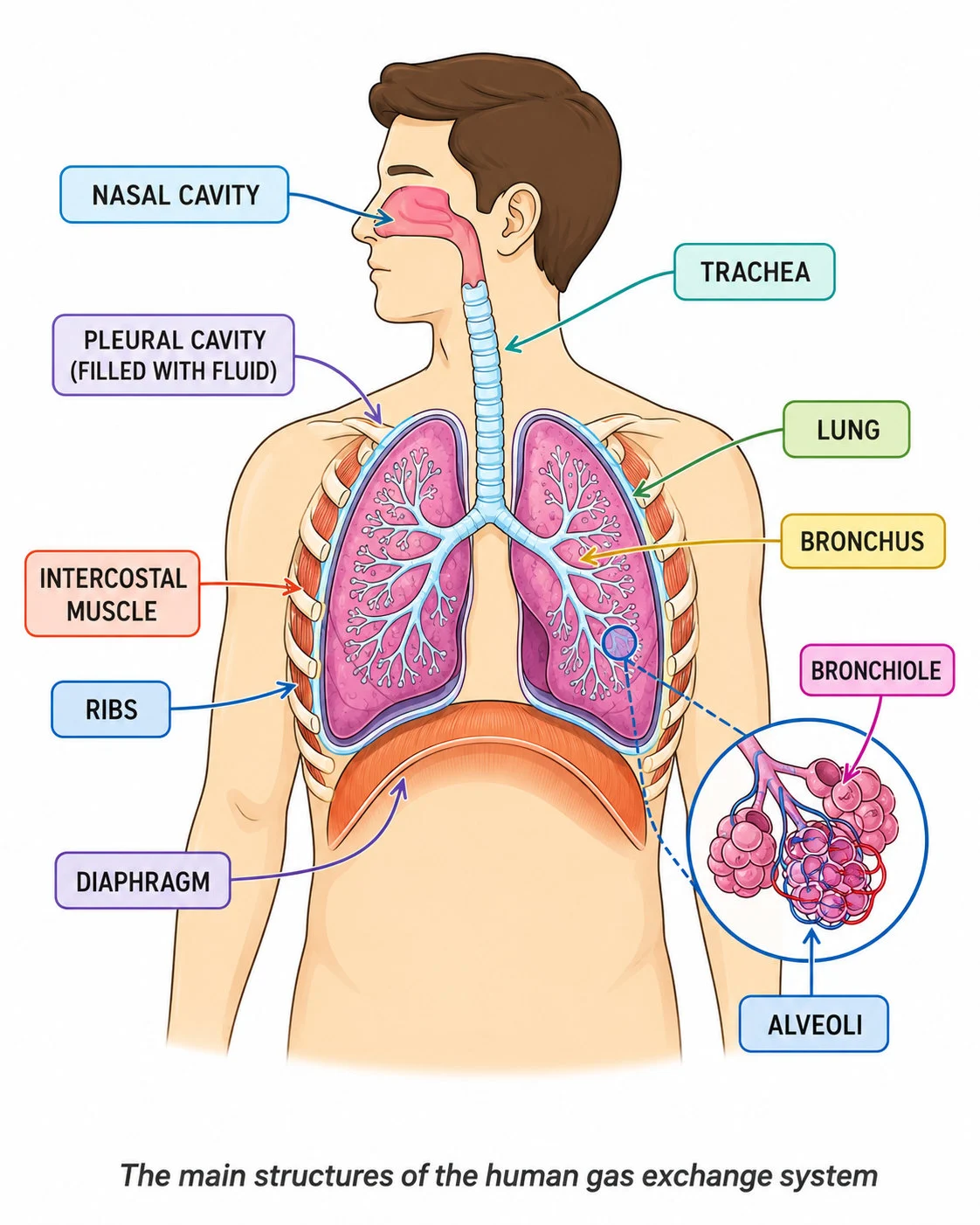
Structures of the human gas exchange system
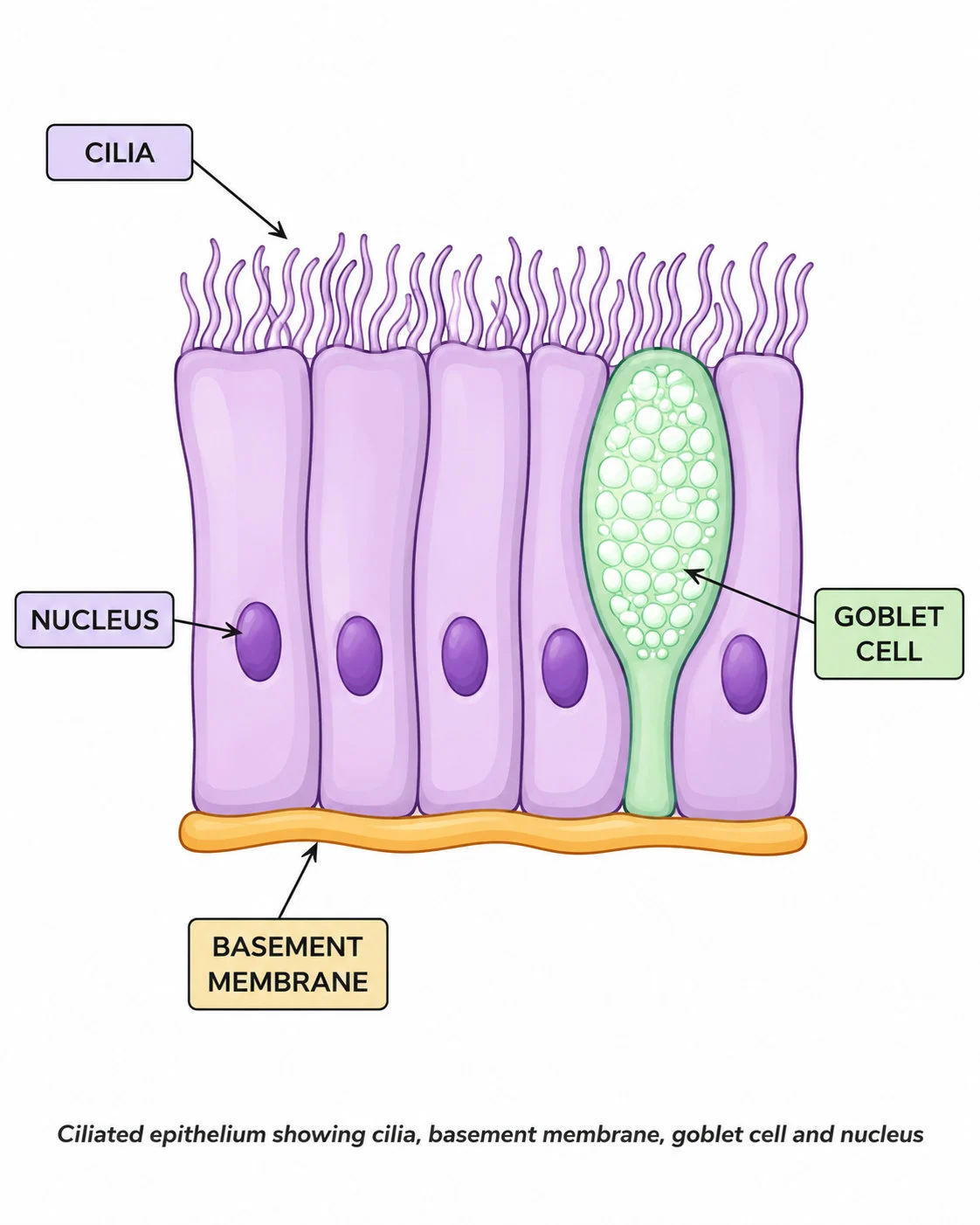
Ciliated epithelium and goblet cells

Humans have a large, active body, so every cell needs a rapid supply of oxygen for aerobic respiration and a way of removing carbon dioxide.

The gas exchange system
The lungs are the organs of gas exchange.
Diaphragm
They sit in the chest (thorax), protected by the rib cage and separated from the abdomen by a sheet of muscle, the diaphragm.
Air passes through the following path:
The gas exchange system
Nose or mouth
The nose or mouth, where air is warmed and moistened.
Trachea
The trachea (windpipe), a tube held open by rings of cartilage.
  • It is lined with ciliated cells and mucus-secreting goblet cells that trap dust and microbes and sweep them away.
Bronchi
Two bronchi (one bronchus for each lung), which divide into many smaller bronchioles.
Alveoli
The bronchioles end in clusters of tiny air sacs called alveoli, where gas exchange takes place.

Section 2 of 9Adaptations of the alveoli

pasted 1782296692486 0
pasted 1782303931483 0
Alveoli: specialised for gas exchange
Alveoli specialised for gas exchange
Diffusion at the alveolar surface

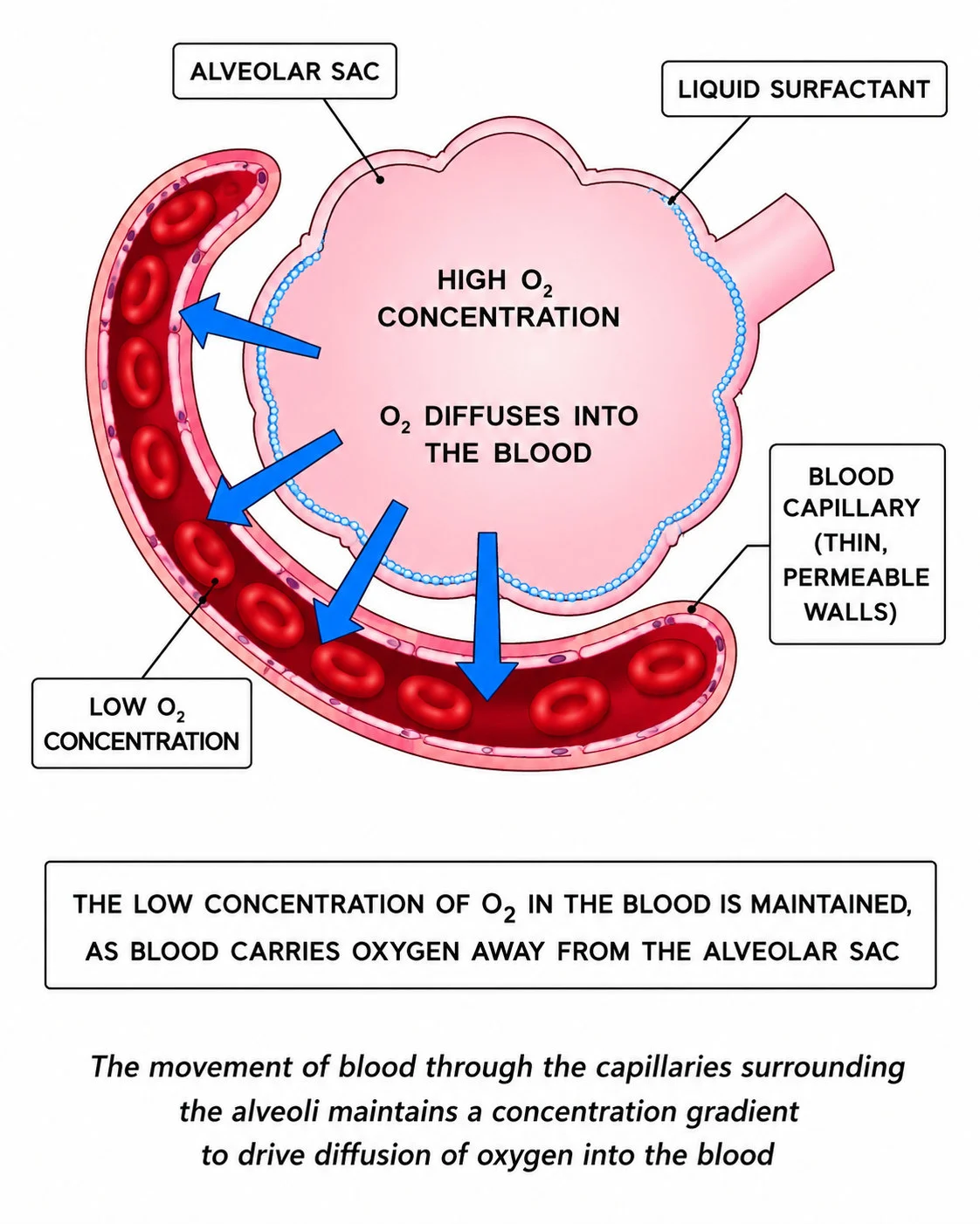
Each lung has millions of alveoli, giving a huge surface area (about 70 m2 in total). Oxygen diffuses from the air in the alveolus, across the thin wall, into the blood in the surrounding capillaries; carbon dioxide diffuses from the blood into the alveolus and is breathed out. The alveoli are adapted in several ways:

Large surface area
a large surface area due to the vast number of alveoli;
Thin wall
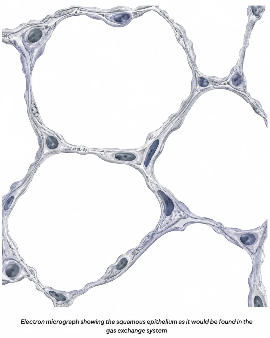
a thin wall, one cell thick, giving a short diffusion distance;
a moist lining so that gases dissolve;
Capillaries
a dense network of capillaries with walls one cell thick, which brings a rich blood supply;
Ventilation
constant ventilation and blood flow keeping a steep concentration gradient: the alveolar air has more oxygen and less carbon dioxide than the blood arriving.
GasMoves fromMoves to
OxygenAlveolar airBlood
Carbon dioxideBloodAlveolar air

Section 3 of 9Ventilation: how breathing works

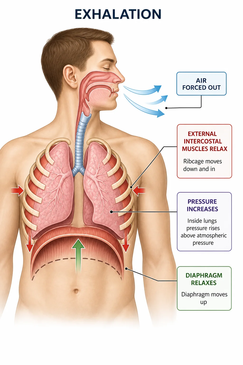
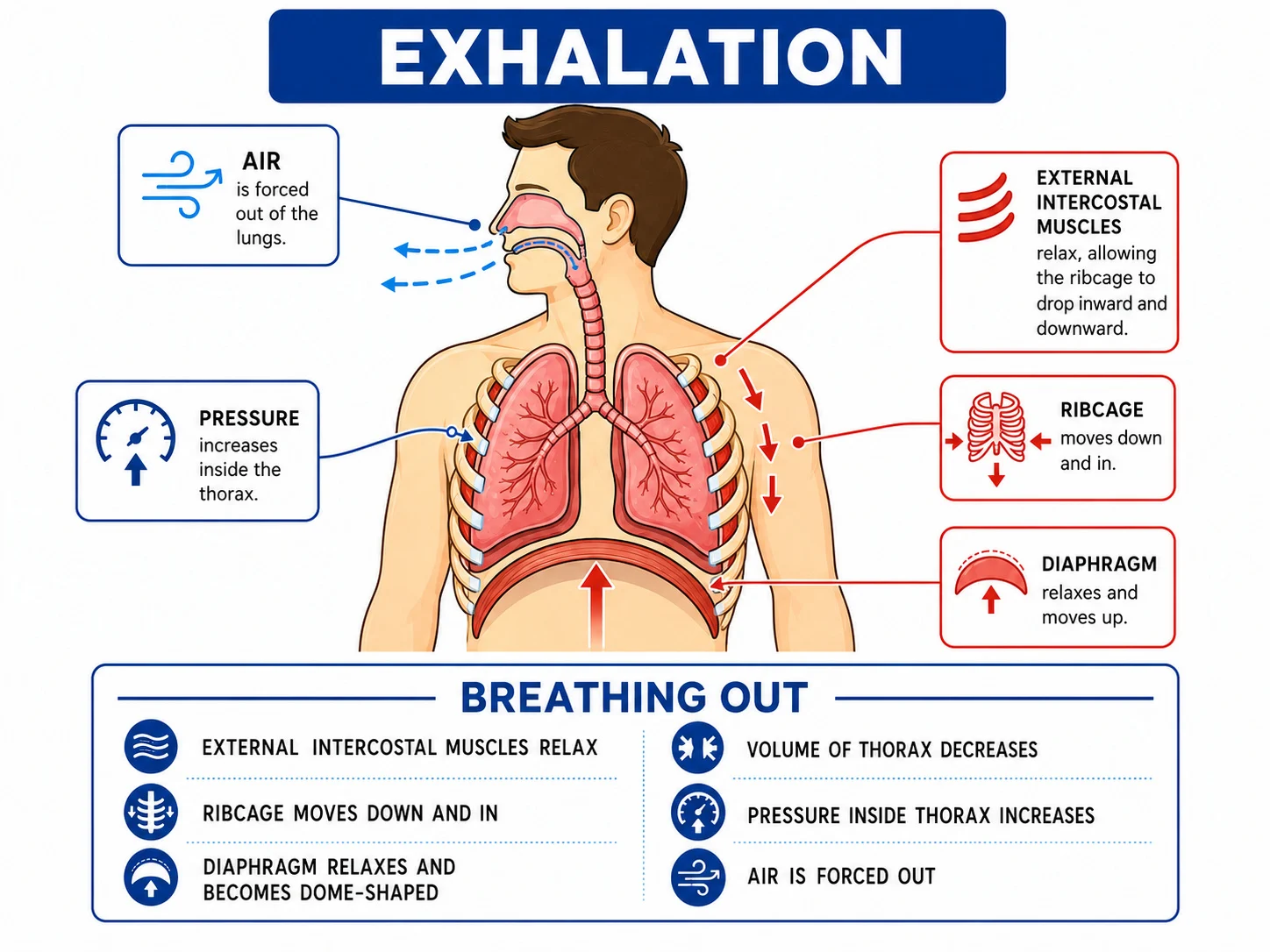
Exhalation: air moves out of the lungs
The Human Thorax
The Human Thorax
Exhalation mechanism in the thorax
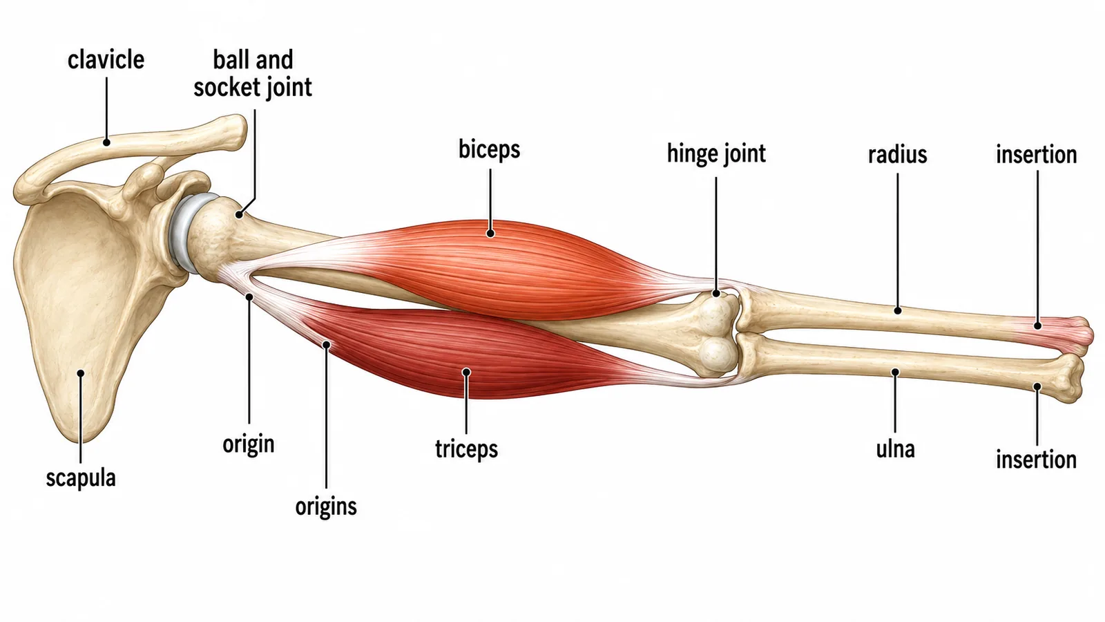
muscle movement
Inhalation vs Exhalation
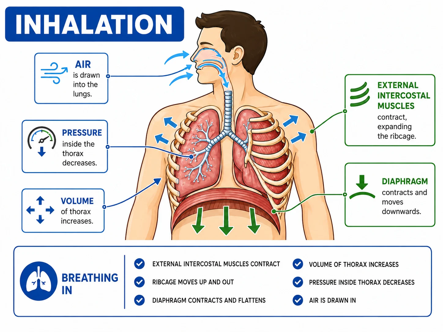
Inhalation: muscle action and pressure
Exhalation: diaphragm relaxation, rib movement and pressure changes
Inhalation: diaphragm contraction and increase in thoracic volume

Breathing is the movement of air in and out of the lungs. It depends on changes in the volume and pressure of the thorax.

Inhalation (breathing in)Exhalation (breathing out)
Intercostal musclesContract, ribs move up and outRelax, ribs move down and in
DiaphragmContracts and flattensRelaxes and curves upward
Volume of thoraxIncreasesDecreases
Pressure in lungsFalls below atmosphericRises above atmospheric
Air movementAir flows inAir flows out

Air always moves from a region of higher pressure to lower pressure, so when the pressure inside the thorax is lower than outside, air flows in. Breathing out at rest is mostly passive, but during exercise the internal intercostal and abdominal muscles can contract to force air out more quickly.

Section 4 of 9Lung volumes and measurements

The tidal volume is the volume of air in one normal breath (about 500 cm3 at rest).

Breathing rate
The breathing rate is the number of breaths per minute (about 12 to 20 at rest).
Ventilation rate
The ventilation rate or minute volume is tidal volume multiplied by breathing rate.
For example, 500 cm3 x 15 breaths per minute = 7500 cm3 per minute.
Vital capacity
The vital capacity is the largest volume that can be exhaled after a deep breath.
During exercise, both breathing rate and tidal volume increase to supply more oxygen.

Section 5 of 9Health and disease

Lung diseases reduce the efficiency of gas exchange.

1
In asthma the bronchioles narrow, which restricts airflow, and inhalers relax the muscles.
2
In emphysema, alveolar walls break down, reducing surface area, so the person is breathless.
3
Lung cancer and bronchitis are linked to smoking: tar damages the cilia, so mucus and microbes accumulate, and carcinogens cause mutation of cells.
4
Fibrosis scars the lungs, thickening the walls so diffusion is slower.

The gas exchange system links to transport in cells (diffusion across the alveolar wall), the circulatory system (blood carries the gases) and respiration (cells use the oxygen to release energy).

Section 6 of 9Interpreting data and the effect of exercise

Experiment: plant without carbon dioxide (soda lime) under bell jar

Questions often present a spirometer trace, which is a graph of lung volume against time.

Interpreting data and the effect of exercise
Each rise and fall is one breath; the height from trough to peak is the tidal volume, and the number of peaks in a minute is the breathing rate.
A trace during exercise shows peaks that are taller and closer together, because both tidal volume and rate have increased.
To calculate breathing rate from a trace, count the peaks in a given time and scale to a minute: 6 peaks in 20 seconds is 18 breaths per minute.

Why does this happen? Muscles respire faster during exercise, producing more carbon dioxide. The rise in carbon dioxide in the blood is detected by the brain, which increases the rate and depth of breathing to remove it and to supply more oxygen. This keeps the concentration gradients at the alveoli steep and so speeds gas exchange.

A model of the lungs using a bell jar, a balloon and a rubber sheet is commonly used to explain pressure changes.

Interpreting data and the effect of exercise
Pulling the rubber sheet down increases the volume inside the jar, which lowers the pressure and makes the balloons inflate.
Its limitations are that the model has no ribs, that it shows only one pair of balloons and does not show the alveoli or blood supply,
  • which you should mention if asked to evaluate it.

Section 7 of 9Exam-style questions

Parts of the breathing system and functions
  1. Describe how air moves into the lungs during inhalation. [3]3 marks

  2. Explain three ways in which alveoli are adapted for efficient gas exchange. [3]3 marks

  3. A person takes 14 breaths per minute with a tidal volume of 450 cm3. Calculate the volume of air breathed in one minute. [2]2 marks

Section 8 of 9Key terms

TermMeaning
AlveolusTiny air sac in the lung where gas exchange occurs.
TracheaWindpipe, a tube that carries air to the bronchi.
BronchusOne of the two tubes that carry air into the lungs.
DiaphragmSheet of muscle below the lungs that helps breathing.
Intercostal musclesMuscles between the ribs.
VentilationMovement of air in and out of the lungs.
Tidal volumeVolume of air in a normal breath.
CiliaTiny hairs that sweep mucus.

Section 9 of 9Exam technique

1
Saying the diaphragm relaxes and rises during inhalation; it contracts and flattens.
2
Saying lungs 'fill with air' because they suck it; say pressure falls so air flows in.
3
Confusing bronchi and bronchioles, or trachea with oesophagus.
4
Writing that oxygen diffuses from the blood into the air; it moves from the air into the blood.
\n