Section 1 of 9Why we use microscopes
Most cells and their parts are too small to see with the naked eye.
Section 2 of 9Light microscopes

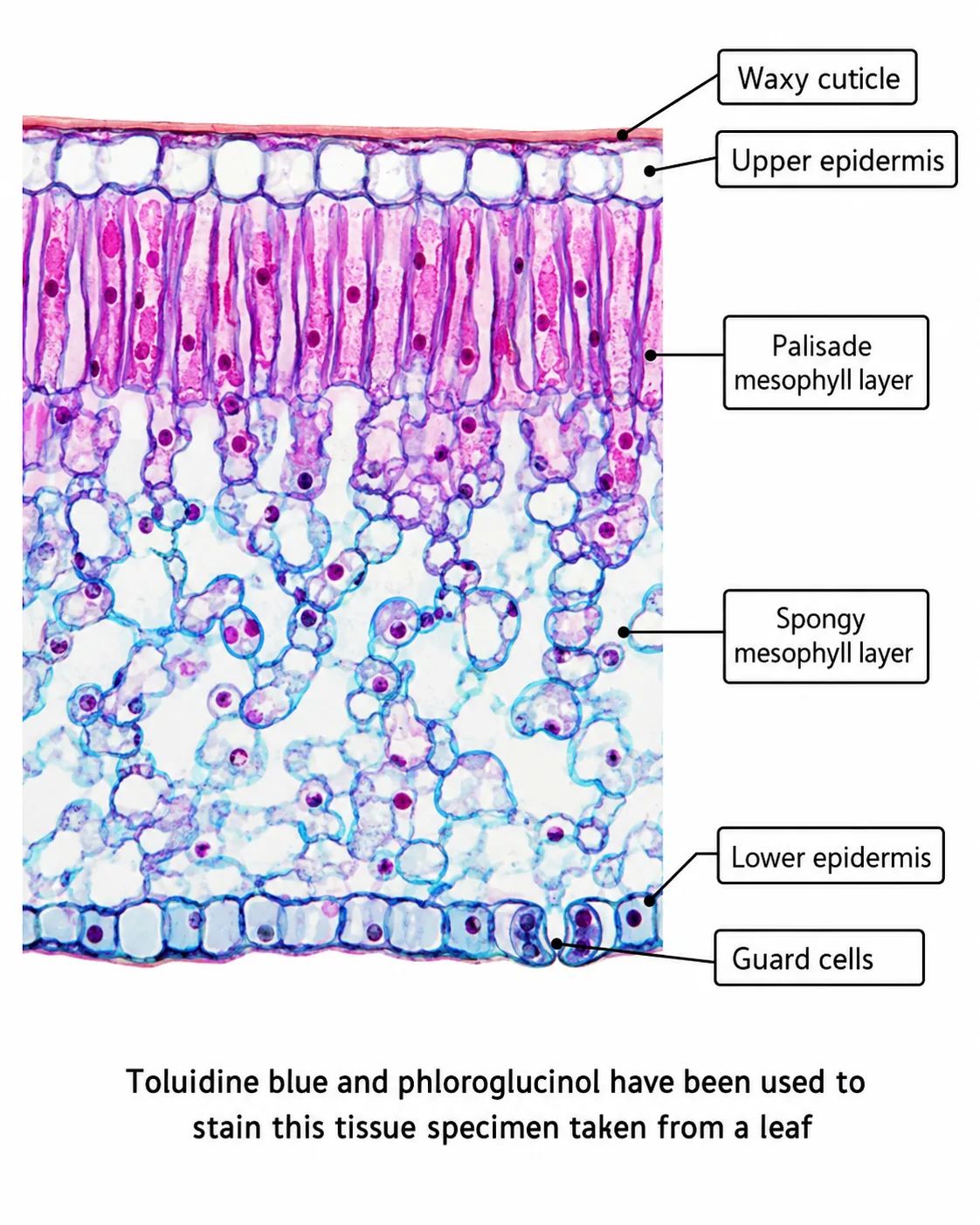
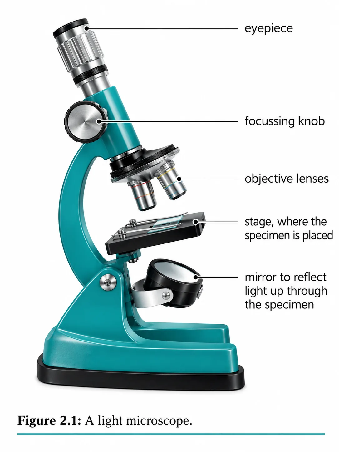
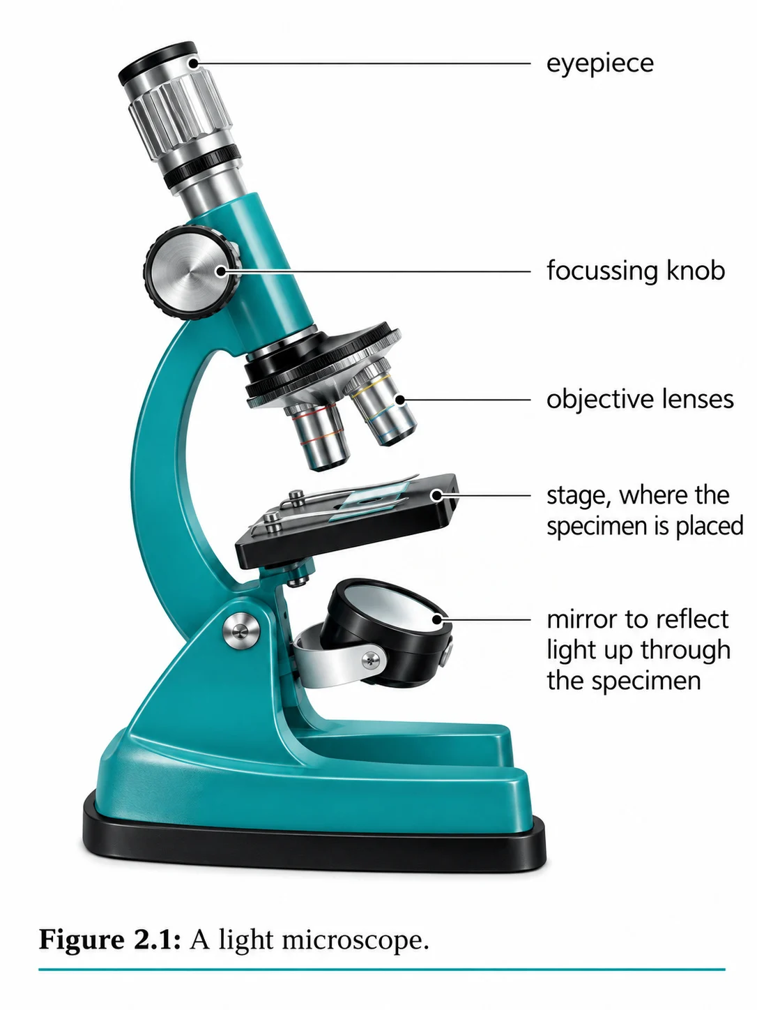
The light microscope shines a beam of light through a thin specimen, and lenses produce an enlarged image.
- and can be used to view living specimens, often with coloured stains to make structures stand out.
Section 3 of 9Electron microscopes
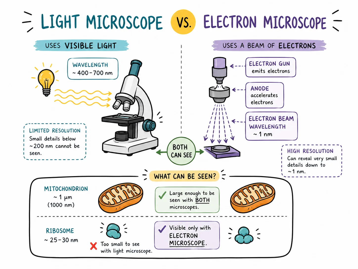

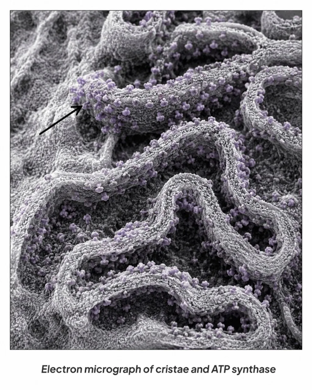

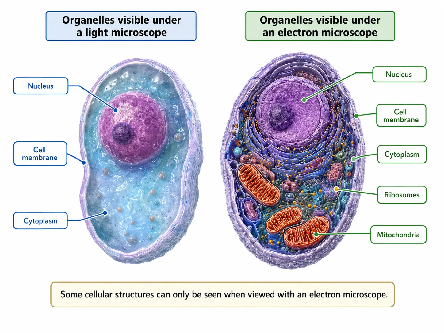


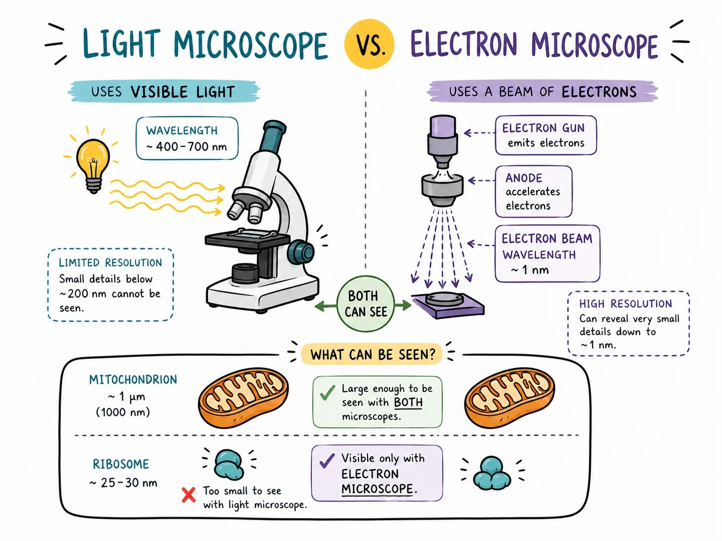
An electron microscope uses a beam of electrons instead of light.
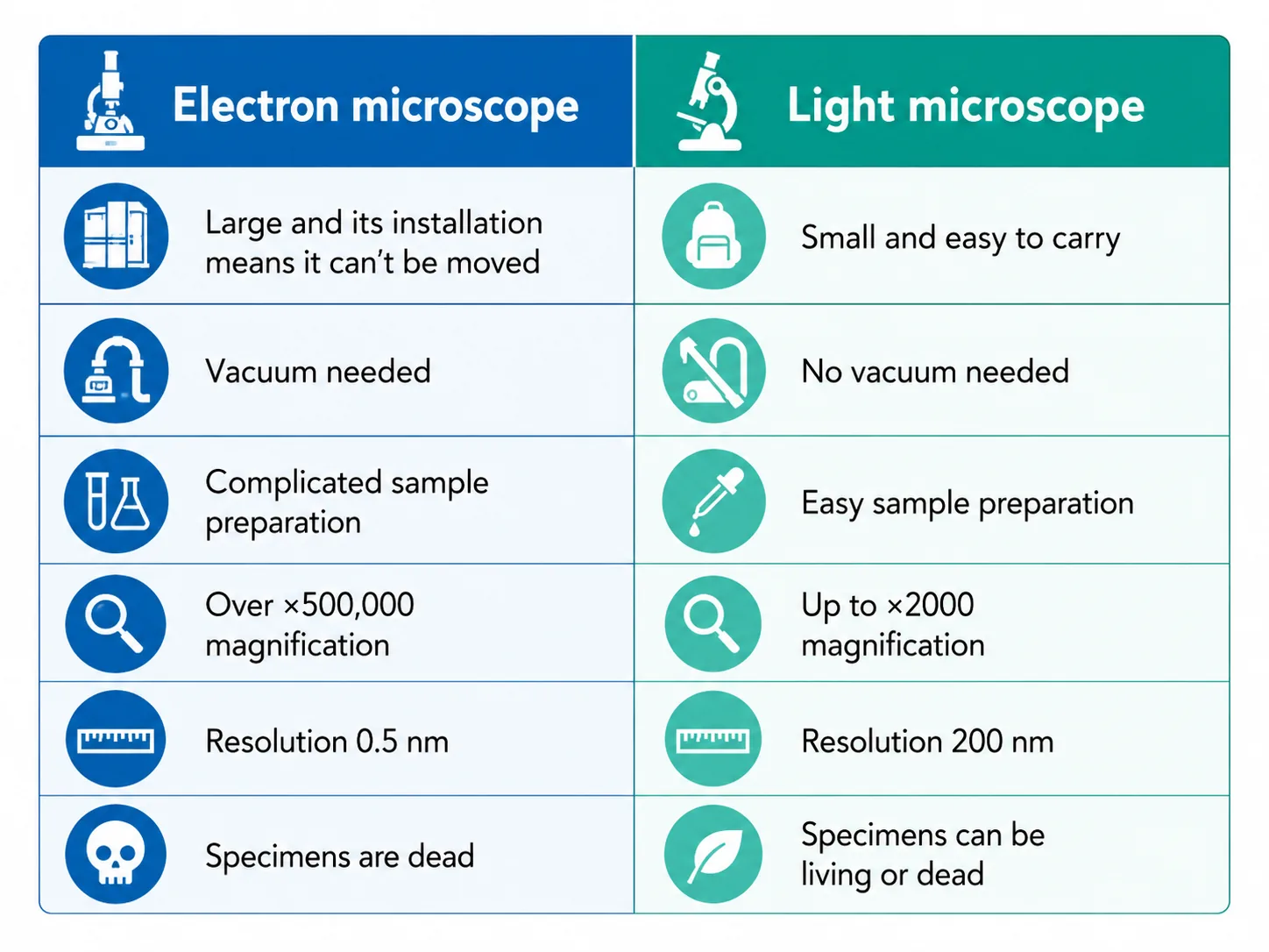
- so the microscope has much higher magnification and much higher resolution (about 0.2 nanometres).
- (over x1 000 000 for some)
- such as the internal structure of mitochondria and chloroplasts, ribosomes and plasmids.
| Feature | Light microscope | Electron microscope |
|---|---|---|
| Radiation used | Light | Electrons |
| Maximum magnification | About x1500 | Over x1 000 000 |
| Resolution | About 200 nm | About 0.2 nm |
| Living specimens | Yes | No |
| Cost and size | Cheap, small | Expensive, large |
Section 4 of 9Magnification calculations

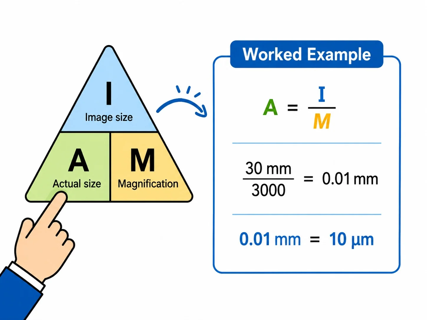
The key equation is:
magnification = image size / real size
Rearrange it to find image size (= magnification x real size) or real size (= image size / magnification).
For a light microscope the total magnification is the eyepiece lens magnification multiplied by the objective lens magnification, so a x10 eyepiece with a x40 objective gives x400.
Section 5 of 9Using the microscope well
Always start with the lowest-power objective lens to find the specimen, then increase power and use only the fine focus. Turn the lens away from the slide when focusing. Draw what you see and record the magnification so that others can work out real sizes. The required practical (next lesson) puts these skills into practice.
Section 6 of 9Units, standard form and the order of magnitude


Questions often give sizes in different units and expect you to convert them. Remember the ladder: millimetre (mm) to micrometre (um) is x1000, and micrometre to nanometre (nm) is another x1000. Expressed in metres: 1 mm = 1 x 10⁻³ m, 1 um = 1 x 10⁻⁶ m and 1 nm = 1 x 10⁻⁹ m.
You may also be asked how many times bigger one thing is than another, an order of magnitude comparison.
A useful way to check any magnification answer is to ask whether it is sensible. A drawing of a cell on an A4 page showing a cell about 100 um wide drawn 10 cm across gives x1000, which is a plausible light-microscope magnification. If you calculate x1 000 000 for a light microscope drawing, you have made a unit error.
Magnification can also be given as a scale bar. Measure the bar with a ruler, and divide by the length it represents to find the magnification, and then measure any structure on the image and divide by that magnification to find its true size.
Section 7 of 9Exam-style questions
A cell has a real width of 40 um. In a photograph the width is 8 cm. Calculate the magnification. [3]3 marks
8 cm = 80 000 um [1]. Magnification = 80 000 / 40 [1] = x2000 [1].
State two advantages of an electron microscope over a light microscope. [2]2 marks
Higher magnification [1]; higher resolution so more detail of sub-cellular structures can be seen [1].
A mitochondrion is drawn 30 mm long at a magnification of x15 000. Calculate its real length in micrometres. [3]3 marks
30 mm = 30 000 um [1]. Real size = image / magnification = 30 000 / 15 000 [1] = 2 um [1].
Section 8 of 9Key terms
| Term | Meaning |
|---|---|
| Magnification | How many times larger an image is than the real object. |
| Resolution | The ability to see two close points as separate. |
| Light microscope | Microscope using light, magnifying up to about x1500. |
| Electron microscope | Microscope using electron beams for very high magnification and resolution. |
| Micrometre (um) | One millionth of a metre. |
| Nanometre (nm) | One billionth of a metre. |
| Sub-cellular structure | A structure within a cell, such as a ribosome. |