8.4.1Exchange and Transport in Animals
The Blood and Blood Vessels
5 min read 9 parts Lesson 6 of 7 1 quiz
Section 1 of 9Components of blood



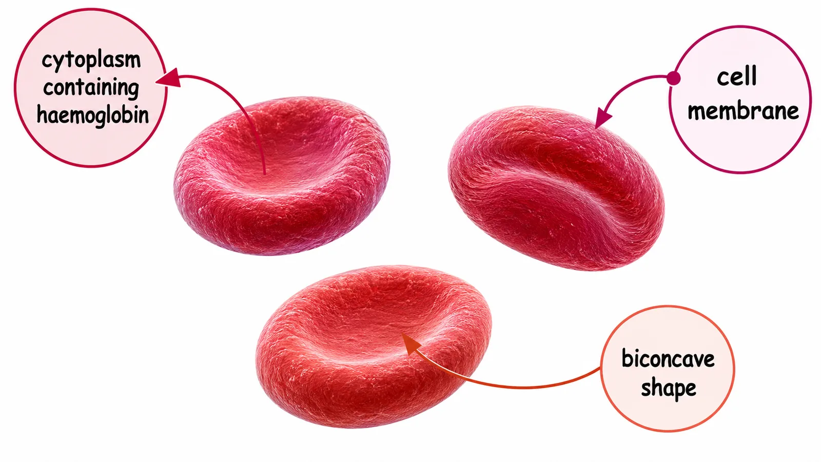

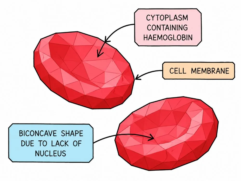
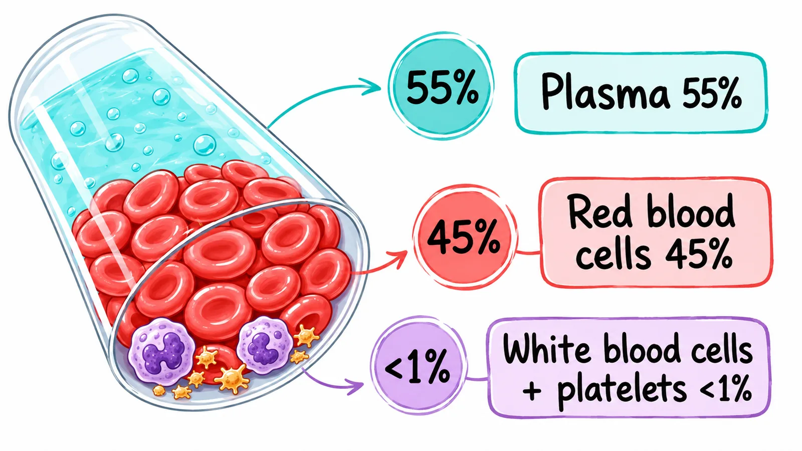
Blood is a tissue made of cells suspended in a liquid, and it transports substances around the body. It has four main components.
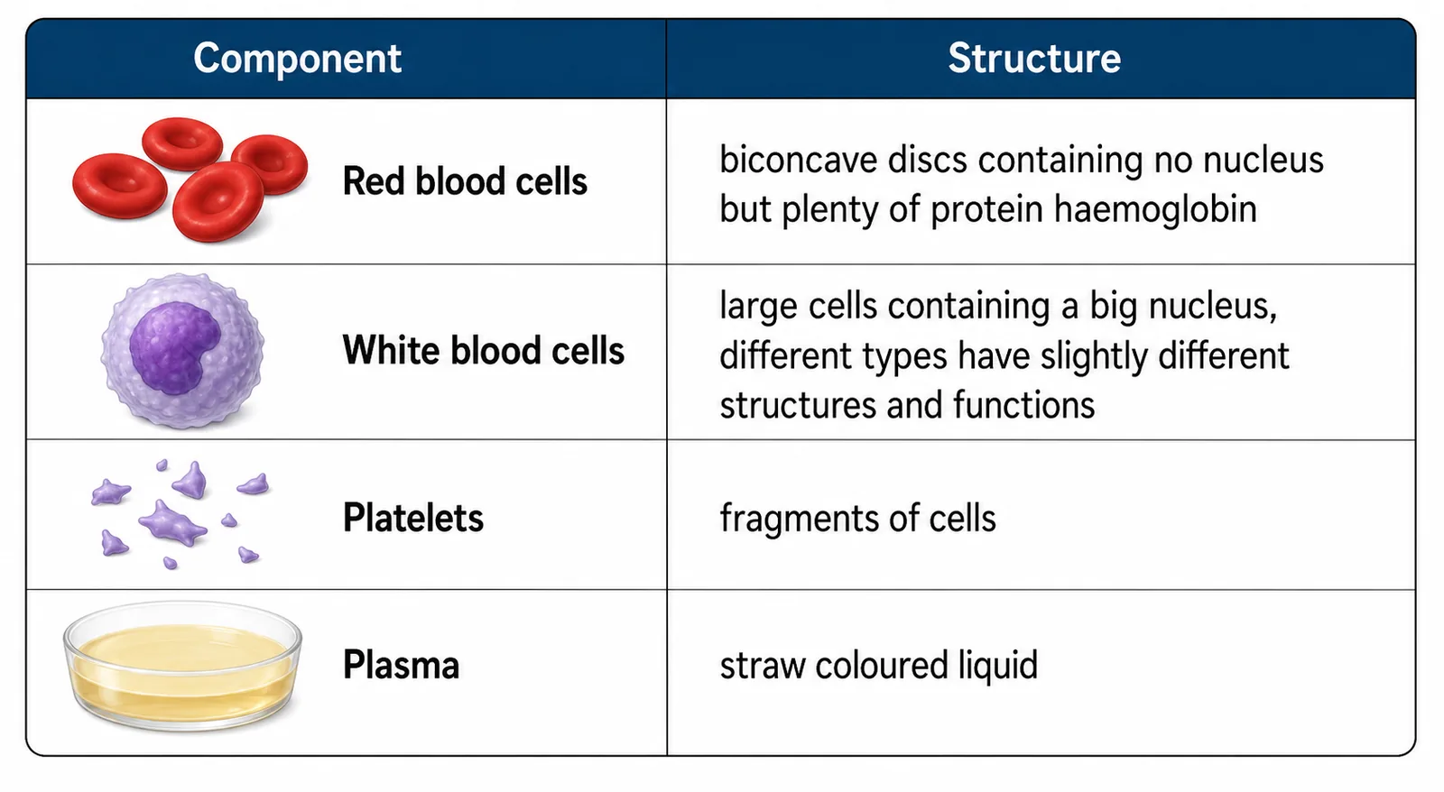
| Component | Description | Function |
|---|---|---|
| Plasma | Pale yellow liquid, about 55% of blood | Transports carbon dioxide, glucose, amino acids, urea, hormones and heat |
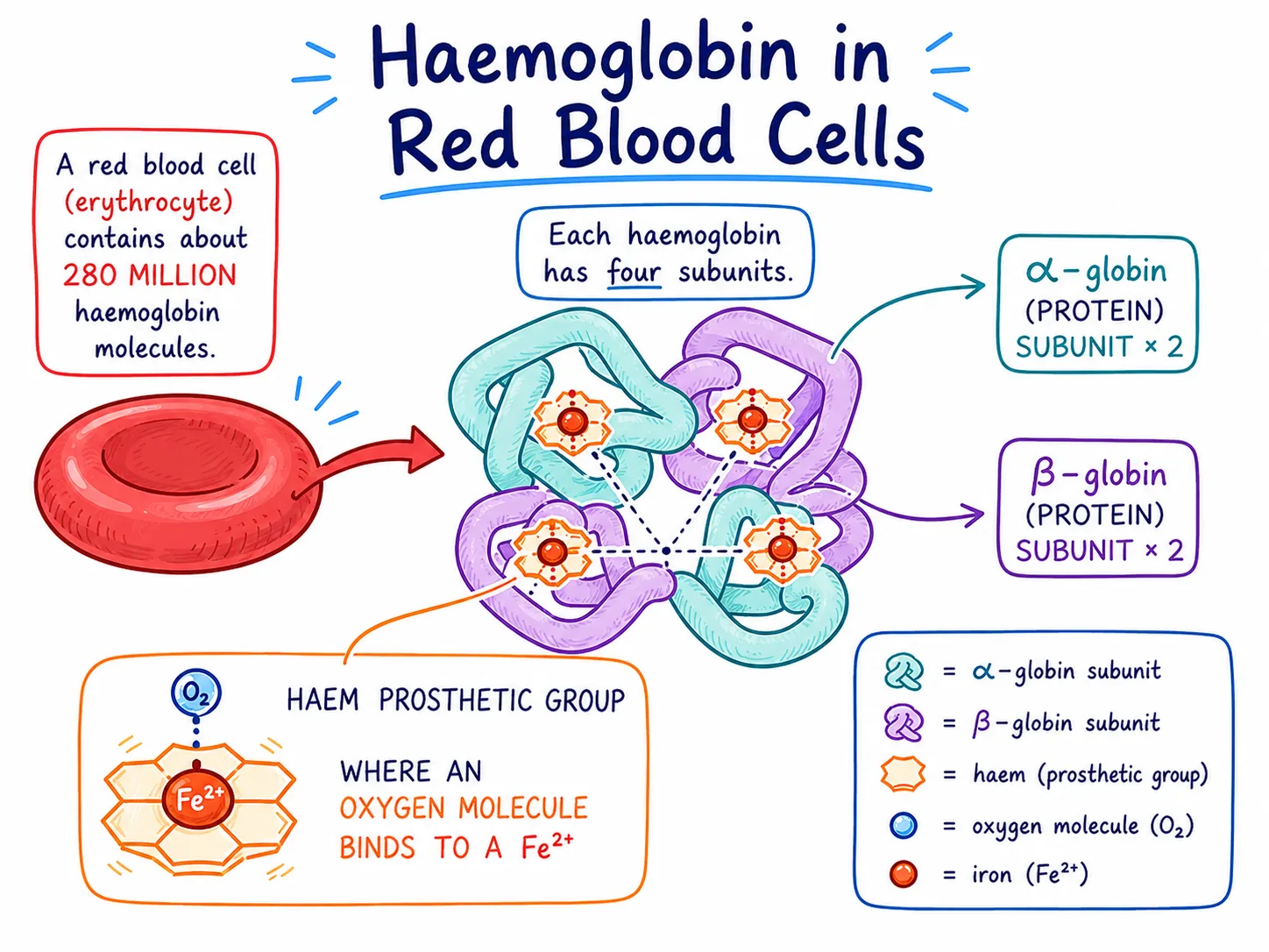
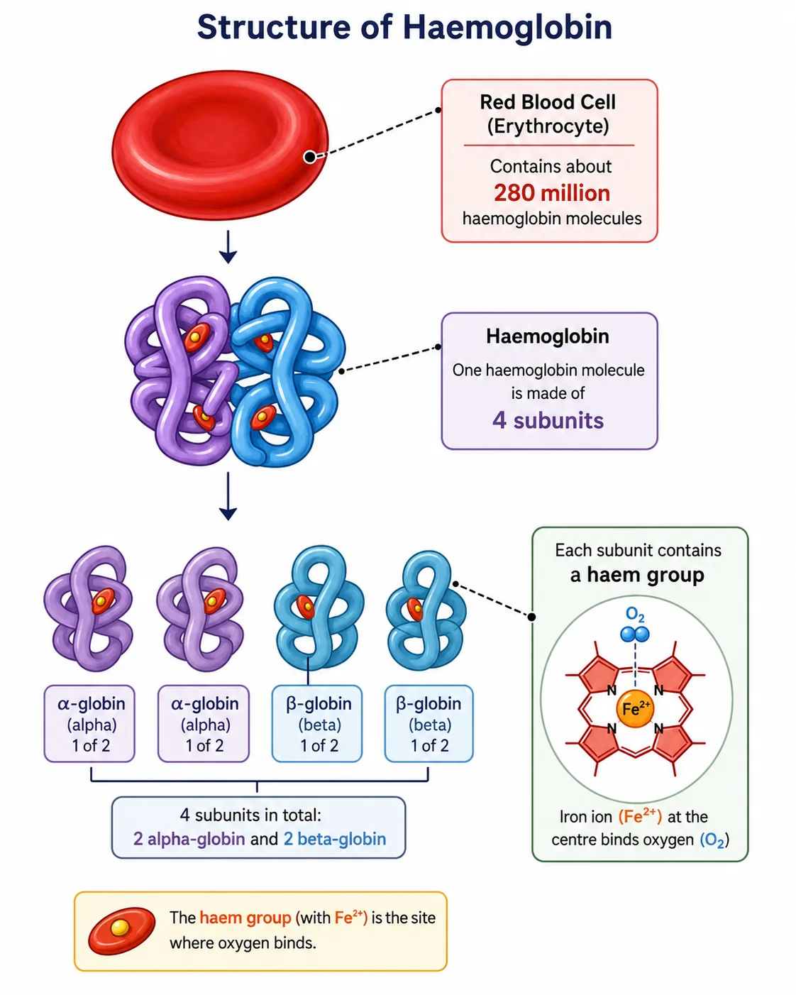
| Red blood cells | Biconcave discs, no nucleus, contain haemoglobin | Carry oxygen as oxyhaemoglobin |
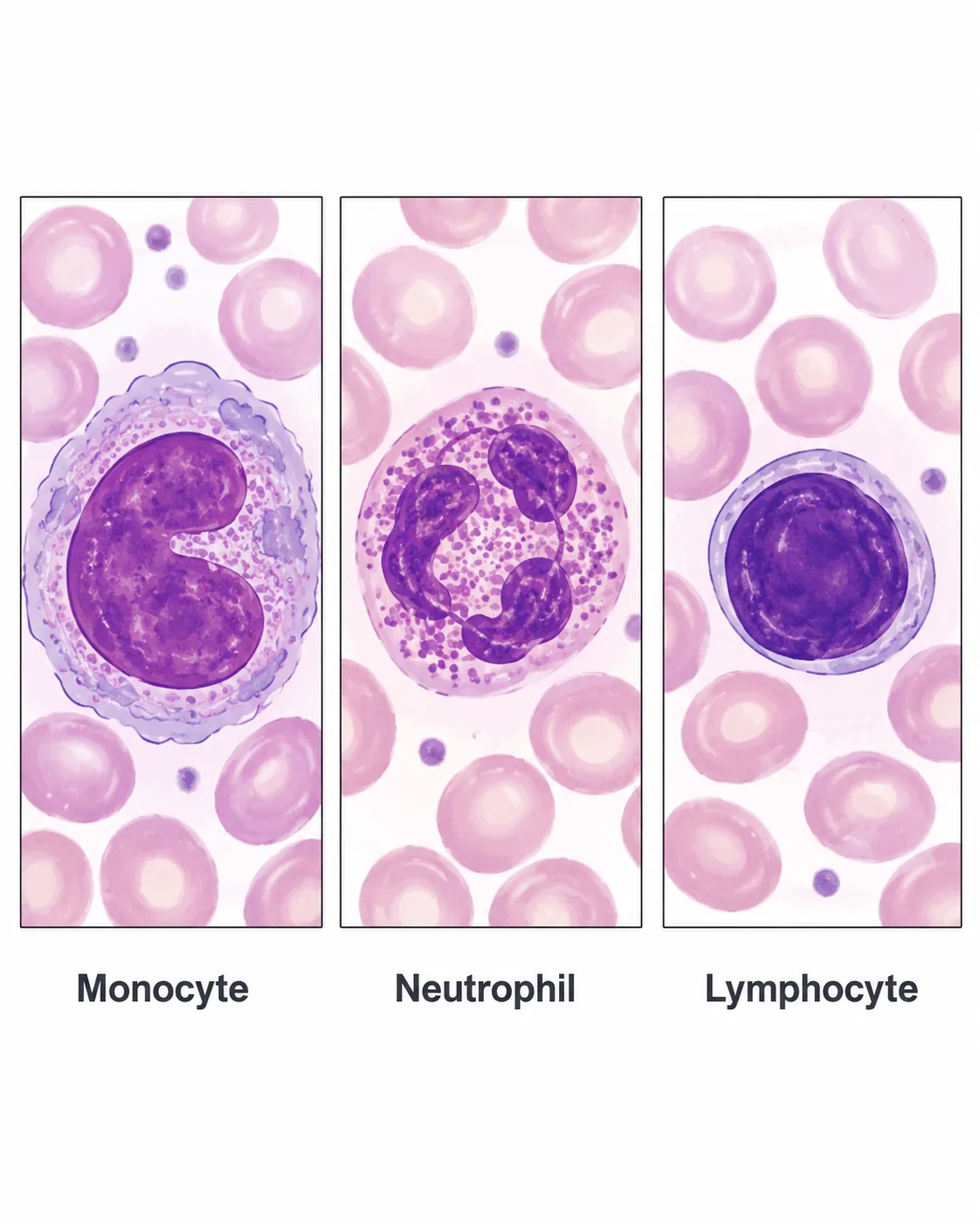
| White blood cells | Have a nucleus; phagocytes and lymphocytes | Defence against pathogens: engulf them or make antibodies |
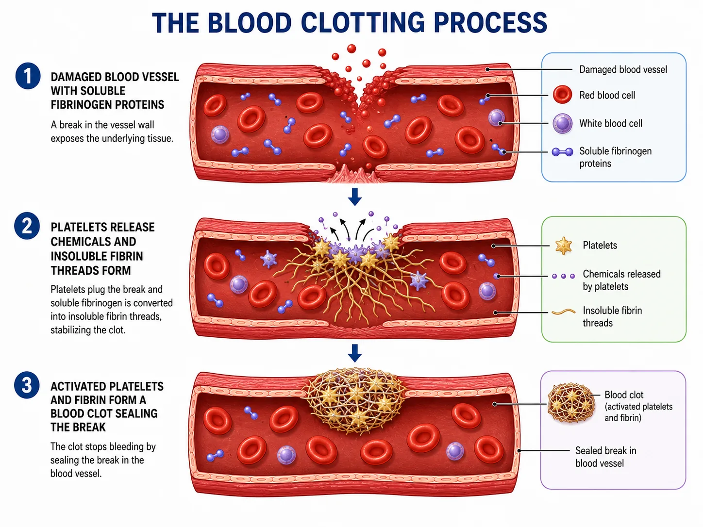
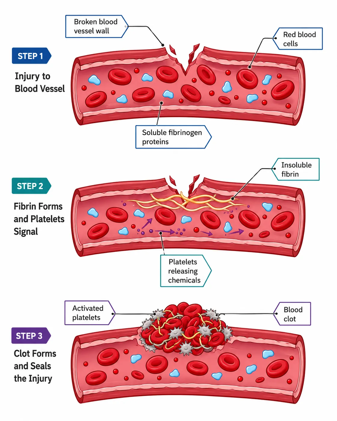
| Platelets | Cell fragments | Help blood clot at wounds |
Red blood cells are adapted for oxygen transport: the biconcave shape gives a large surface area to volume ratio, the lack of a nucleus gives more room for haemoglobin, and they are flexible to squeeze through capillaries. Haemoglobin combines with oxygen in the lungs (high oxygen concentration) and releases it in the tissues (low oxygen concentration).
Section 2 of 9Arteries and veins
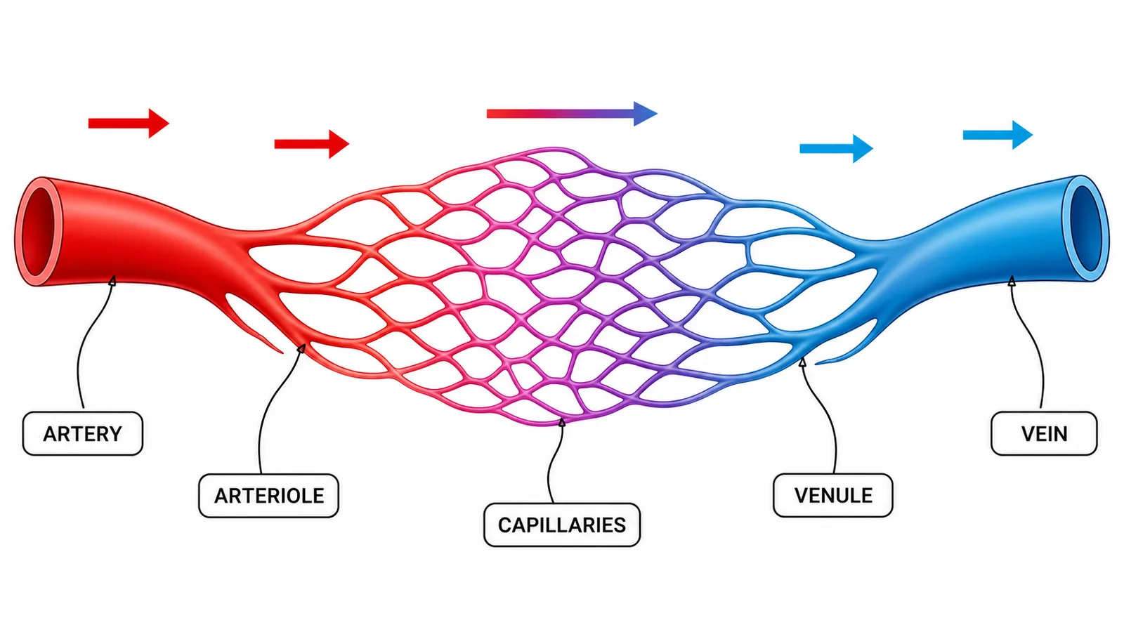
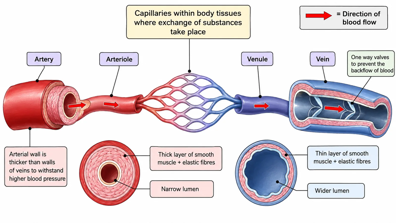

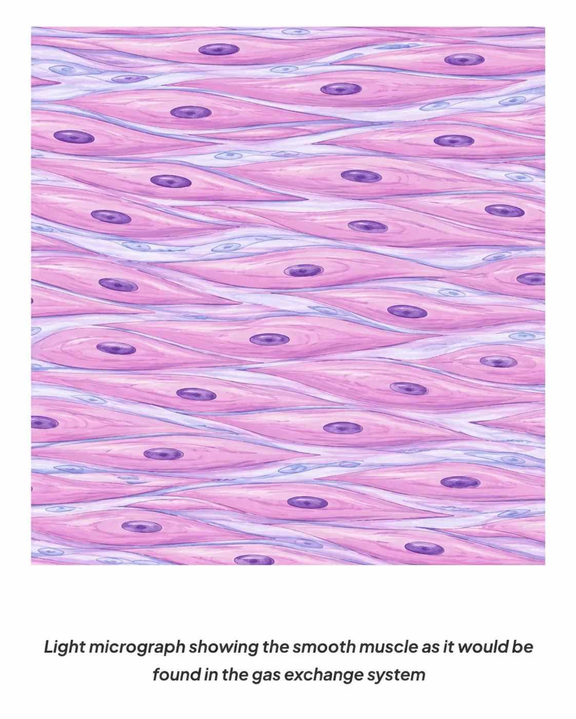
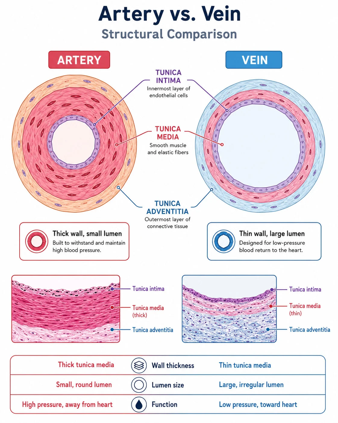
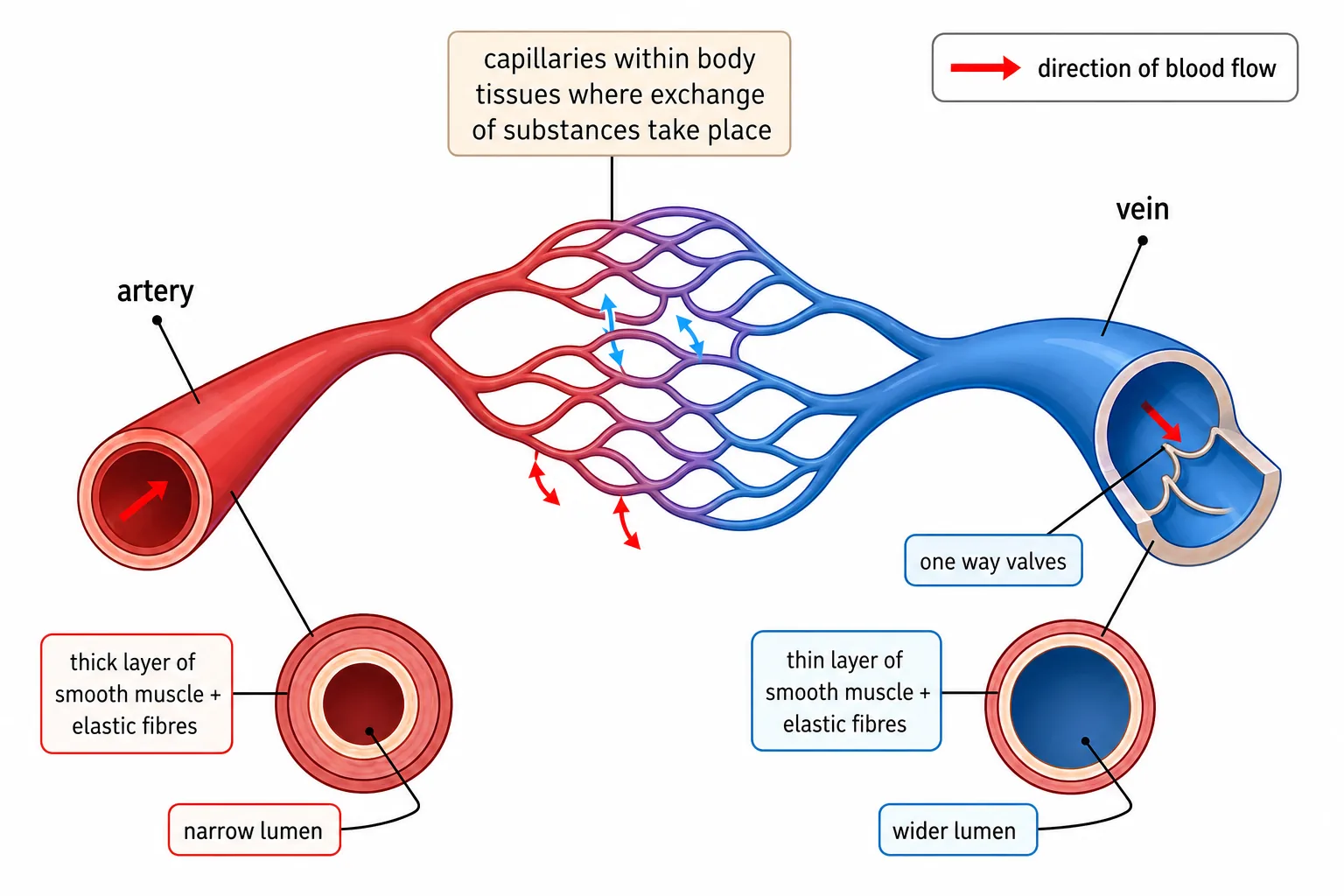
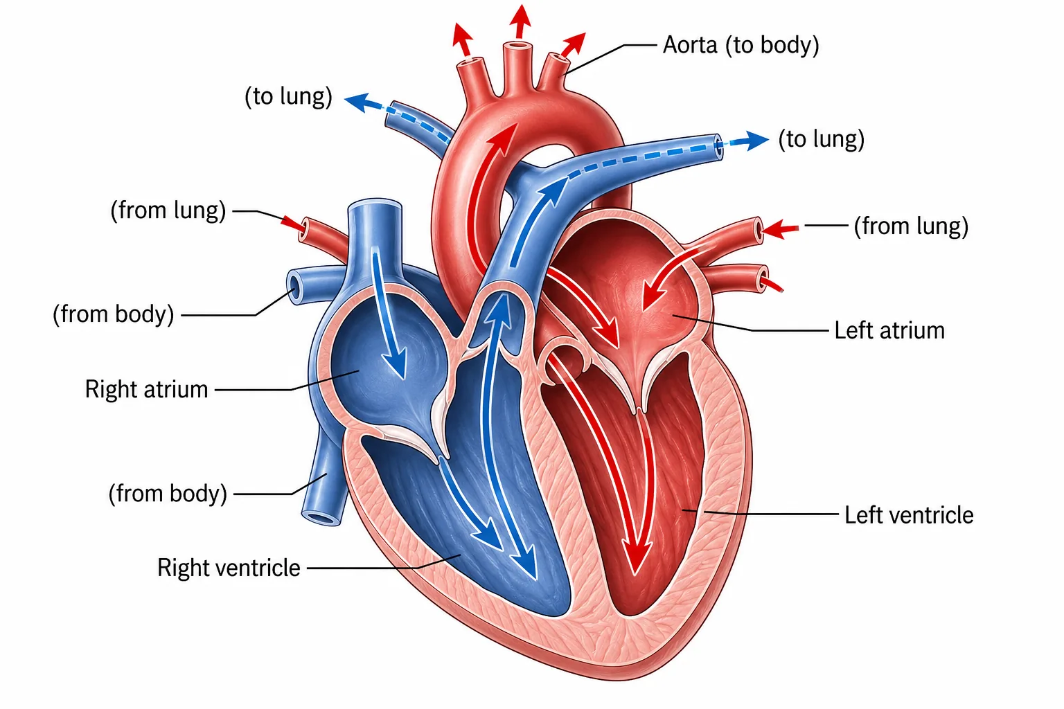
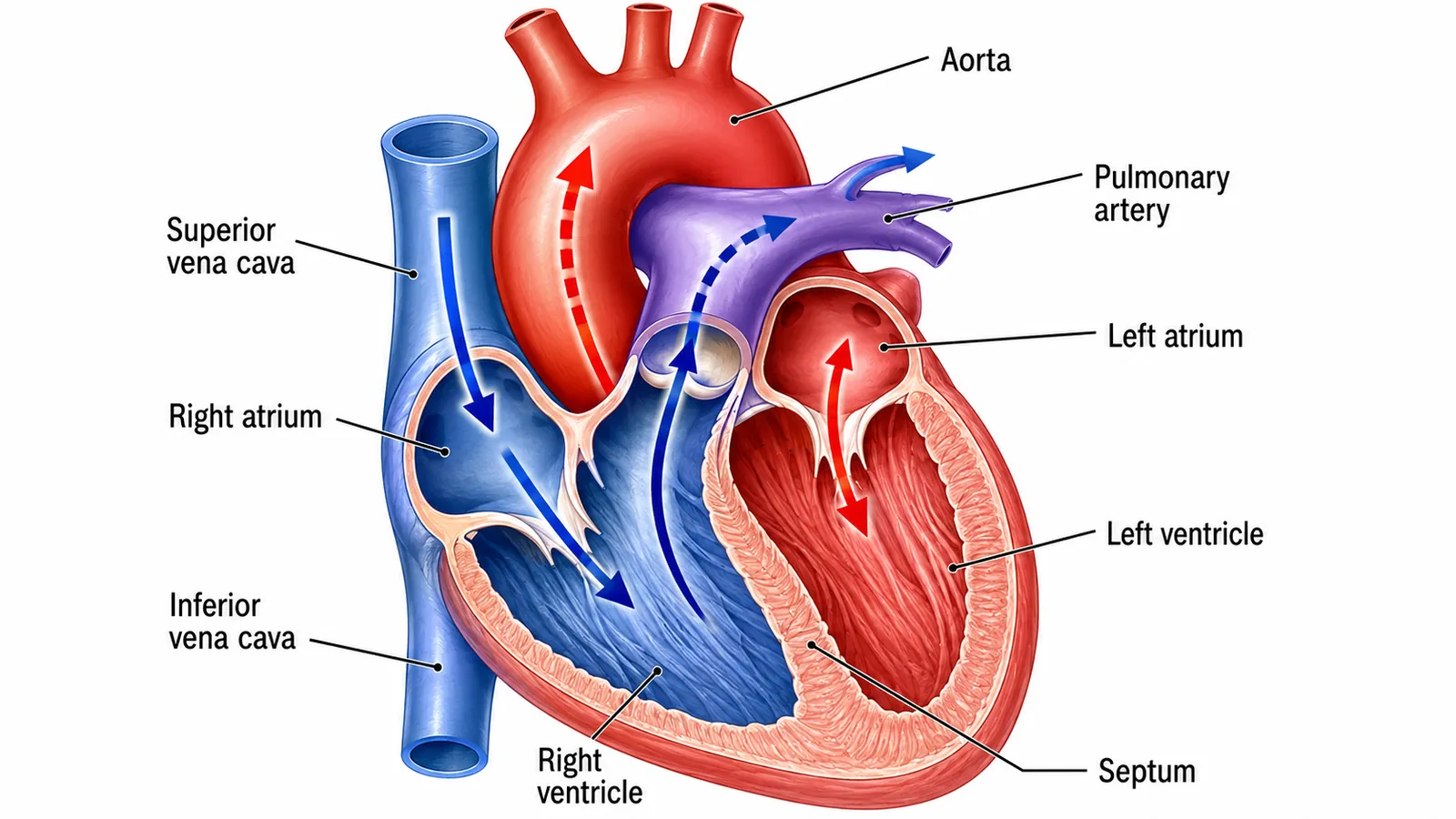
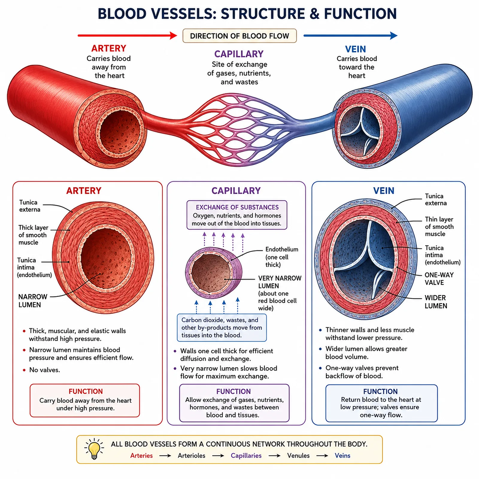
Arteries carry blood away from the heart, usually oxygenated (except the pulmonary artery).
Veins carry blood towards the heart, usually deoxygenated (except the pulmonary vein). The pressure is low, so the walls are thinner with less muscle and elastic tissue, and the lumen is wide. Valves prevent backflow of blood, which is needed because the pressure is low. Muscles squeezing the veins as we move help return the blood to the heart.
Section 3 of 9Capillaries

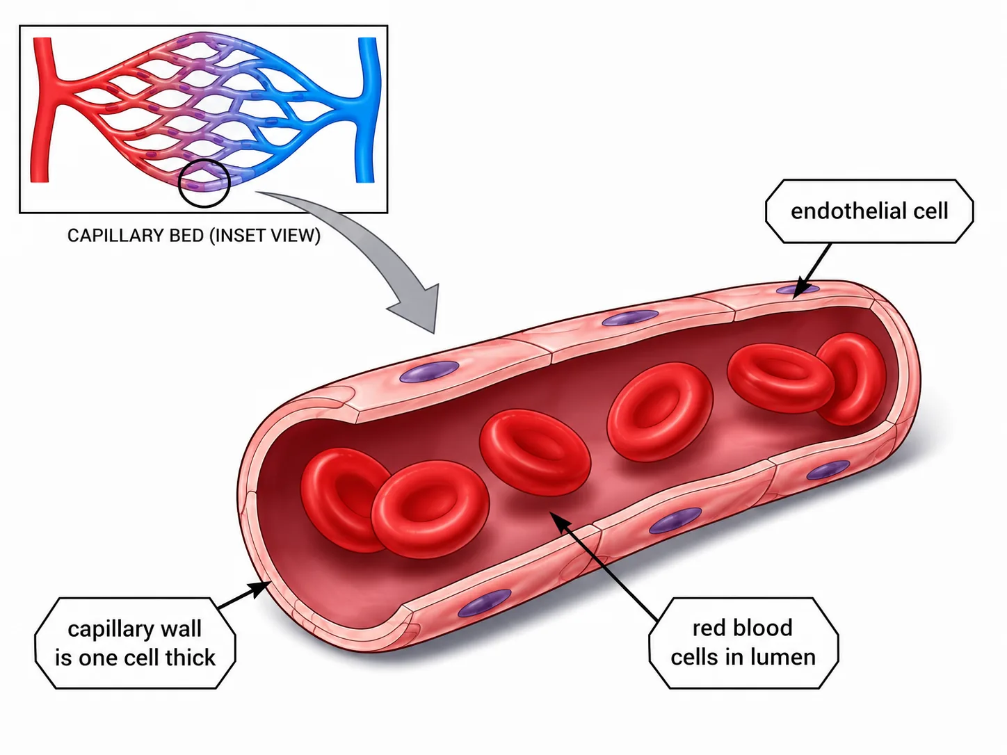
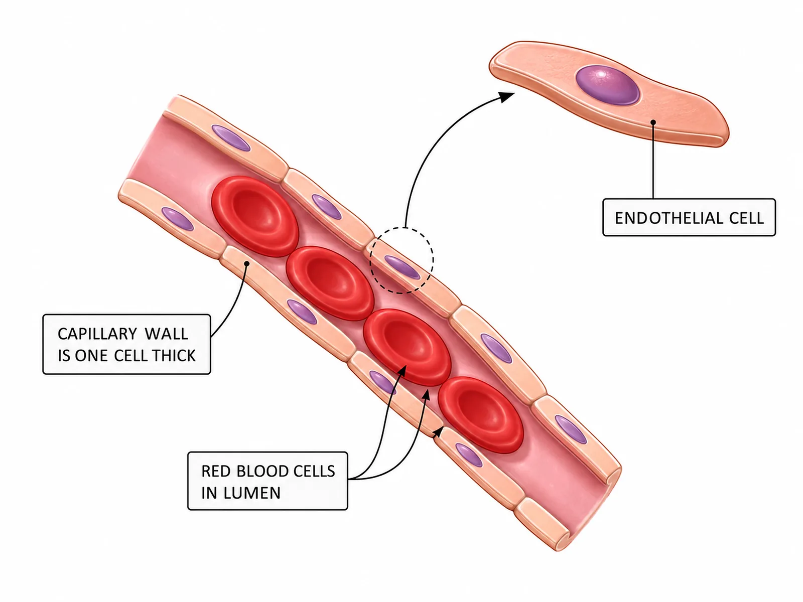
Capillaries are tiny vessels that link arteries and veins and run through all tissues.
- giving a very short diffusion distance, and they are permeable,
- allowing exchange of substances with the tissue fluid.
- giving more time for exchange of oxygen, glucose, carbon dioxide and urea with cells.
| Feature | Artery | Vein | Capillary |
|---|---|---|---|
| Direction | Away from heart | To heart | Between arteries and veins |
| Wall | Thick, muscle and elastic | Thin | One cell thick |
| Lumen | Narrow | Wide | Very narrow |
| Valves | No | Yes | No |
| Blood pressure | High | Low | Falling |
Section 4 of 9Blood flow and exchange with tissues


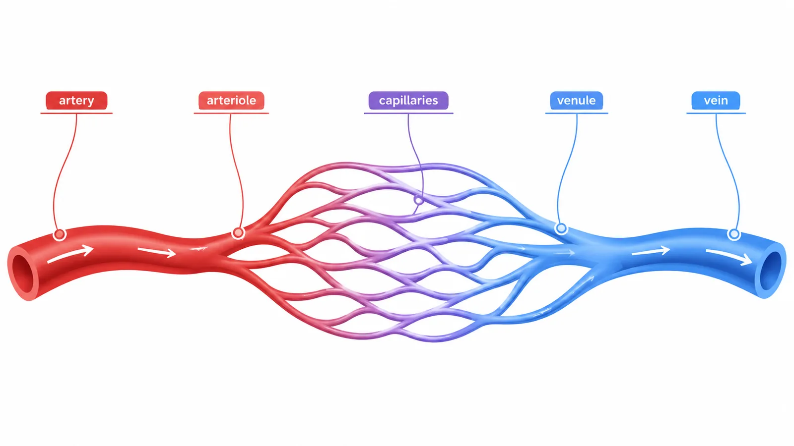
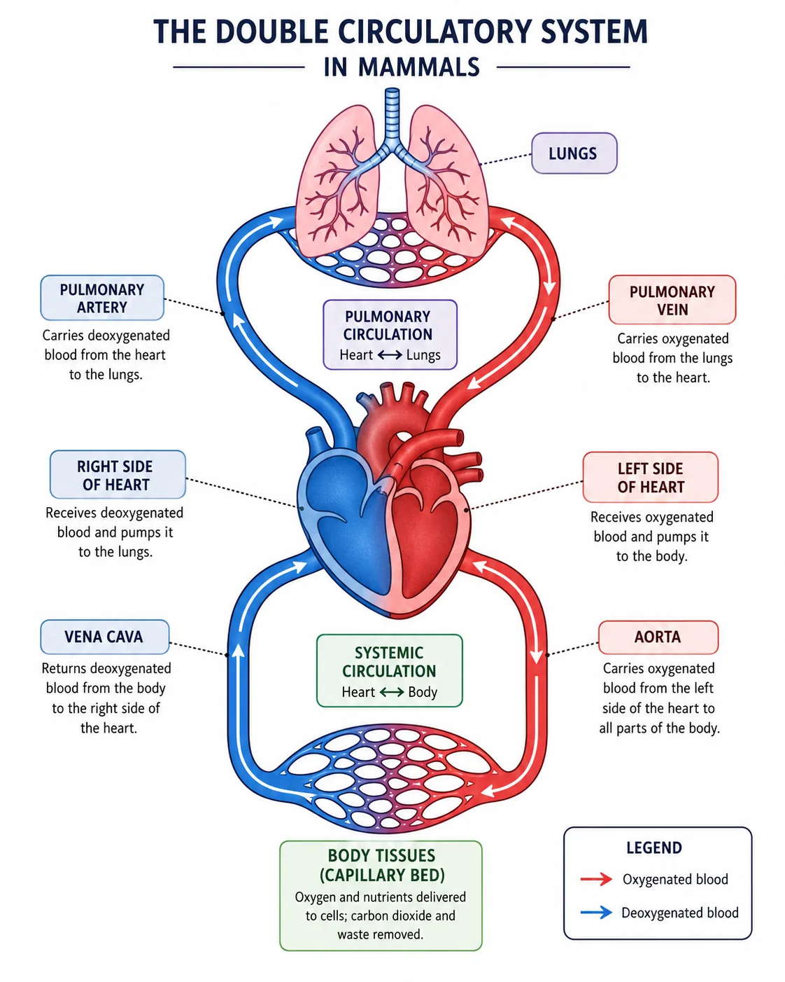
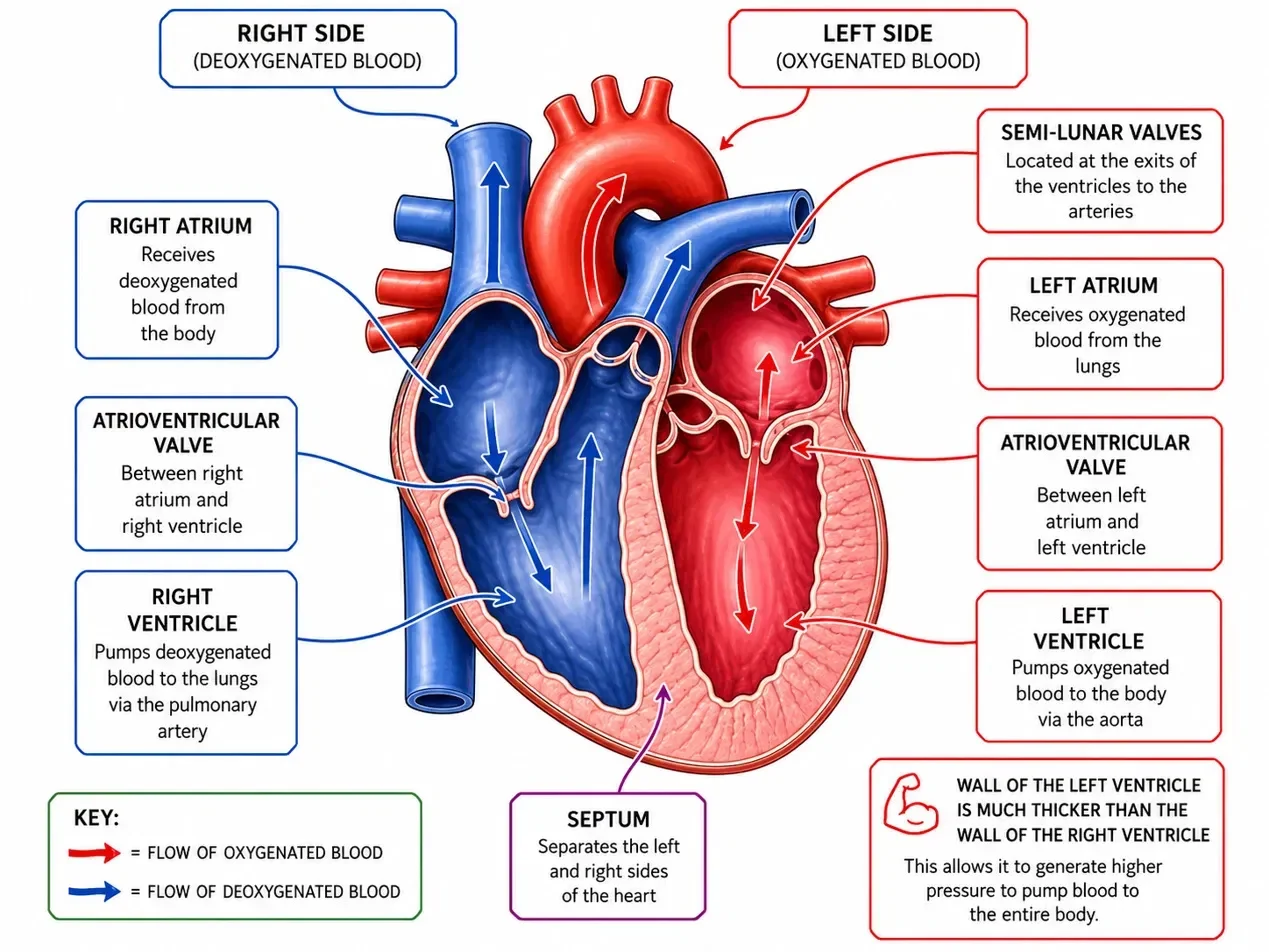
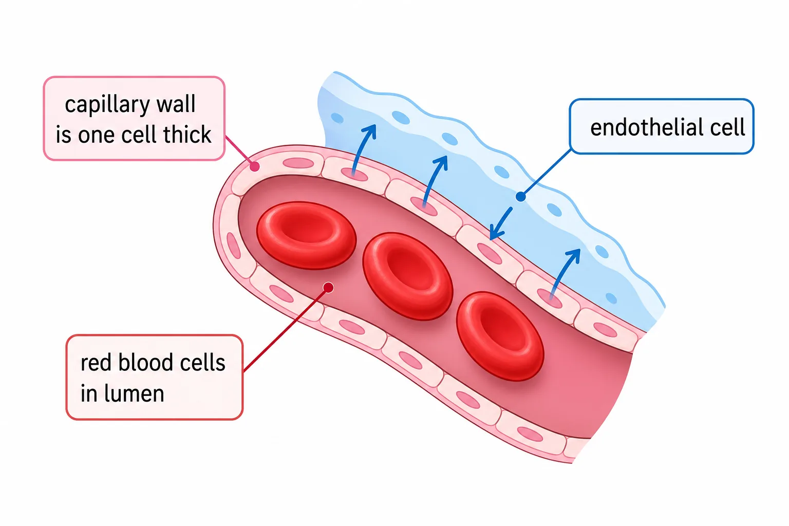
As blood flows through the capillaries, plasma leaks out through the thin walls to form tissue fluid, which bathes the cells.
- and carbon dioxide and urea diffuse from the cells into the tissue fluid and then into the blood.
Section 5 of 9Blood in health and disease


Anaemia is a condition in which blood carries too little oxygen, usually because of too few red blood cells or too little haemoglobin; iron is needed to make haemoglobin, so a lack of iron in the diet is a common cause.
Section 6 of 9Interpreting data and calculations
Questions may give a diagram of a vessel with a measured wall thickness and lumen diameter, asking which vessel type it is and why.
- Remember to use the same units.
Section 7 of 9Exam-style questions
Explain how the structure of an artery is related to its function. [3]3 marks
Arteries carry blood at high pressure, so they have thick walls [1] containing elastic fibres that stretch and recoil to cope with pressure surges [1] and muscle that helps control blood flow, with a narrow lumen to maintain pressure [1].
Describe how red blood cells are adapted to transport oxygen. [3]3 marks
They contain haemoglobin that binds oxygen [1]. The biconcave shape gives a large surface area to volume ratio for diffusion [1]. They have no nucleus, so there is more space for haemoglobin [1].
Explain why capillaries have walls that are only one cell thick. [2]2 marks
This gives a short diffusion distance [1], so oxygen and nutrients can rapidly diffuse from the blood to cells and waste products can move into the blood [1].
Section 8 of 9Key terms
| Term | Meaning |
|---|---|
| Artery | Vessel carrying blood away from the heart at high pressure. |
| Vein | Vessel carrying blood back to the heart at low pressure, with valves. |
| Capillary | Microscopic vessel with walls one cell thick, where exchange occurs. |
| Plasma | Liquid part of blood. |
| Haemoglobin | Protein in red blood cells that binds oxygen. |
| Platelet | Cell fragment involved in blood clotting. |
| Valve | Flap that prevents backflow of blood. |
| Phagocyte | White blood cell that engulfs pathogens. |