8.4.2Exchange and Transport in Animals
The Circulatory System
5 min read 9 parts Lesson 7 of 7 1 quiz
Section 1 of 9A double circulatory system


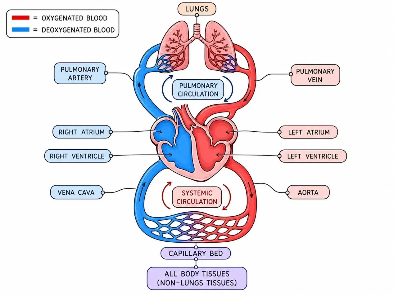
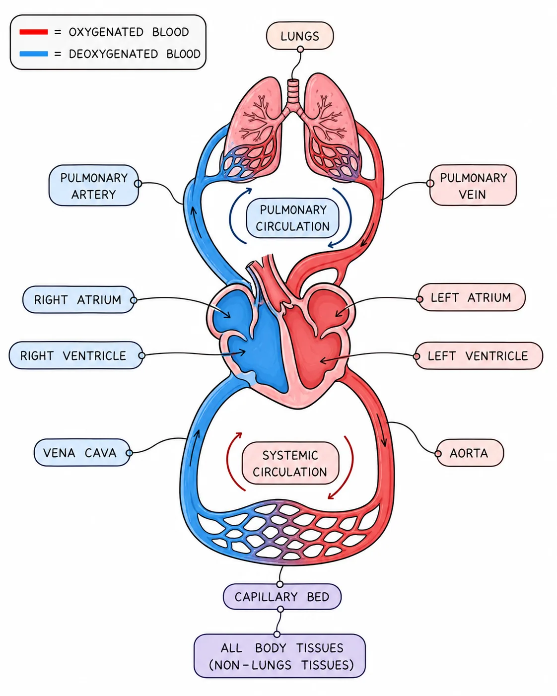

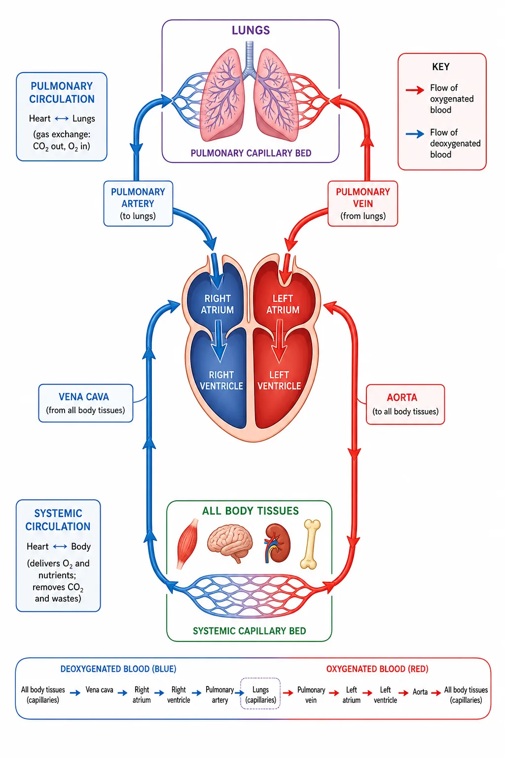


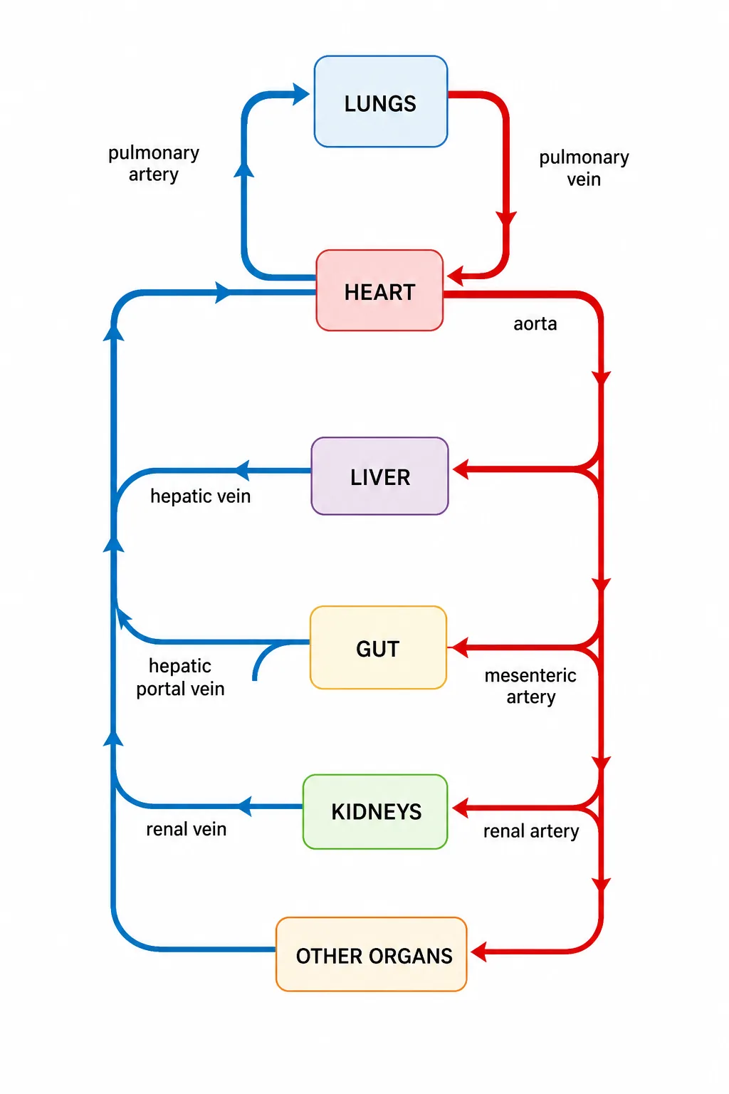
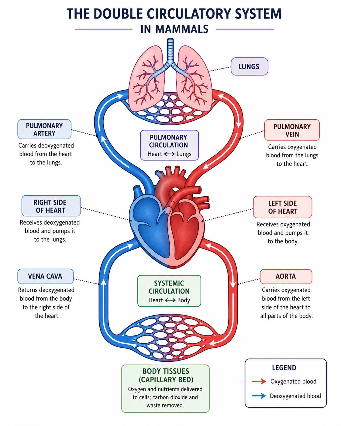
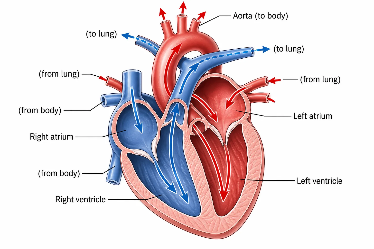
Humans have a double circulatory system: blood passes through the heart twice in each complete circuit of the body.
This is more efficient than a single circulation because the blood returns to the heart to be pumped again after the lungs, which reduces its pressure; the heart can then raise the pressure to supply the body at a higher pressure, so oxygen is delivered more quickly. It also keeps oxygenated and deoxygenated blood separate.
Section 2 of 9Structure of the heart


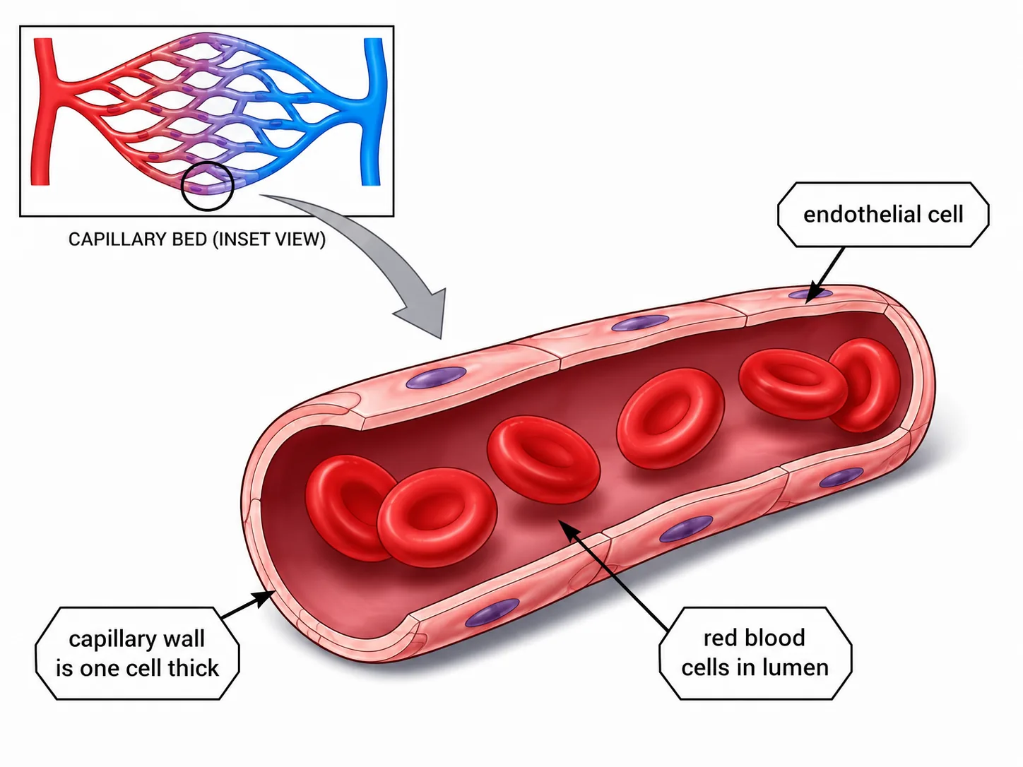


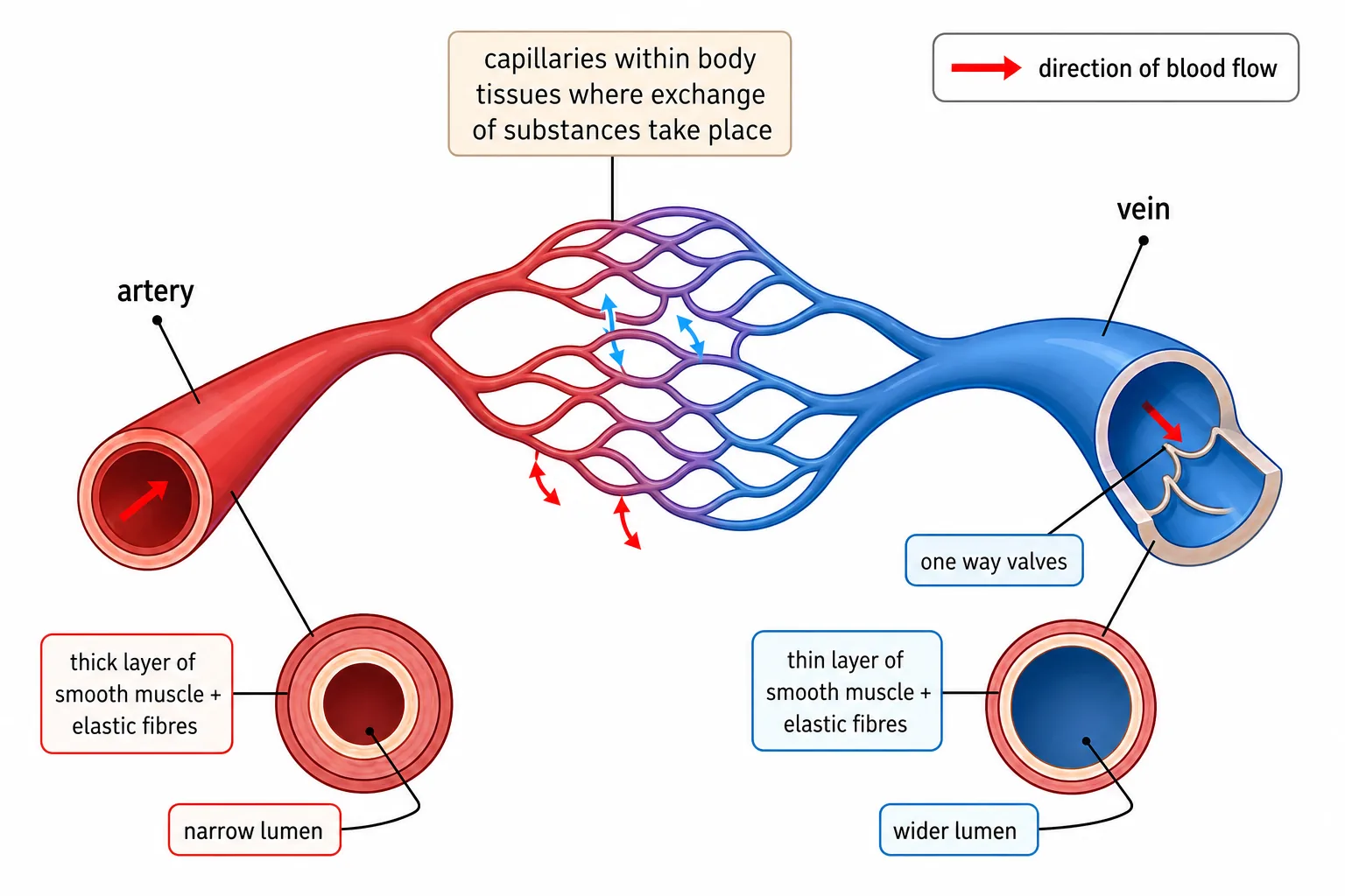


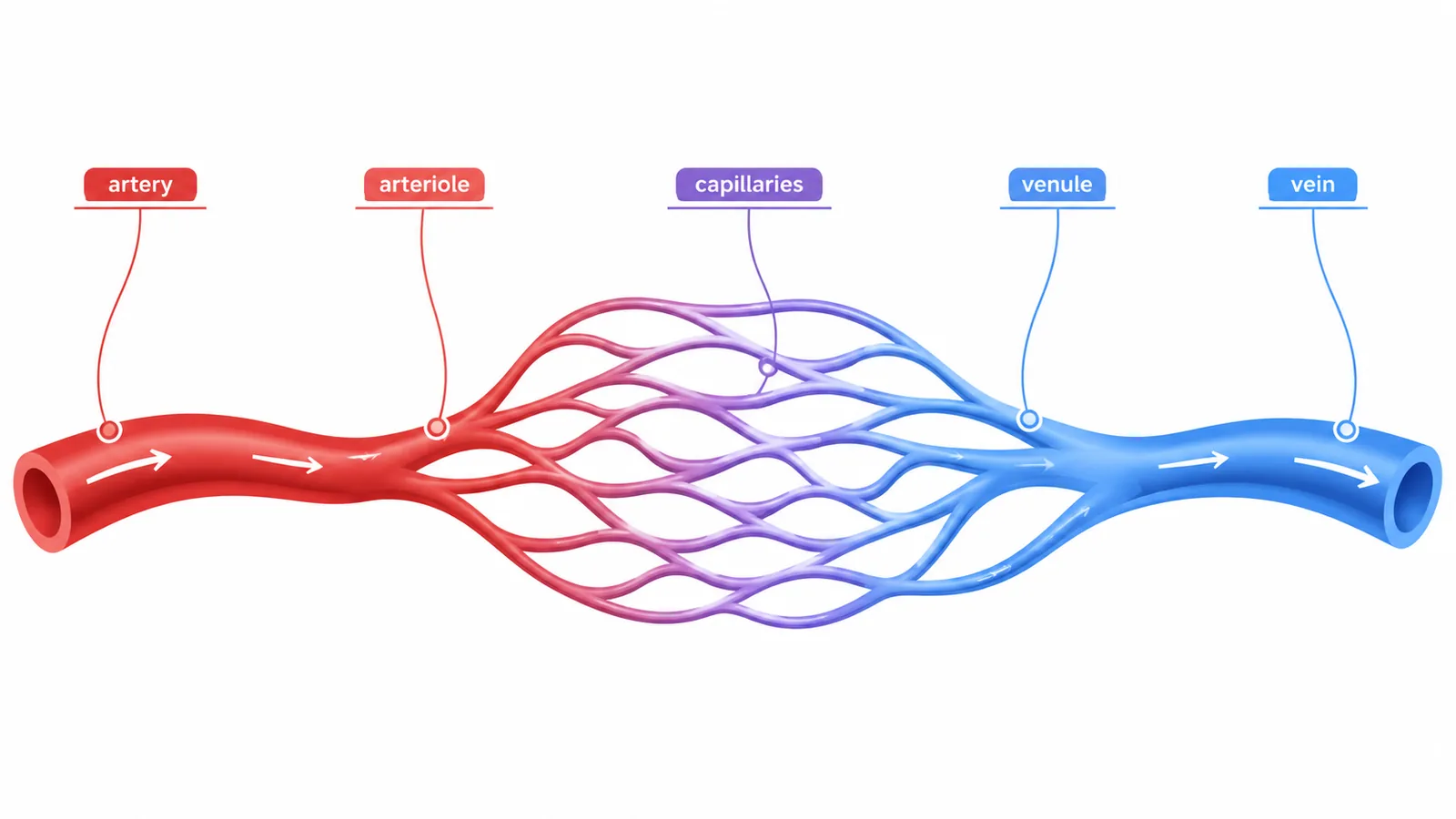


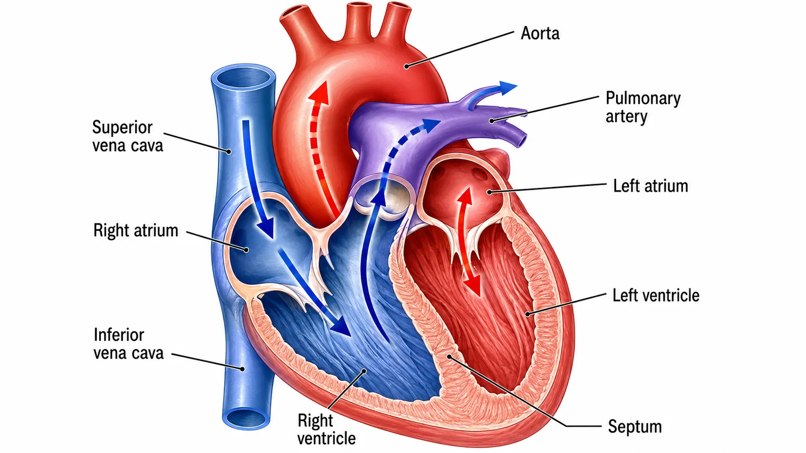
The heart is a muscular organ made of cardiac muscle, with four chambers: two atria (upper, thin-walled, receive blood) and two ventricles (lower, thick-walled, pump blood out). A wall called the septum separates the left and right sides. Valves between the atria and ventricles and in the arteries leaving the heart prevent backflow.
| Blood vessel | Carries blood | Oxygenation |
|---|---|---|
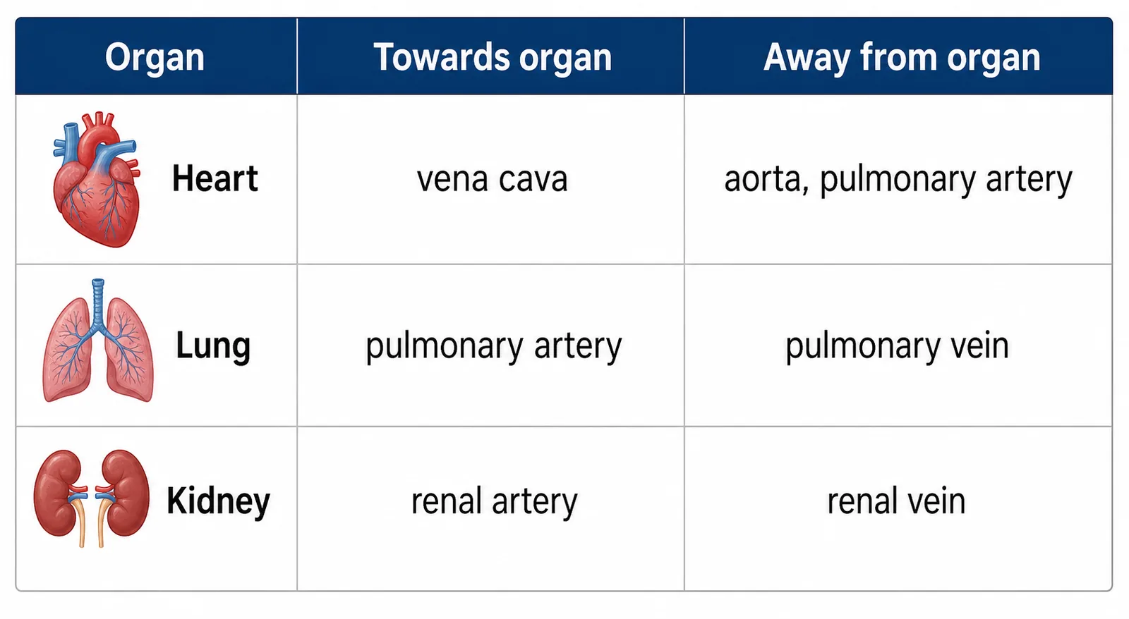
| Vena cava | From the body to the right atrium | Deoxygenated |
| Pulmonary artery | From the right ventricle to the lungs | Deoxygenated |
| Pulmonary vein | From the lungs to the left atrium | Oxygenated |
| Aorta | From the left ventricle to the body | Oxygenated |
The left ventricle has a thicker muscular wall than the right ventricle because it pumps blood at high pressure around the whole body, whereas the right ventricle pumps blood only to the nearby lungs, where lower pressure is needed to avoid damaging the delicate capillaries.
Section 3 of 9Path of the blood
The coronary arteries on the surface of the heart supply the heart muscle itself with oxygen and glucose.
Section 4 of 9Control of heart rate

A group of cells in the right atrium wall called the pacemaker (sinoatrial node) sets the natural rhythm of the heart by sending out electrical impulses that cause the atria to contract and then the ventricles.
Section 5 of 9Heart disease and treatments


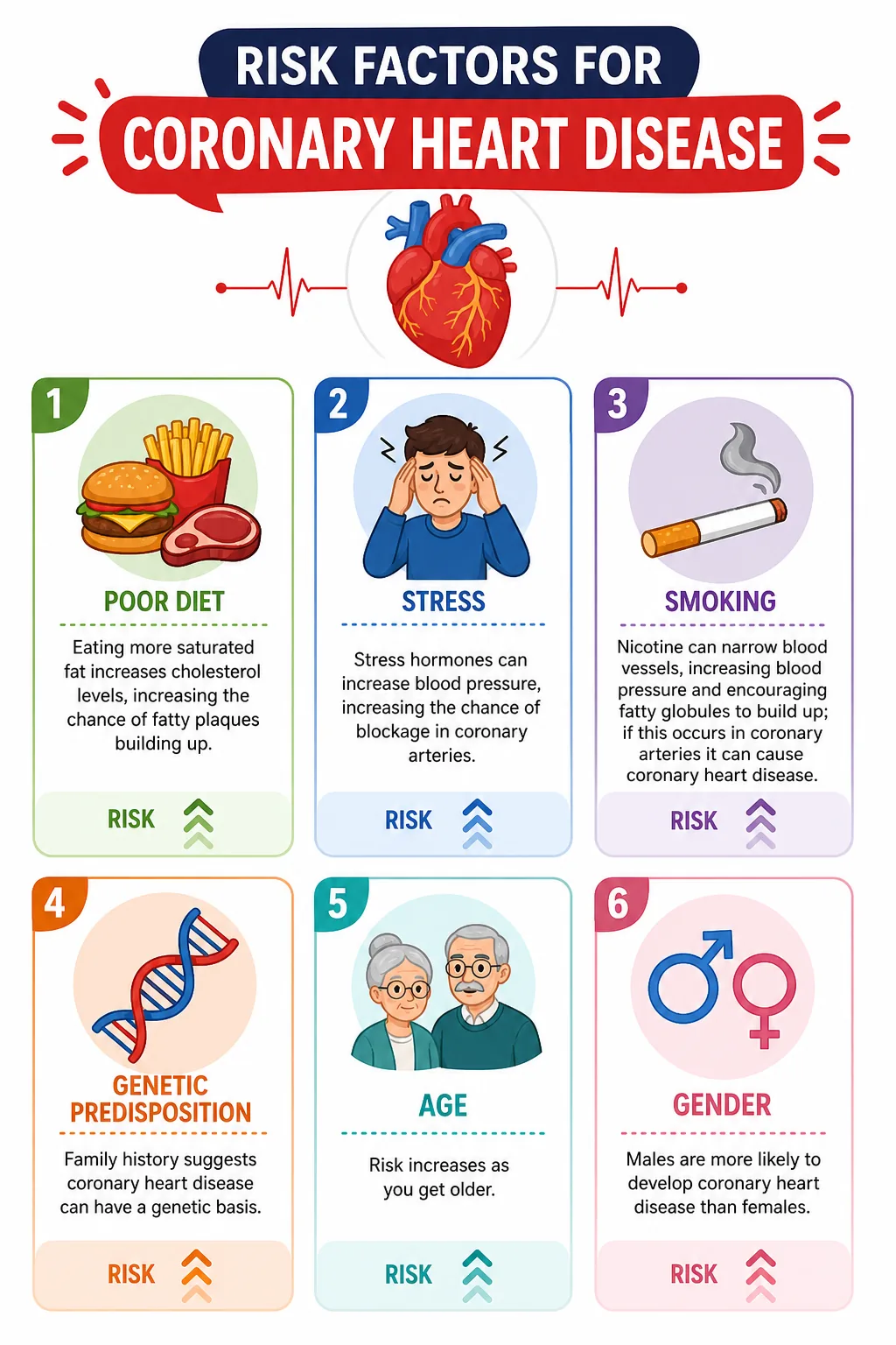


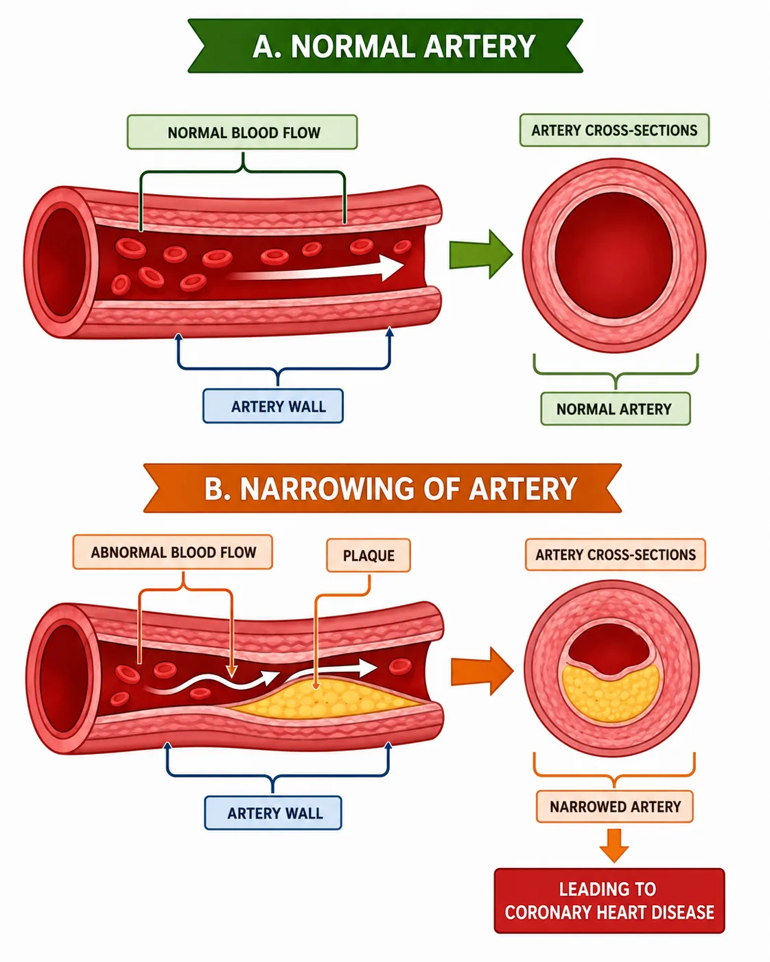

In coronary heart disease, fatty deposits build up in the coronary arteries, narrowing them and reducing blood flow to the heart muscle.
- and medicines to reduce blood pressure.
Section 6 of 9Heart valves and the heart cycle and using data

The atrioventricular valves between the atria and ventricles close when the ventricles contract, which prevents blood flowing back into the atria; the semilunar valves in the aorta and pulmonary artery close when the ventricles relax.
- then the ventricles contract and force blood into the arteries, and finally the whole heart relaxes while the atria refill.
Heart rate and stroke volume (the volume of blood pumped each beat) are used to calculate cardiac output: cardiac output = stroke volume x heart rate.
Section 7 of 9Exam-style questions


Describe the path taken by blood from the right ventricle to the left atrium. [3]3 marks
Blood leaves the right ventricle through the pulmonary artery to the lungs [1]. It is oxygenated in the lung capillaries [1] and returns to the left atrium through the pulmonary vein [1].
Explain why the wall of the left ventricle is thicker than the wall of the right ventricle. [2]2 marks
The left ventricle pumps blood around the whole body, which needs high pressure [1], while the right ventricle pumps only to the lungs, where high pressure would damage the capillaries [1].
A man has a stroke volume of 80 cm3. His heart rate rises from 70 to 150 beats per minute during exercise. Calculate the increase in cardiac output. [3]3 marks
At rest: 80 x 70 = 5600 cm3 per minute [1]. Exercise: 80 x 150 = 12000 cm3 per minute [1]. Increase = 6400 cm3 per minute [1].
Section 8 of 9Key terms
| Term | Meaning |
|---|---|
| Double circulation | System in which blood passes through the heart twice per circuit of the body. |
| Atrium | Upper heart chamber that receives blood. |
| Ventricle | Lower heart chamber that pumps blood out. |
| Aorta | Main artery carrying oxygenated blood from the left ventricle. |
| Pacemaker | Group of cells that controls the heart rate. |
| Coronary artery | Artery supplying the heart muscle with blood. |
| Statin | Drug that lowers blood cholesterol. |
| Cardiac output | Volume of blood pumped by the heart per minute. |