2.10.9Structure and Function in Living Organisms
The Human Eye: Structure
5 min read 9 parts Lesson 52 of 55 1 quiz
Section 1 of 9Structure and function of the eye

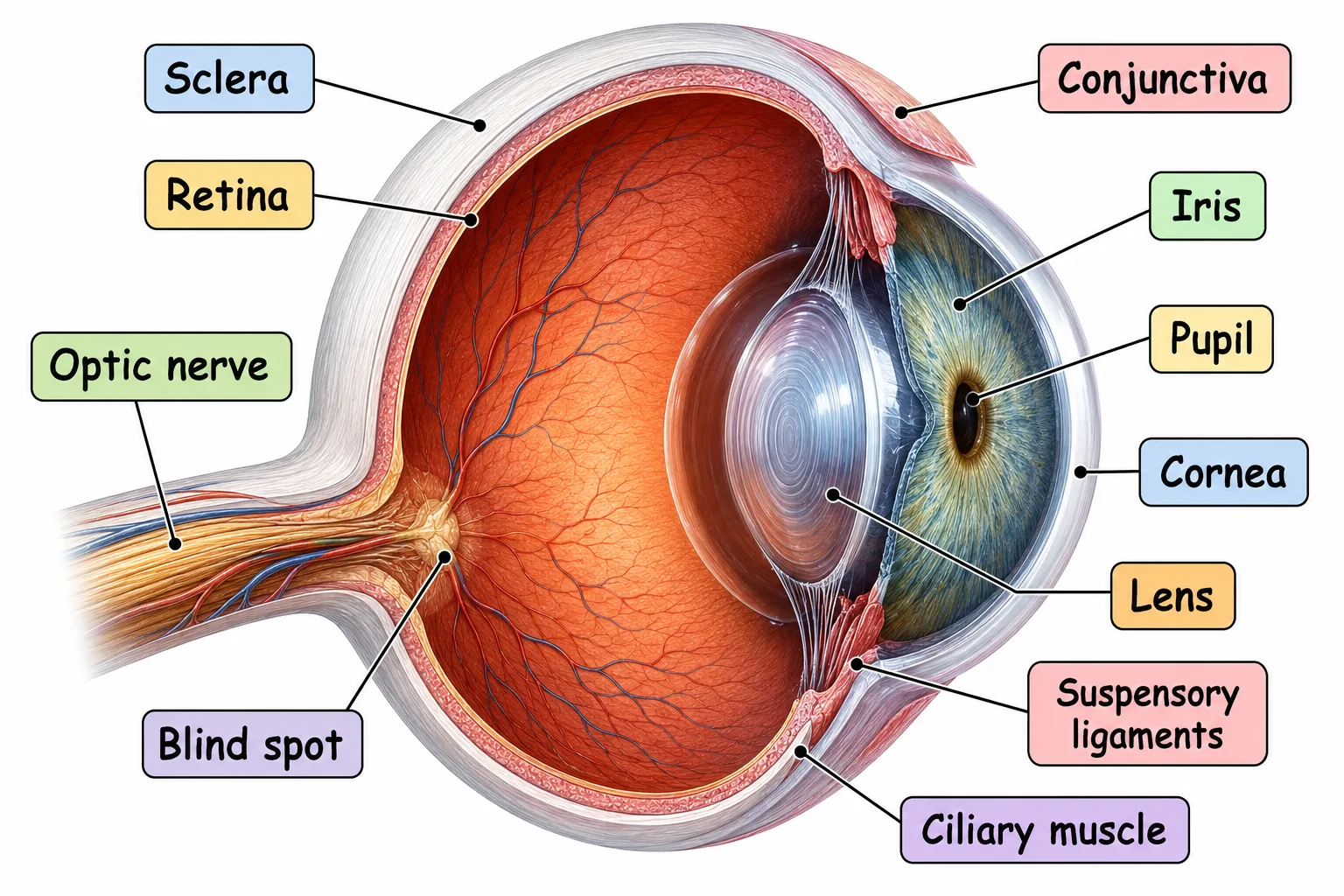
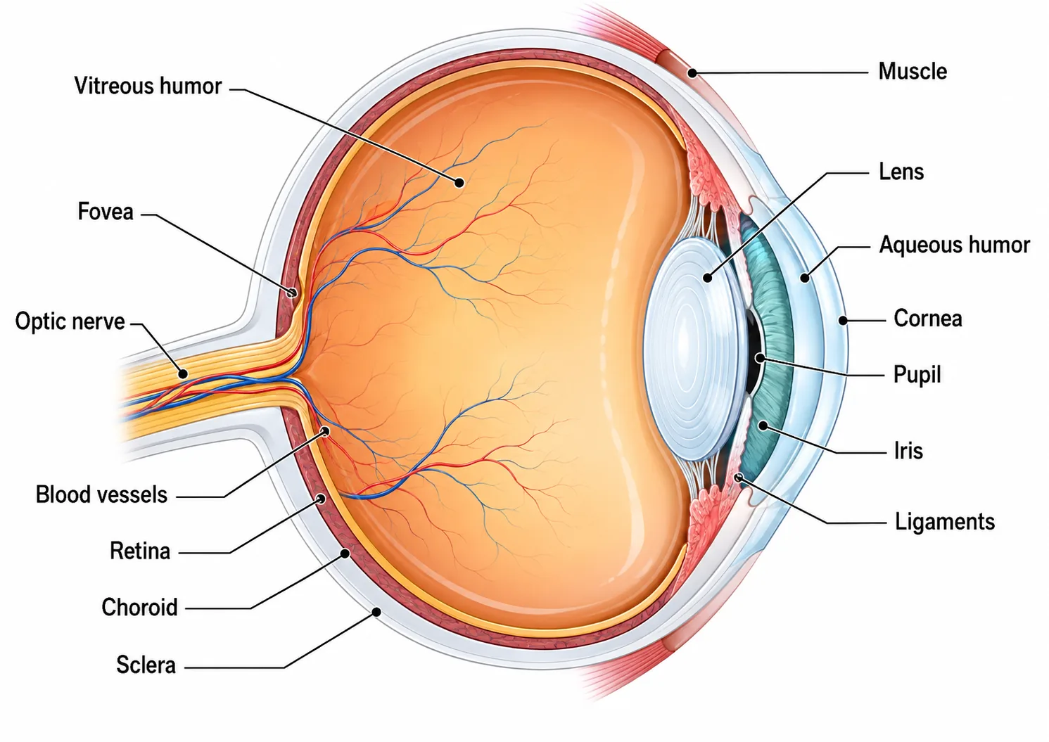
| Structure | Function |
|---|---|
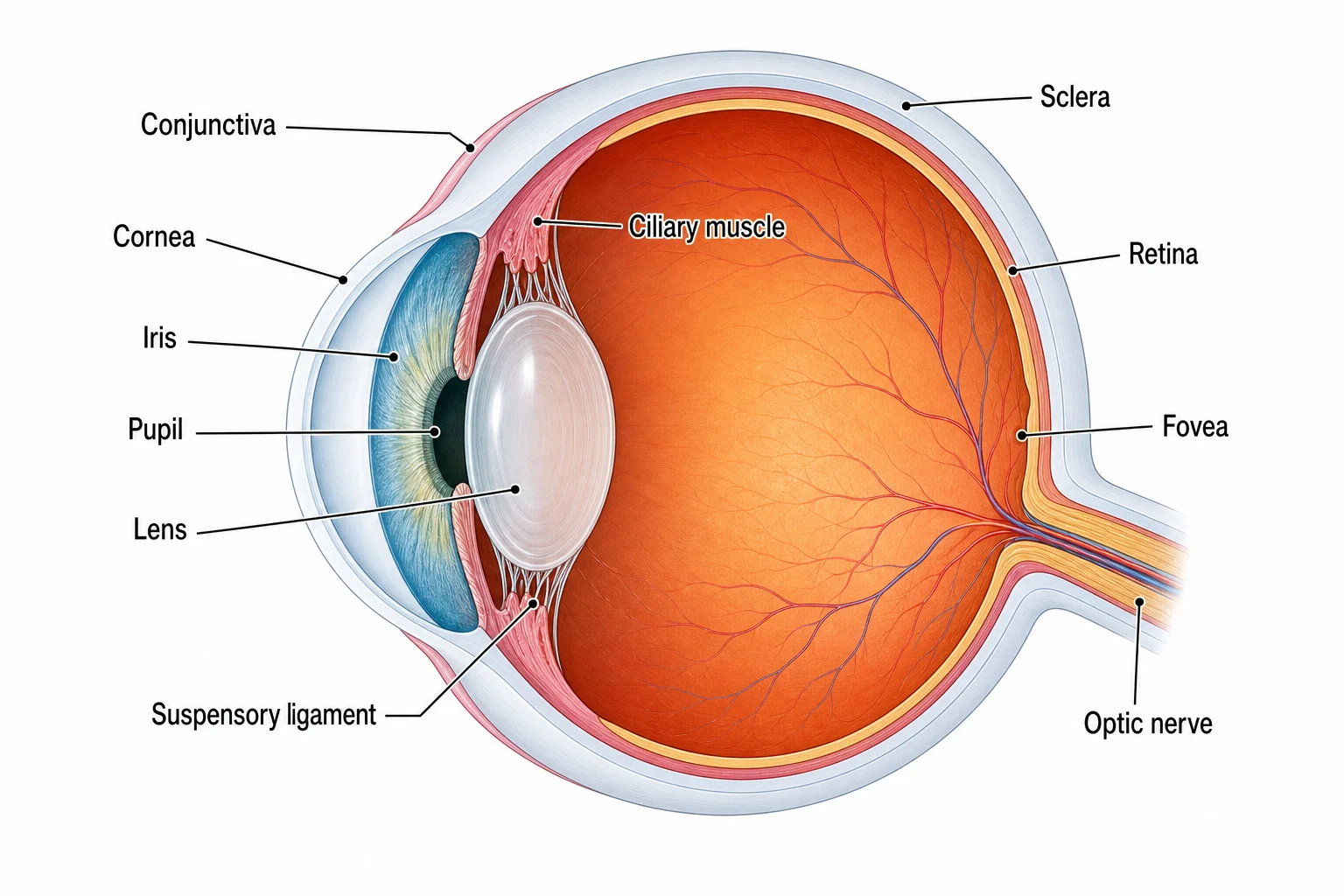
| Cornea | Transparent front surface; refracts (bends) light, doing most of the focusing |
| Iris | Ring of muscle that controls the size of the pupil and so how much light enters |
| Pupil | Hole through which light enters |
| Lens | Transparent, flexible; refracts light and is adjusted to focus on the retina |
| Ciliary muscles | Ring of muscle that changes the shape of the lens |
| Suspensory ligaments | Hold the lens in place and transmit the pull on it |
| Retina | Layer containing the light receptors (rods and cones) |
| Optic nerve | Carries impulses from the retina to the brain |
| Sclera | Tough white outer layer that protects and gives shape |
Light enters through the cornea, passes through the pupil and lens, and is focused onto the retina, where it forms an inverted (upside-down) image. The brain interprets the signals from the optic nerve and turns the image the right way up.
Section 2 of 9Rods and cones
The retina contains two types of light receptor. Rod cells are sensitive to dim light but give black-and-white vision with less detail. Cone cells work in bright light, give colour vision, and are most concentrated in the fovea, the region of sharpest vision. There are three types of cone, sensitive to red, green and blue light, and colour perception depends on the combination of cones stimulated.
Section 3 of 9Controlling the amount of light: the pupil reflex
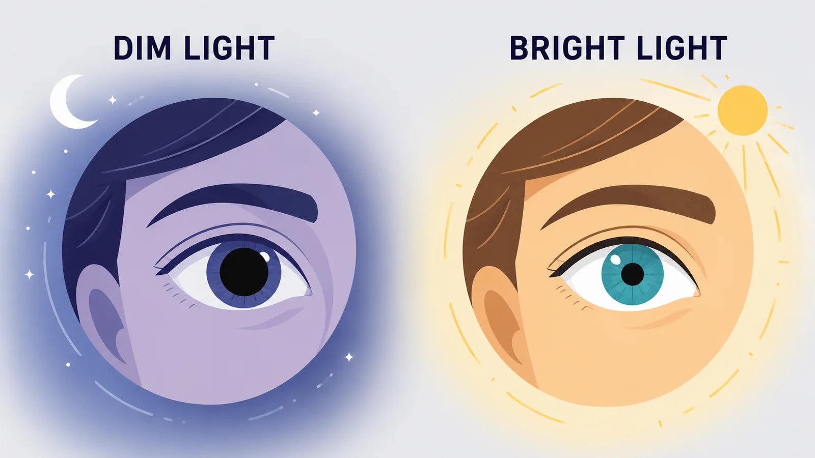

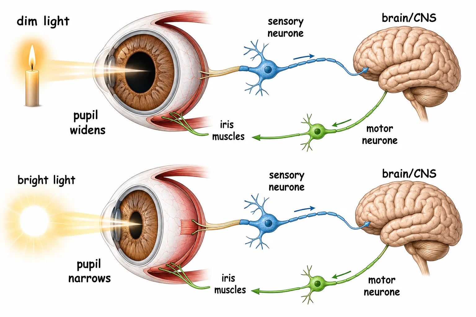
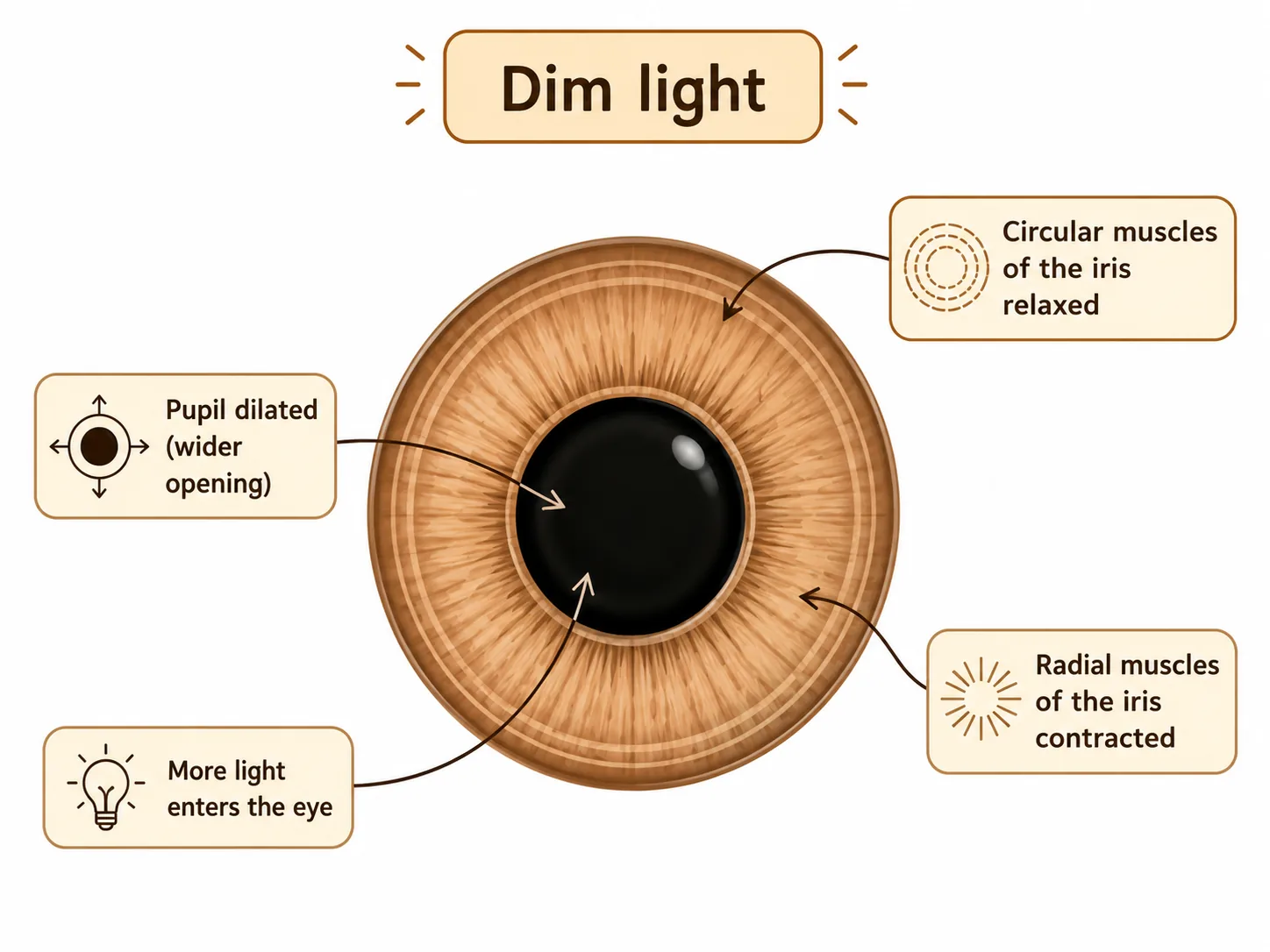

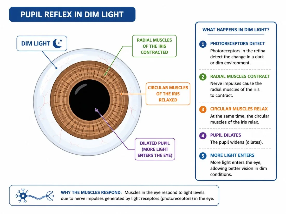
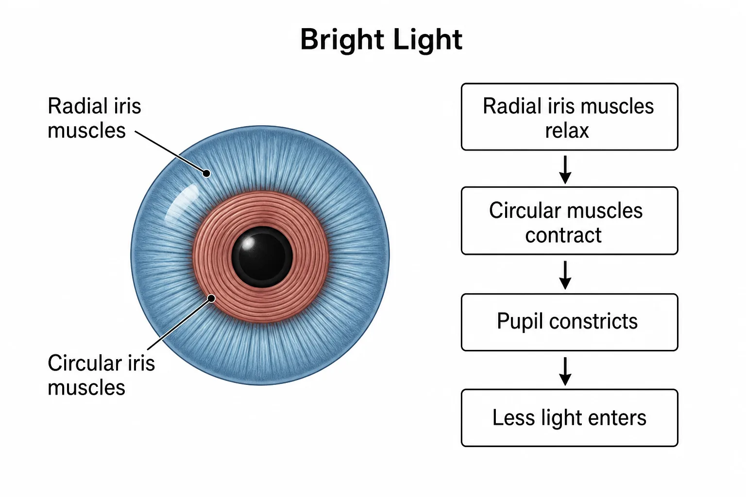


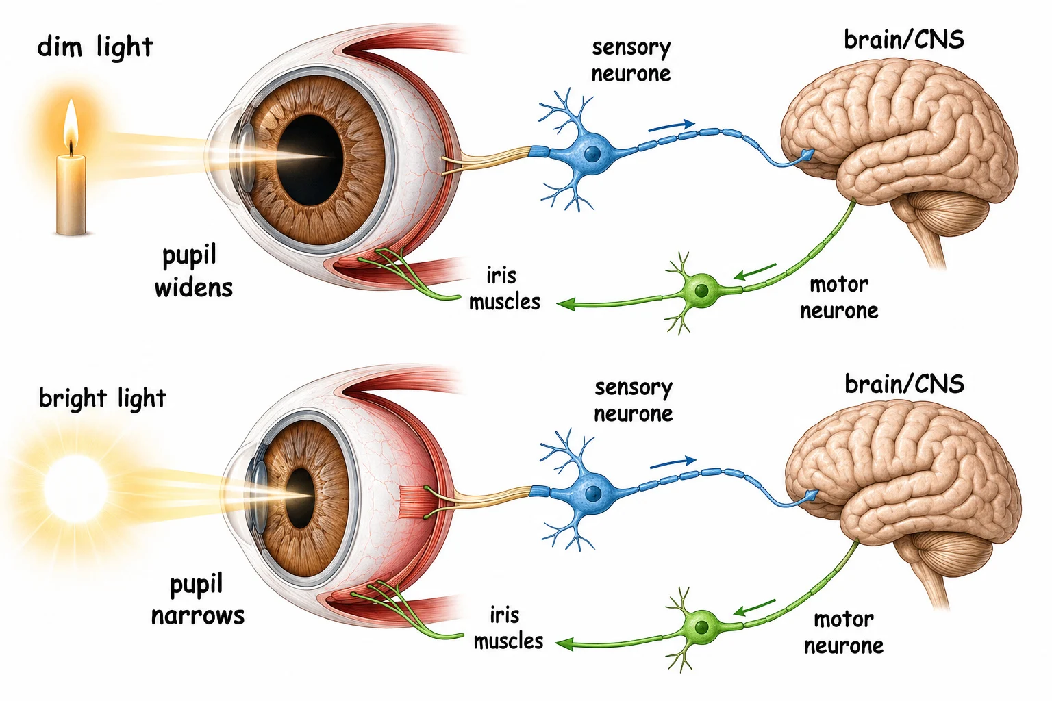
Too much light would damage the retina, and too little would give poor vision, so the pupil size is adjusted by the iris, which has two sets of muscles.
This is a reflexreceptors in the retina detect the brightness, and the response is automatic, via the brain, to the muscles of the iris.
Section 4 of 9Focusing: accommodation
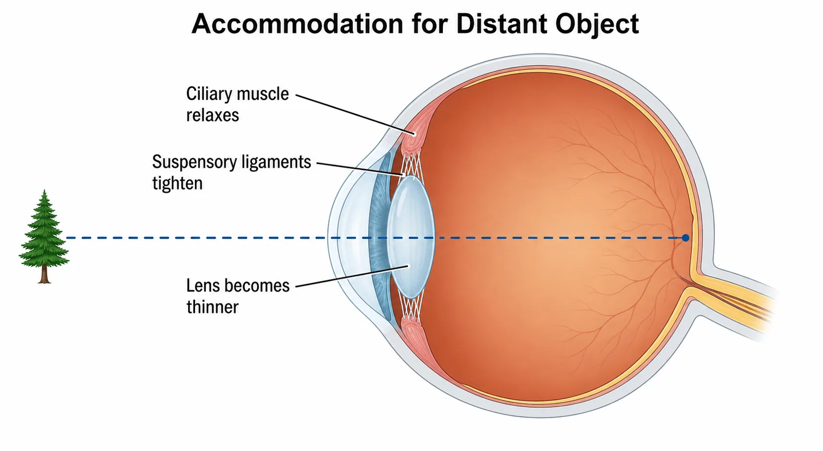


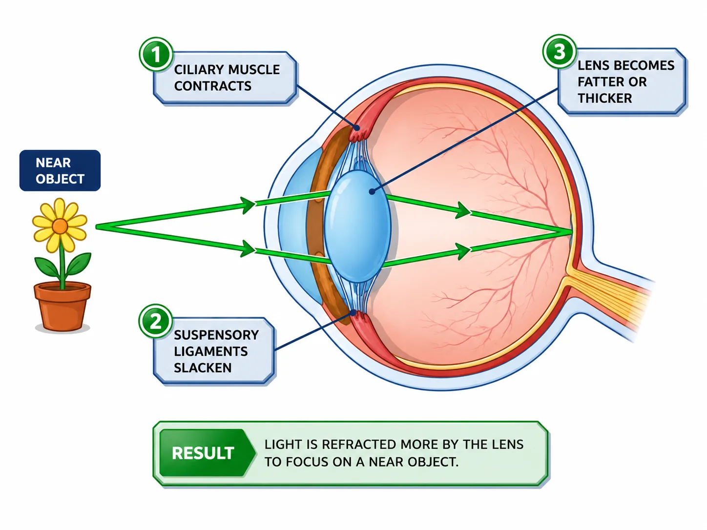
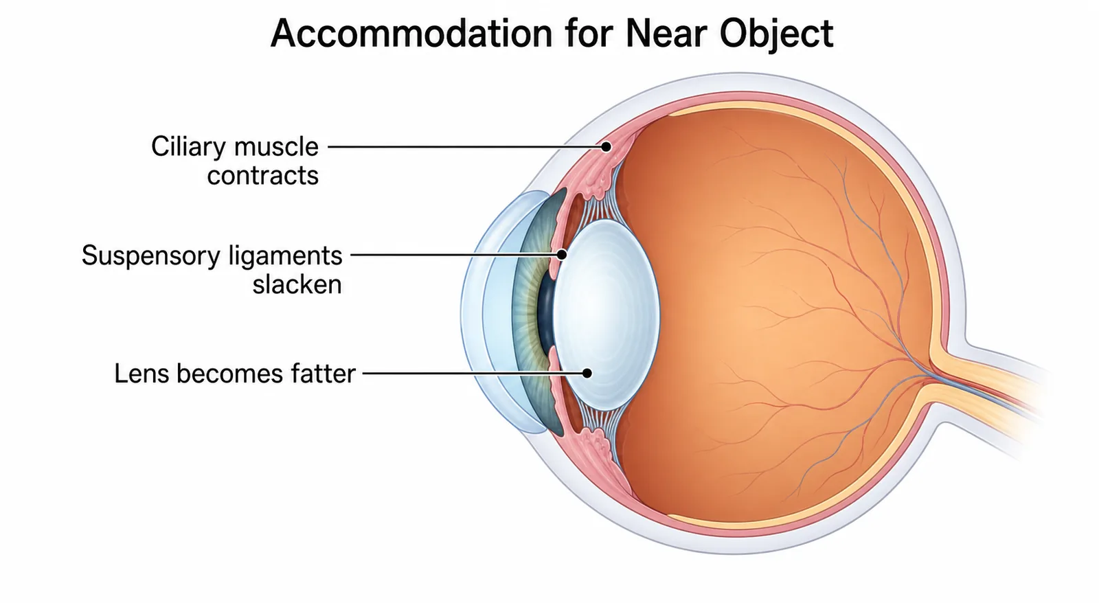
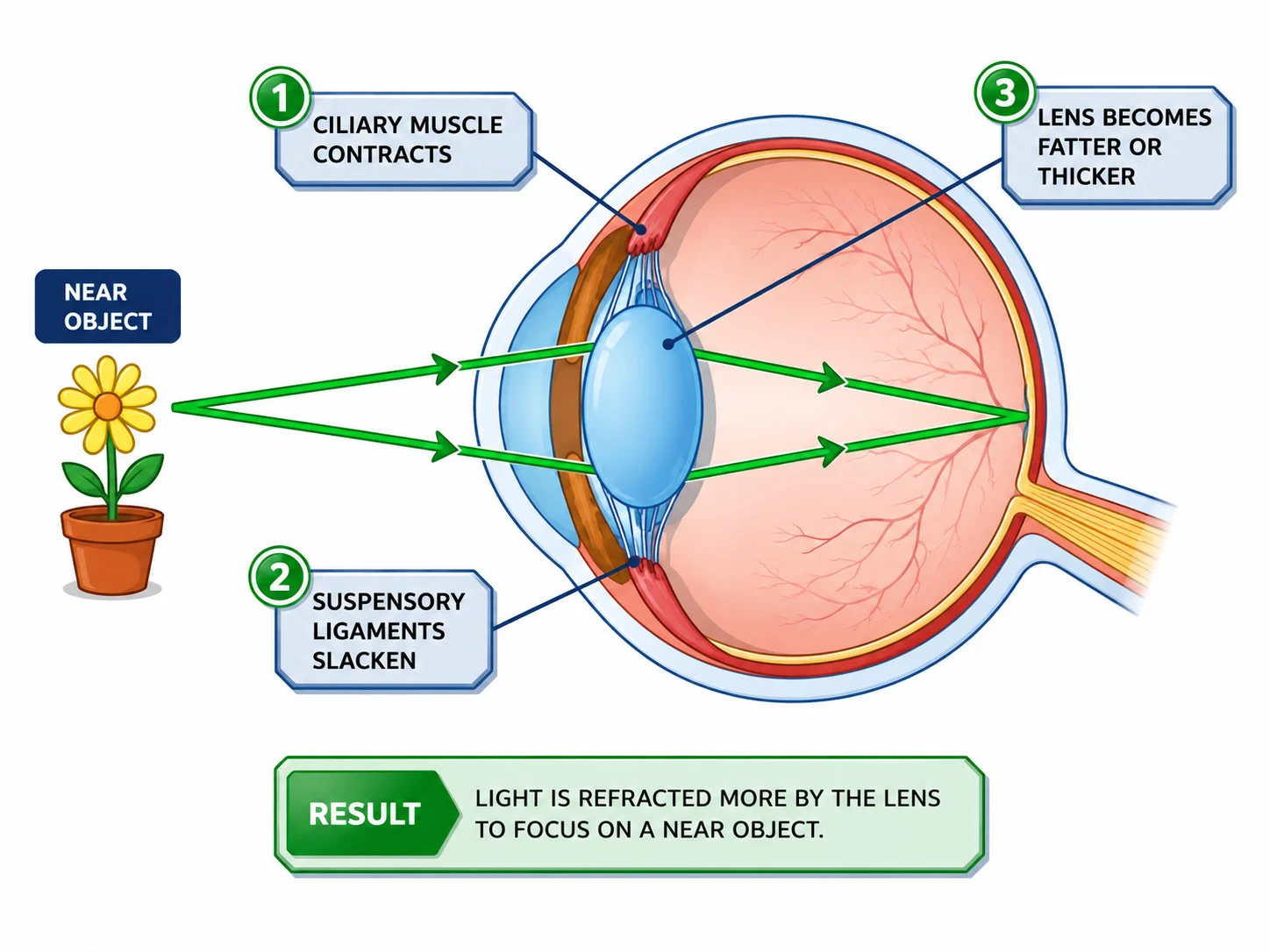
To see clearly, light from an object must be focused on the retina. The lens changes shape to focus on objects at different distances, a process called accommodation.
| Looking at a near object | Looking at a distant object | |
|---|---|---|
| Ciliary muscles | Contract | Relax |
| Suspensory ligaments | Loosen (slack) | Pulled tight |
| Lens shape | Thicker, more curved | Thinner, less curved |
| Light refraction | Greater | Less |
A common error is to reverse the ciliary muscle and ligament states. A helpful way to remember: for near vision the ciliary muscle contracts, which makes the ring smaller, so the ligaments go slack and the elastic lens bulges.
Section 5 of 9Nerve pathway and links
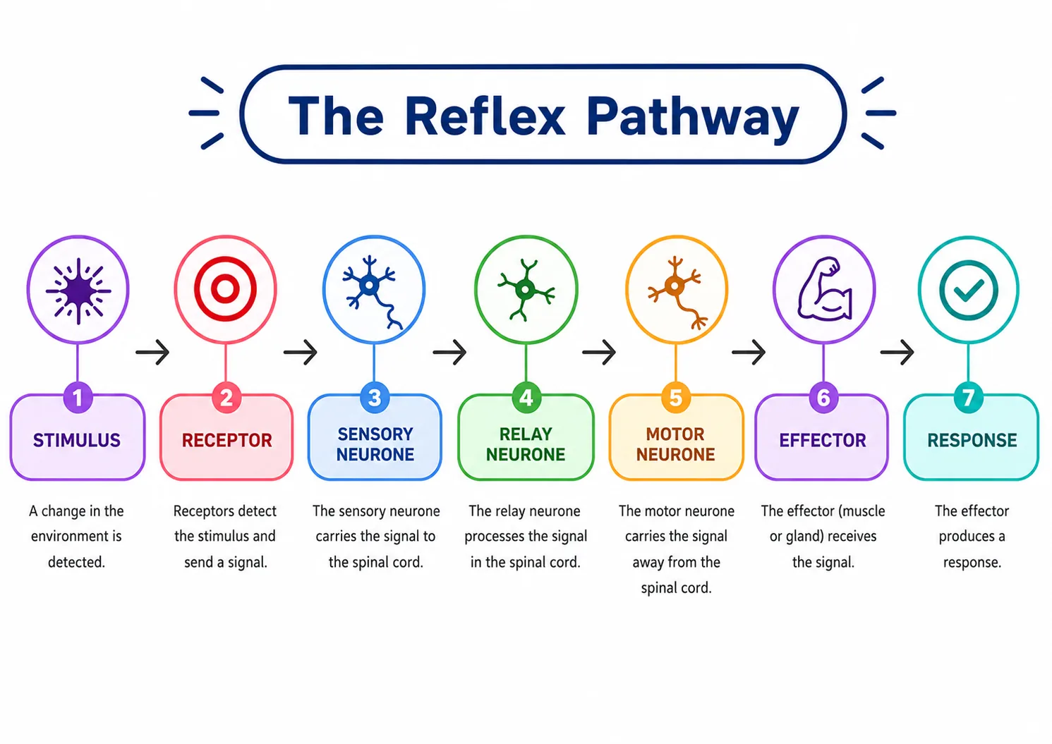
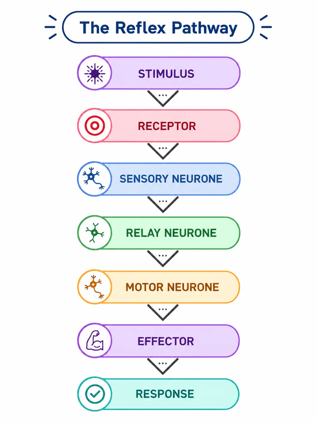
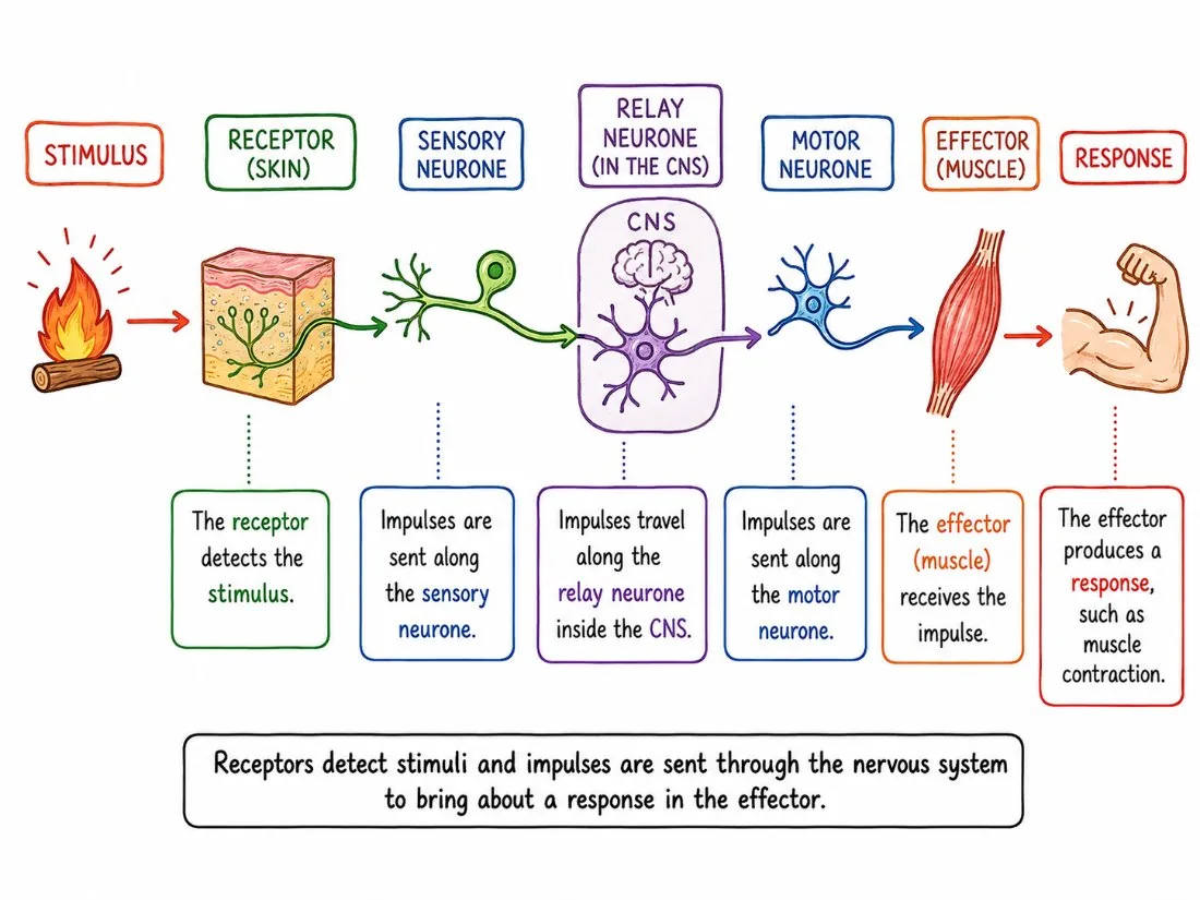
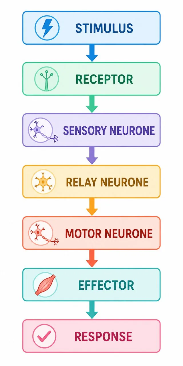
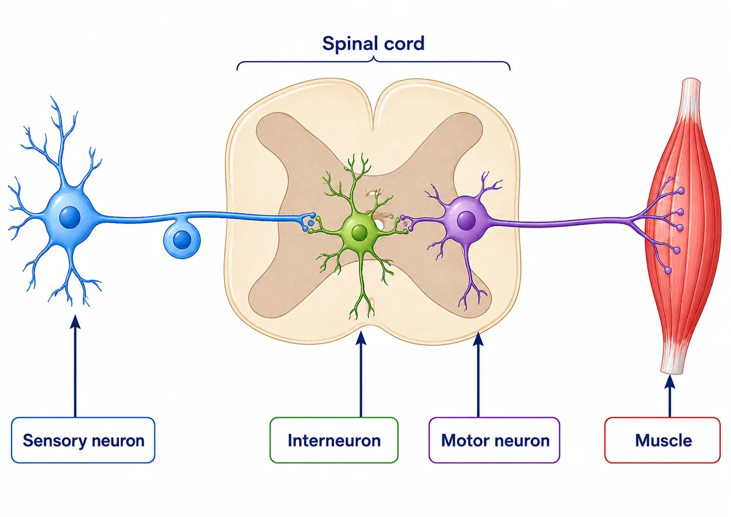
The eye is a sense organits receptors convert light into impulses, which travel in the optic nerve to the visual cortex at the back of the brain.
The eye also demonstrates the same principle as the reflex arc, since the pupil response protects delicate receptor cells without conscious thought.
Section 6 of 9How the eye detects light and sends information


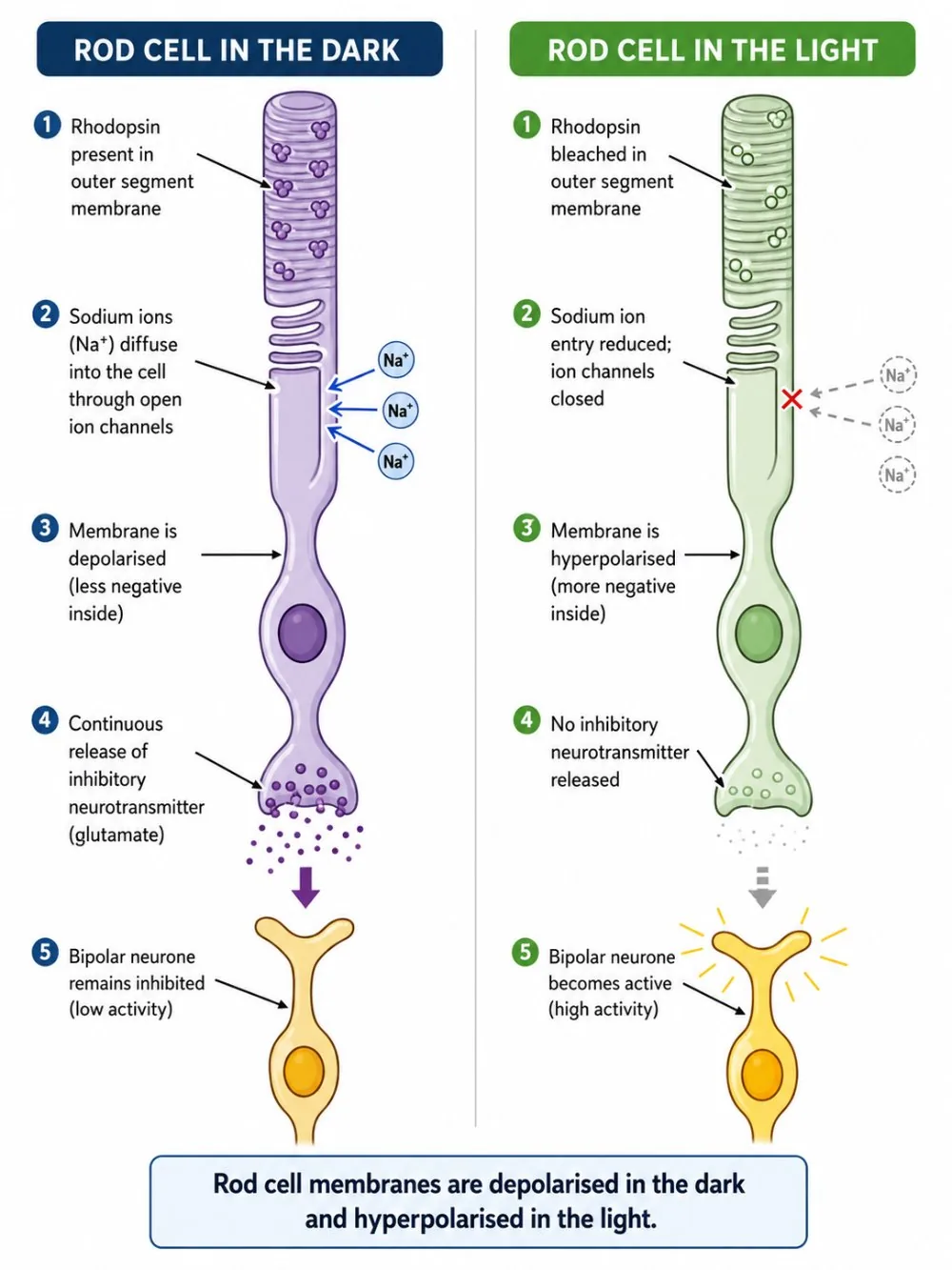
When light reaches the retina it is absorbed by pigments in the rods and cones, which triggers electrical impulses in the receptor cells.
The eye can adapt to dim conditions. After moving from bright light to a dark room, vision is poor at first, then improves over several minutes as the rod pigment is regenerated. In dim light we see mostly in black and white because cones need brighter light to work.
Eyes also contain fluids
- the aqueous humour in front of the lens and the vitreous humour behind it.
- These support the shape of the eyeball and keep the pressure inside constant, helping the lens and cornea to refract light properly.
Section 7 of 9Exam-style questions
Describe how the eye changes when moving from looking at a distant tree to reading a book. [4]4 marks
Ciliary muscles contract [1]; suspensory ligaments become slack [1]; the lens becomes thicker (more curved) [1]; light is refracted more so that it focuses on the retina [1].
Explain how the pupil changes in bright light and why this is useful. [3]3 marks
Circular muscles of the iris contract and radial muscles relax [1], making the pupil smaller [1]. This reduces the light entering and protects the retina from damage [1].
Explain why cones give better colour vision than rods. [2]2 marks
There are three types of cone that respond to different wavelengths (colours) of light [1], whereas rods are all of one type and only detect light intensity [1].
Section 8 of 9Key terms
| Term | Meaning |
|---|---|
| Cornea | The transparent front of the eye that refracts light. |
| Iris | The coloured muscle that controls pupil size. |
| Lens | A flexible, transparent structure that focuses light on the retina. |
| Retina | The layer at the back of the eye containing light receptors. |
| Optic nerve | The nerve carrying impulses from the retina to the brain. |
| Accommodation | The change in the shape of the lens to focus on near or distant objects. |
| Ciliary muscle | A ring of muscle that changes the shape of the lens. |
| Fovea | The region of the retina with the highest density of cones. |