Skip to content
Dabour HubLEARN · PRACTICE · EXPLORESign inStudent

2.2.2Structure and Functions in Living Organisms: Part 1

Animal and Plant Cells: Similarities and Differences

5 min read 9 parts Lesson 4 of 36 1 quiz

0%read

Section 1 of 9Sub-cellular structures
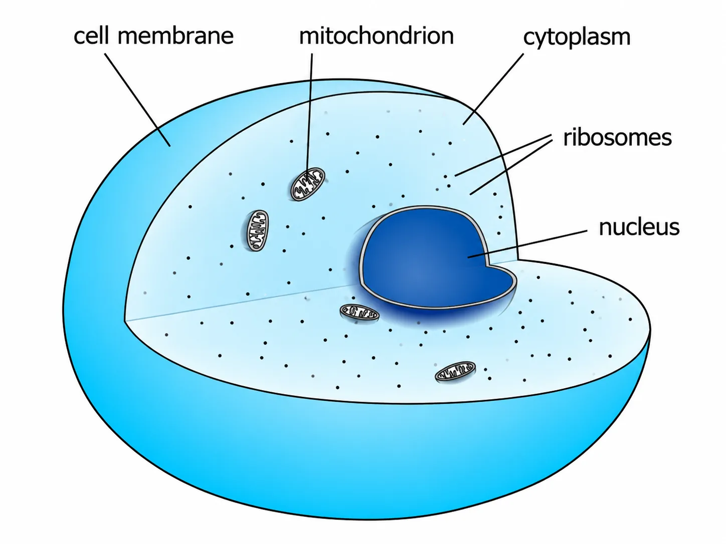
Animal cell 3D section labelled
Animal cell 3D labelled
ribosome structure
Key Structures of the Respiratory System
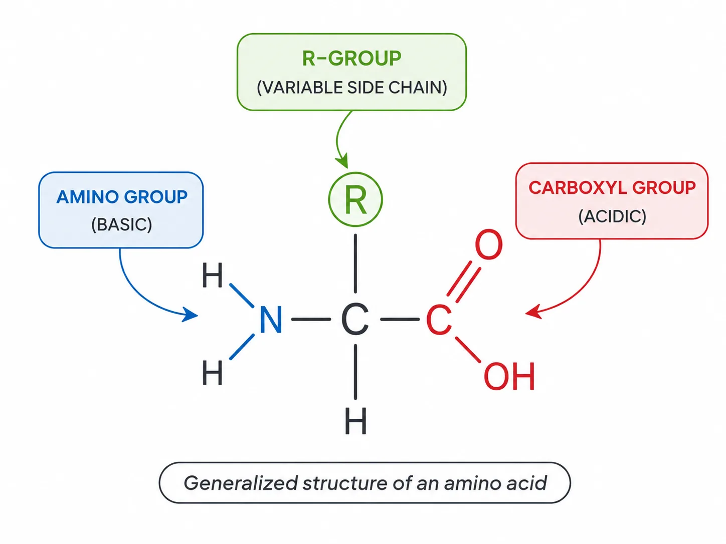
generalized amino acid structure
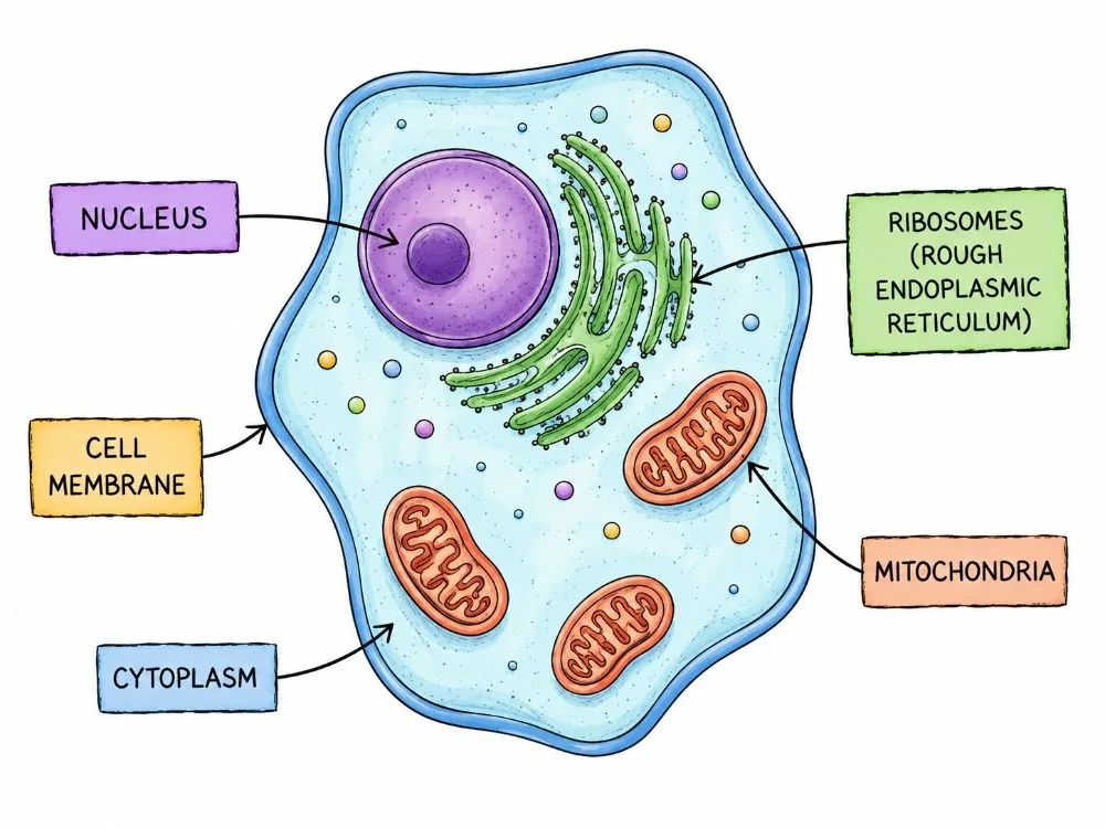
Animal cell labelled (nucleus, mitochondria, ribosomes)
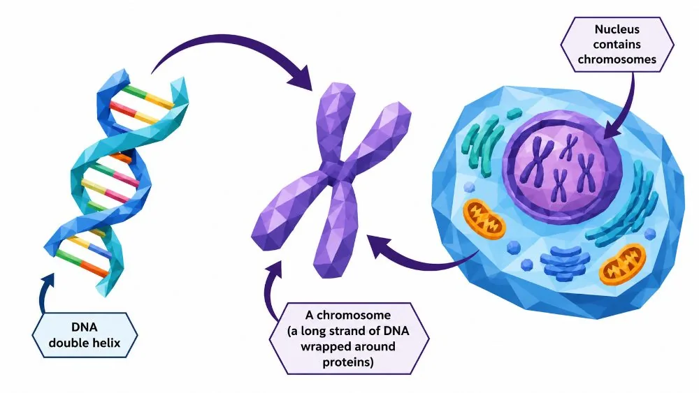
Nucleus, Chromosome and DNA
Animal cell (detailed labelled)
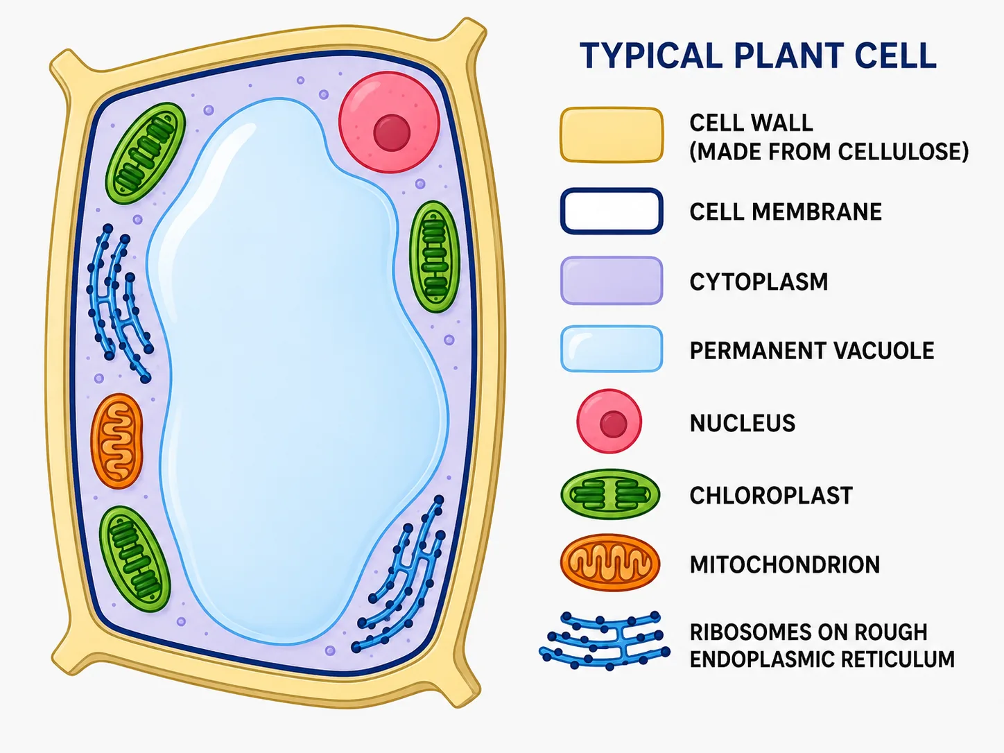
Typical plant cell (labelled with key)
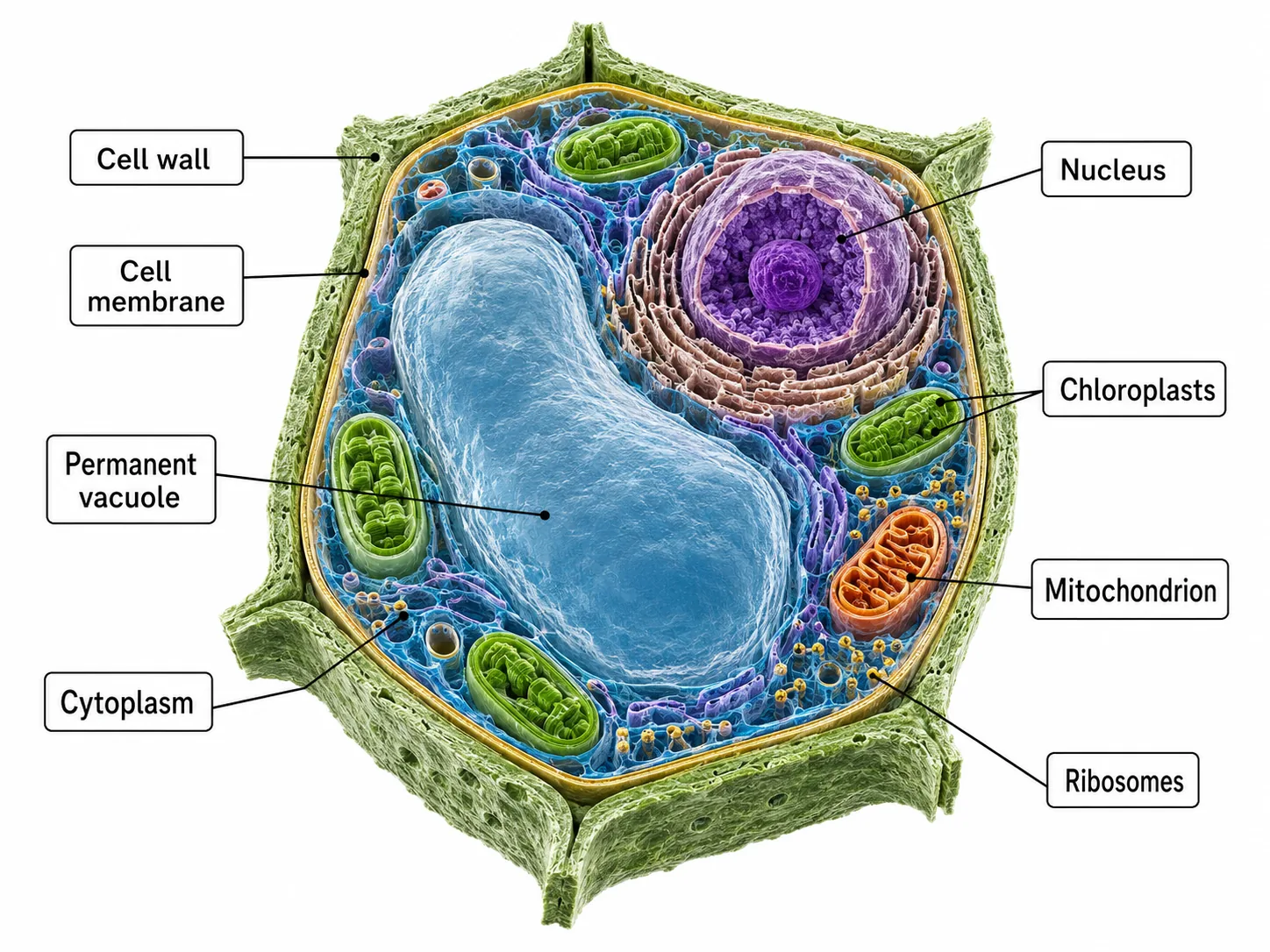
Plant cell 3D labelled
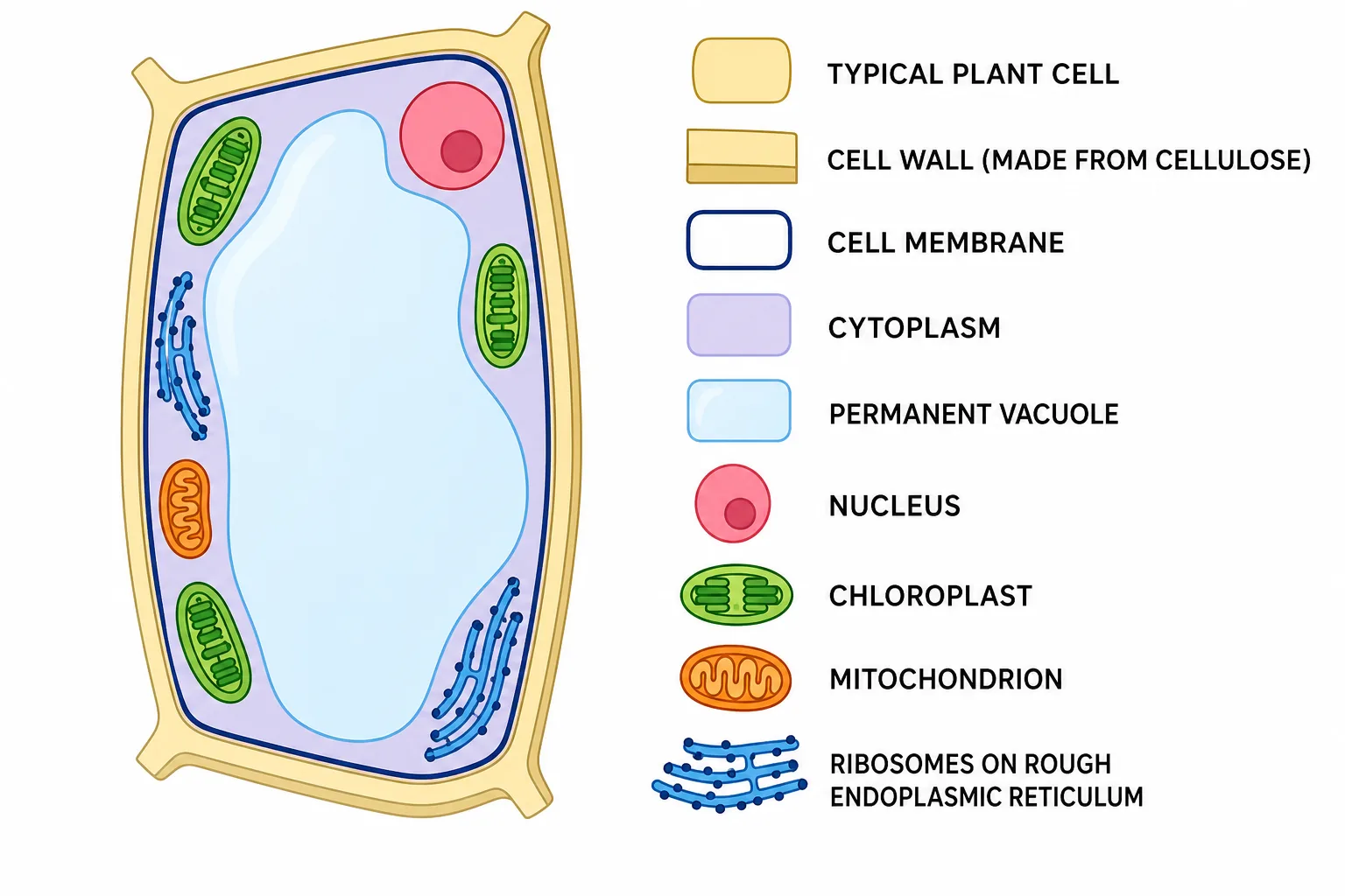
Typical Plant Cell
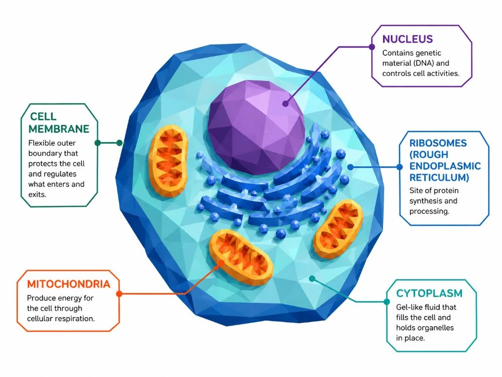
Animal cell labelled (nucleus, rough ER, mitochondria)
Structures of animal and plant cells compared

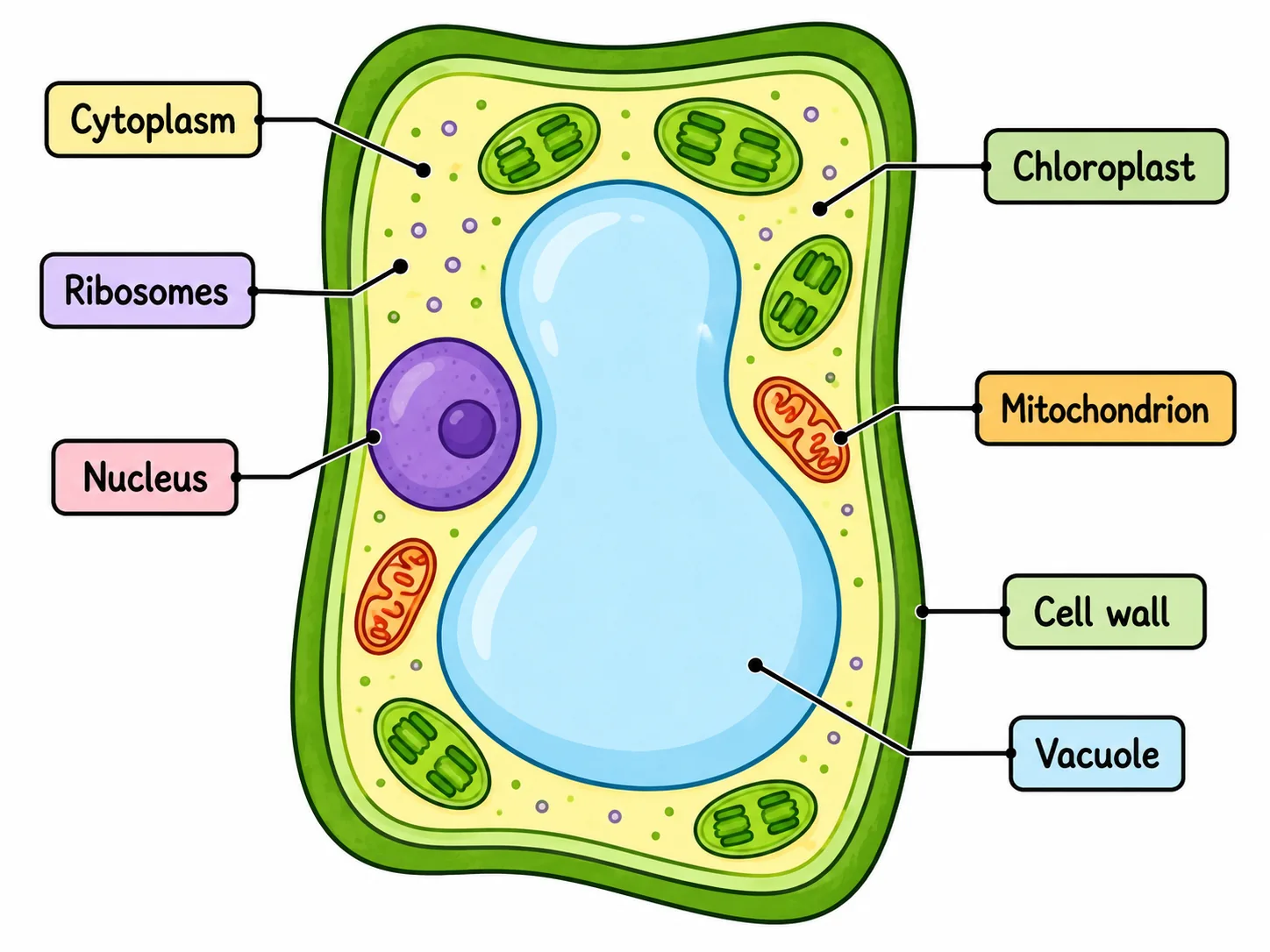
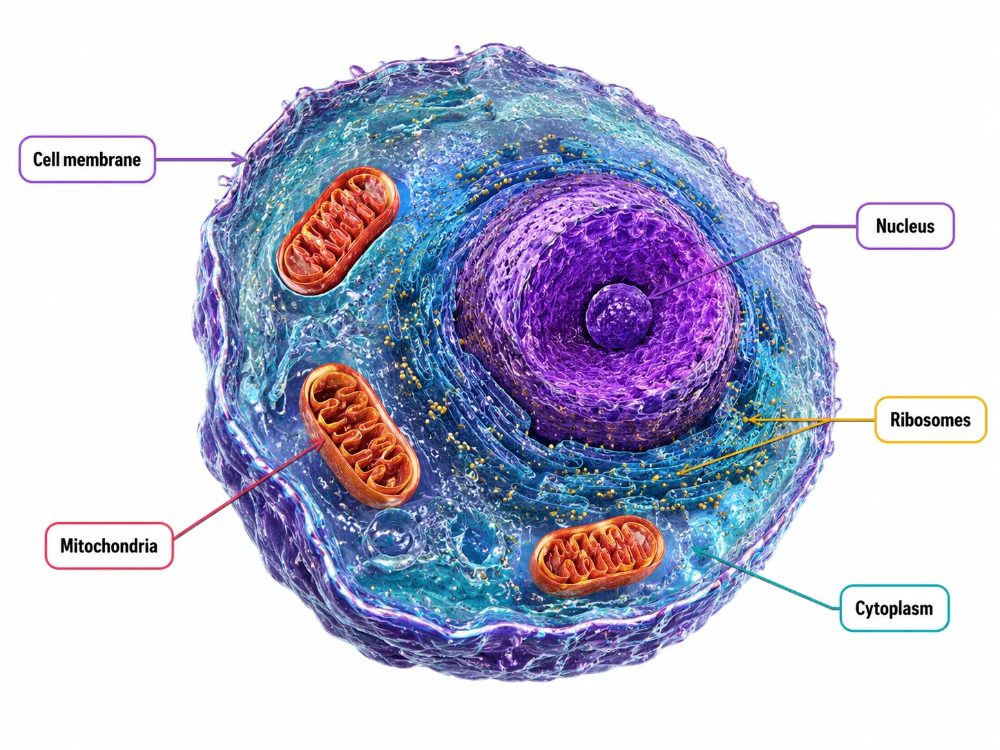
Animal and plant cells are both eukaryotic, so both contain a nucleus, cytoplasm, cell membrane, mitochondria and ribosomes. Looking at what each part does lets you explain how a cell is adapted for its job.

Sub-cellular structures
Nucleus
Nucleus holds the DNA as chromosomes, which carry the genes that control cell activity and inheritance.
Cytoplasm
Cytoplasm the site of many chemical reactions, including the first stage of respiration.
Cell membrane
Cell membrane a thin, partially permeable layer that controls the movement of substances in and out.
Mitochondria
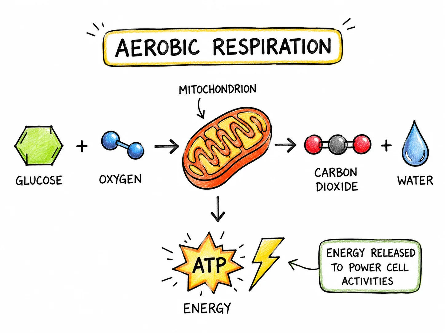
Mitochondria release energy from glucose in aerobic respiration. Cells with high energy demands, such as muscle cells, contain many.
Ribosomes
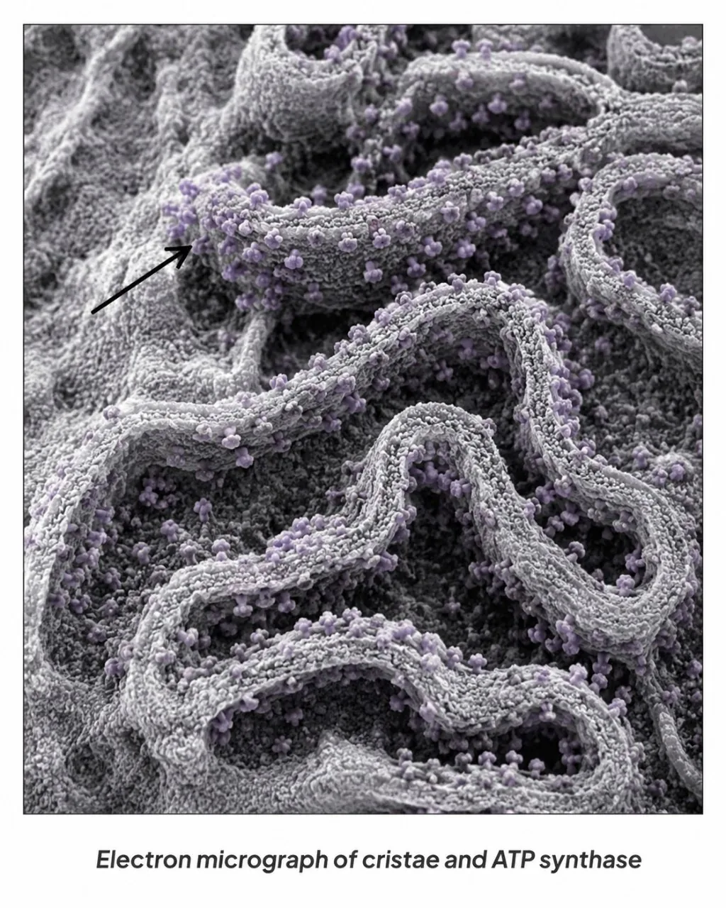
Ribosomes tiny structures that assemble amino acids into proteins following instructions copied from the DNA.

Section 2 of 9Extra structures in plant cells

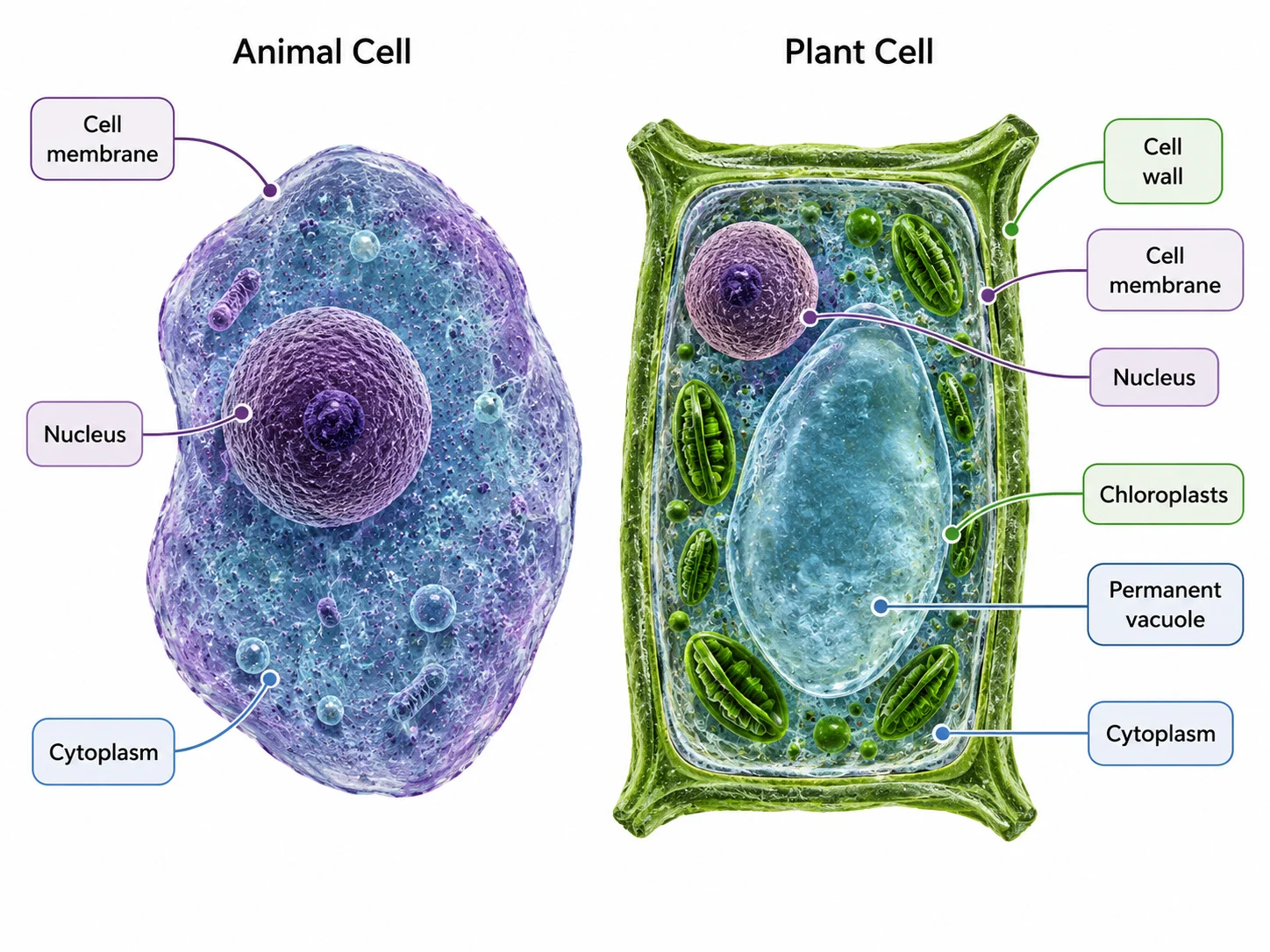
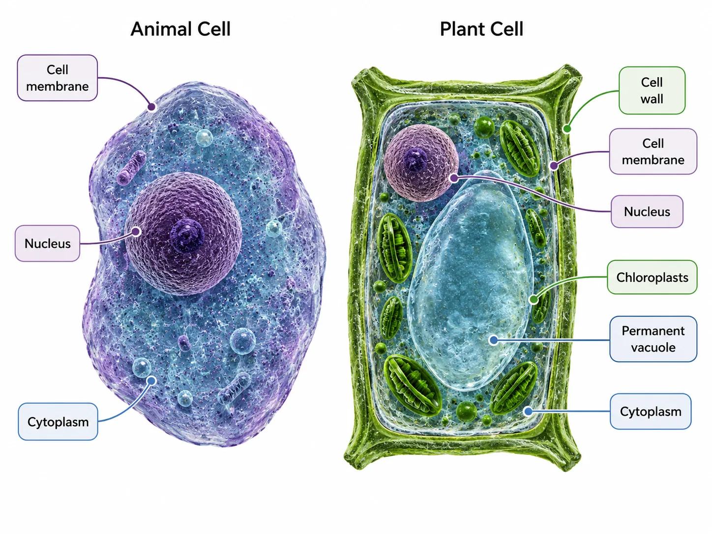
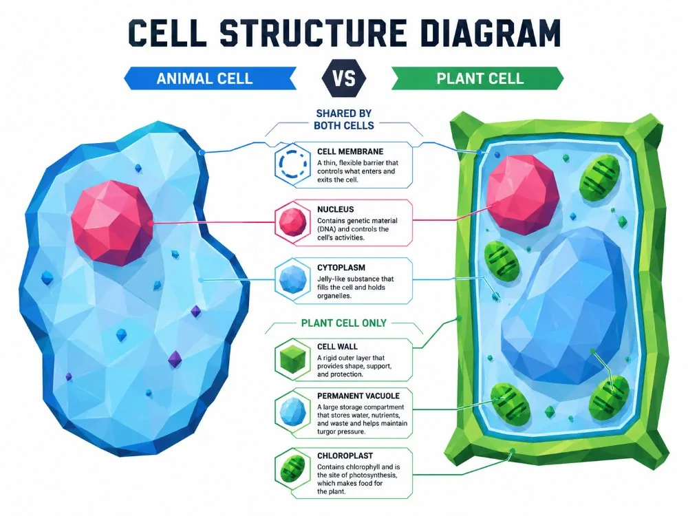
Animal cell vs plant cell labelled
Animal Cell and Plant Cell
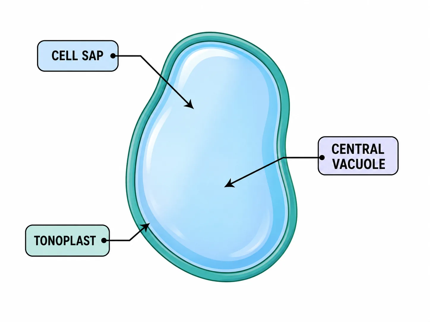
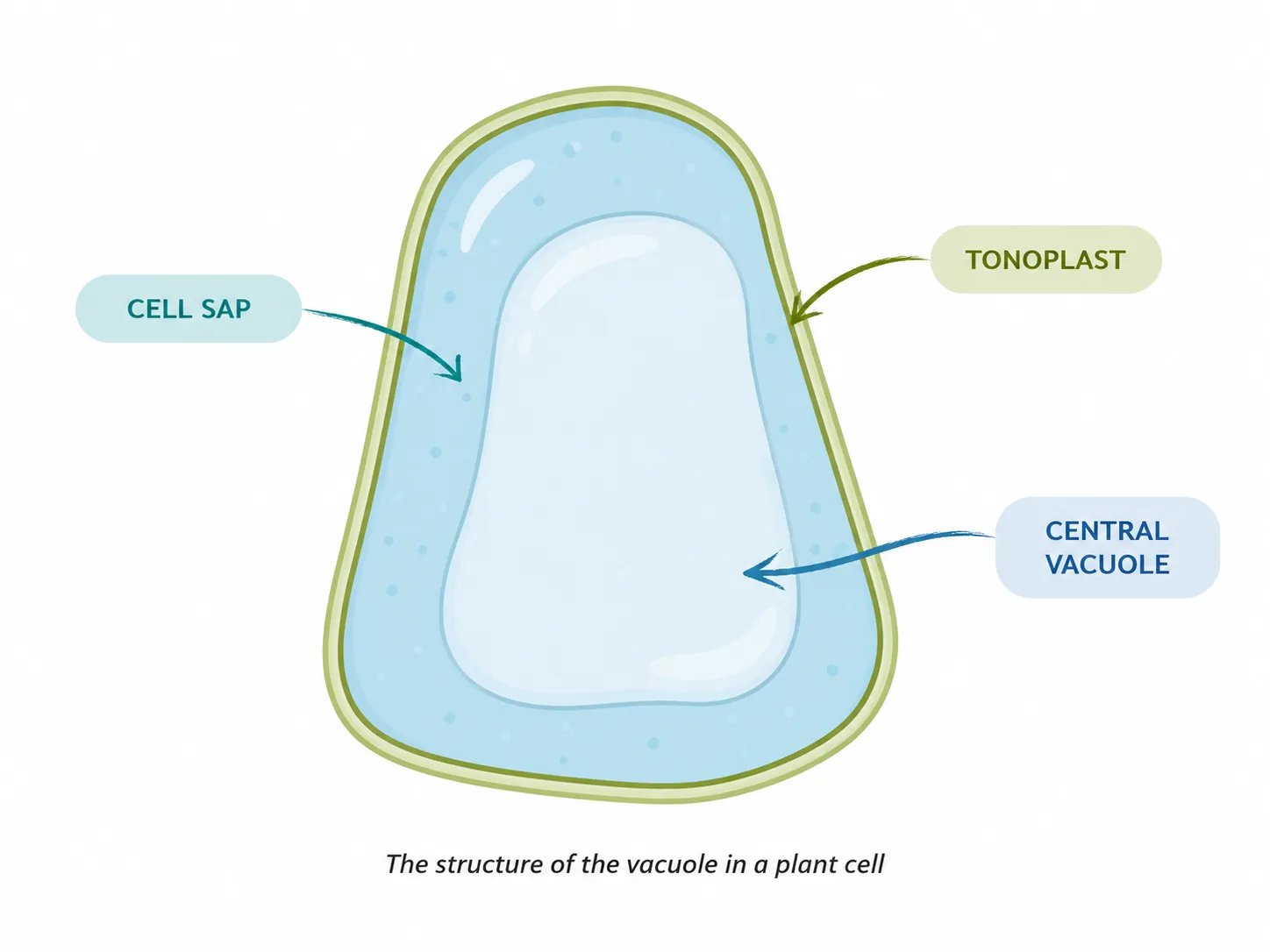
Plant vacuole showing cell sap and tonoplast
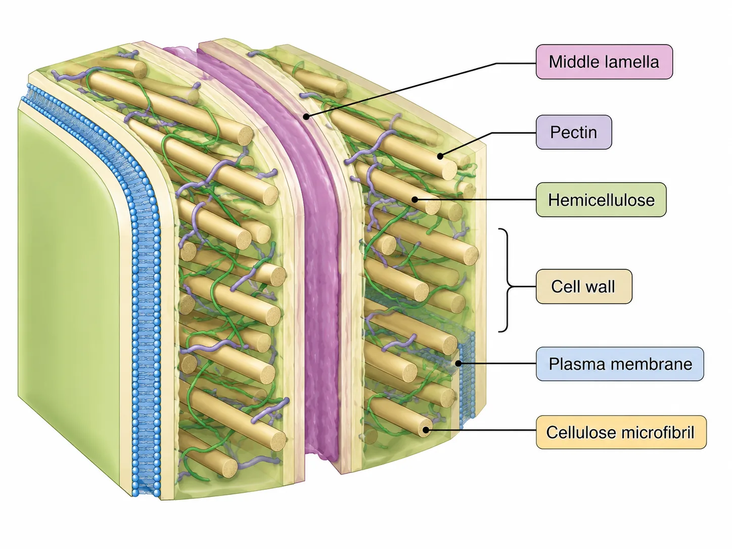
Plant Cell Wall Structure
plant cell vacuole
plant cell wall structure
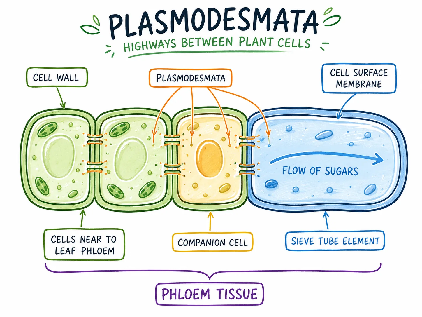
Plasmodesmata — Highways Between Plant Cells (Click-by-Click Lesson)
Animal Cell and Plant Cell
Plant Cell Vacuole — Interactive Diagram
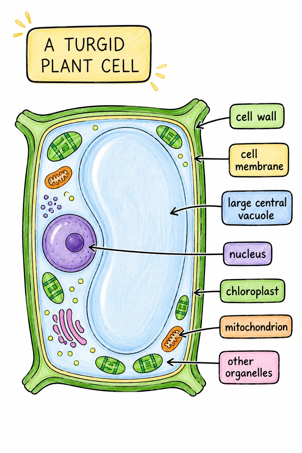
r099-turgid-plant-cell
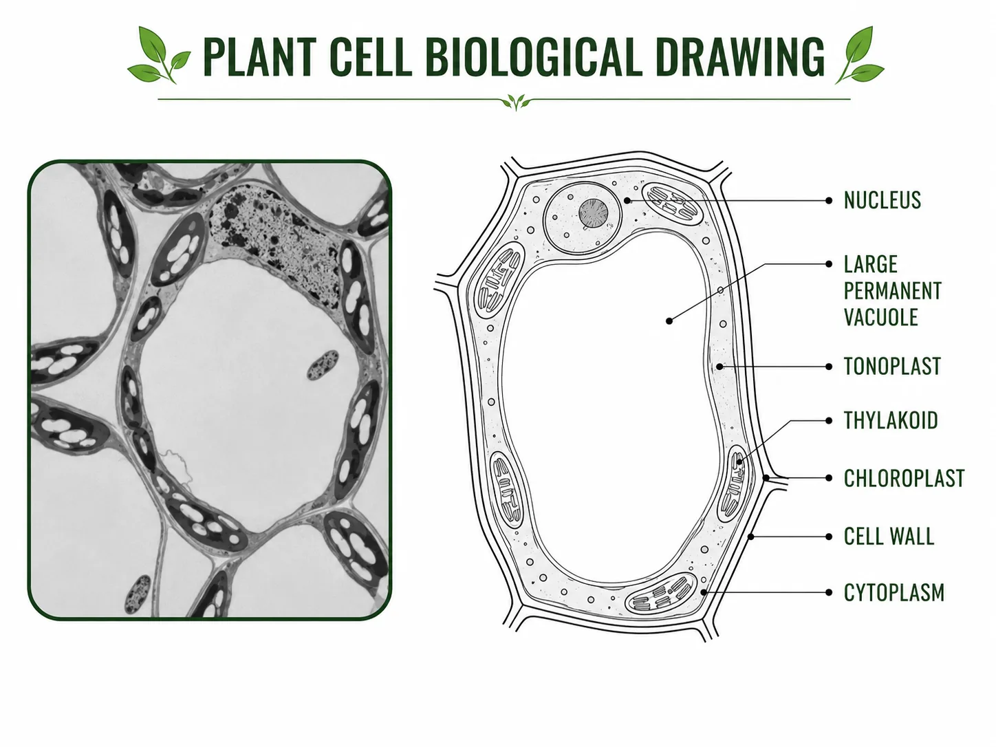
Plant Cell Biological Drawing — Click-by-Click Animation
Cell structure: animal vs plant
pasted 1782543983359 0
Plant cell (detailed labelled)
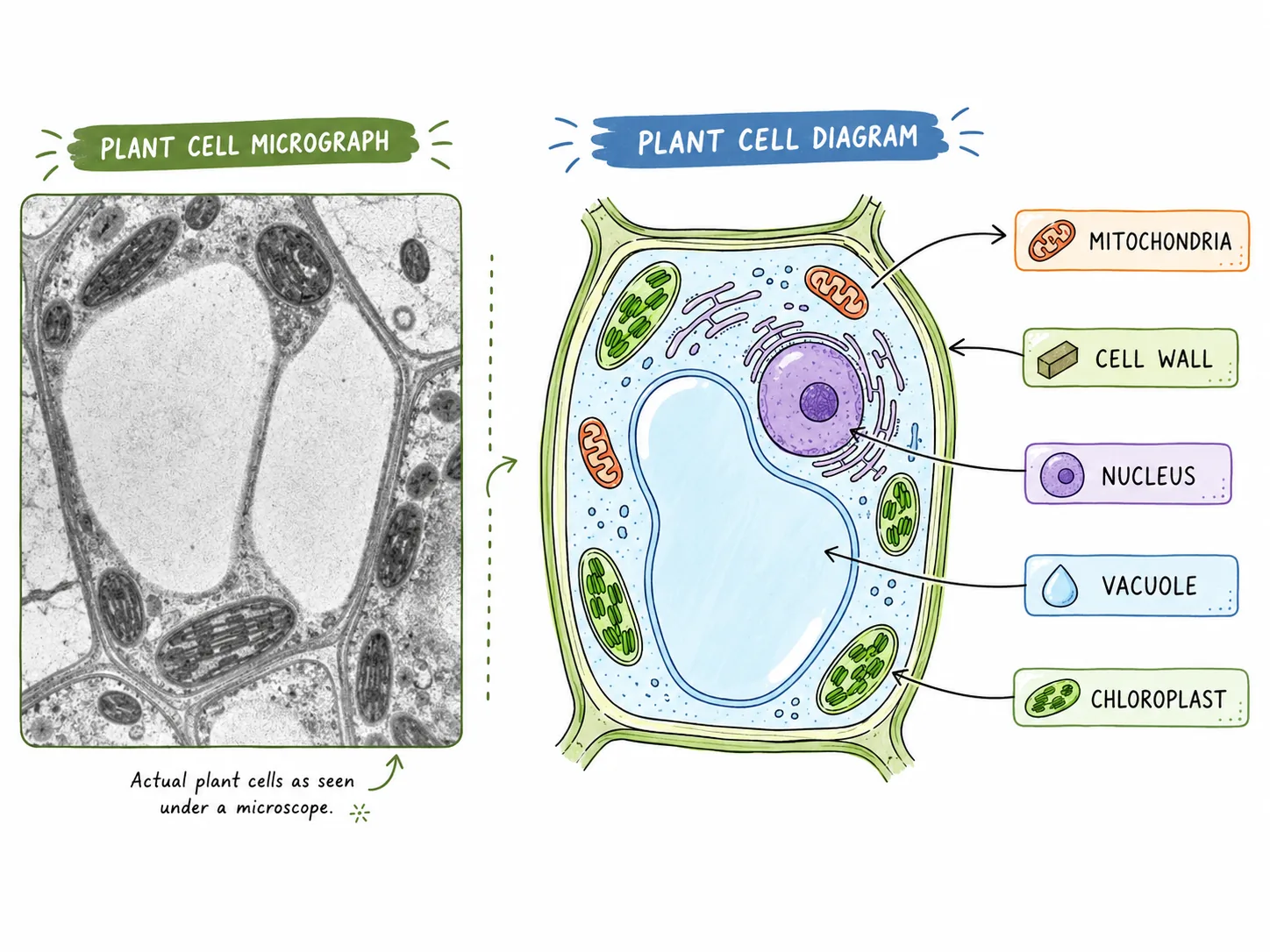
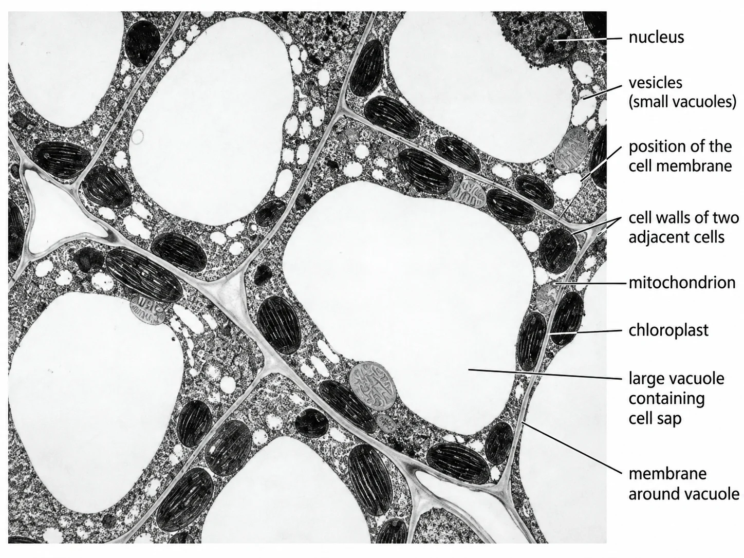
Plant Cell — Micrograph and Diagram
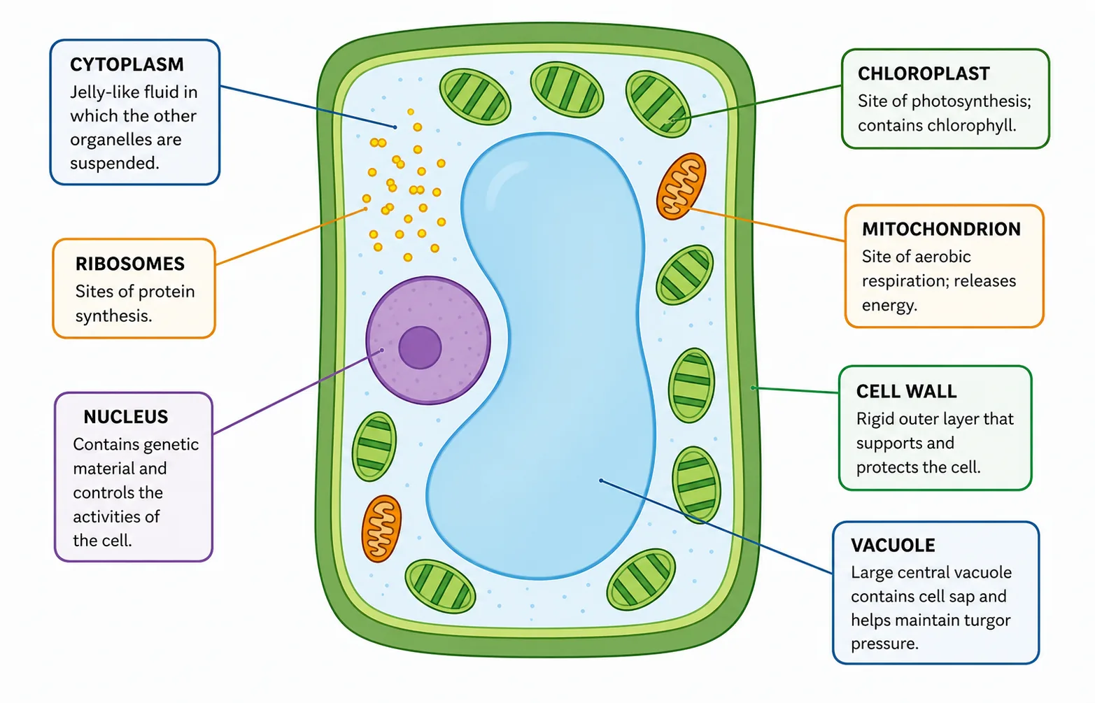
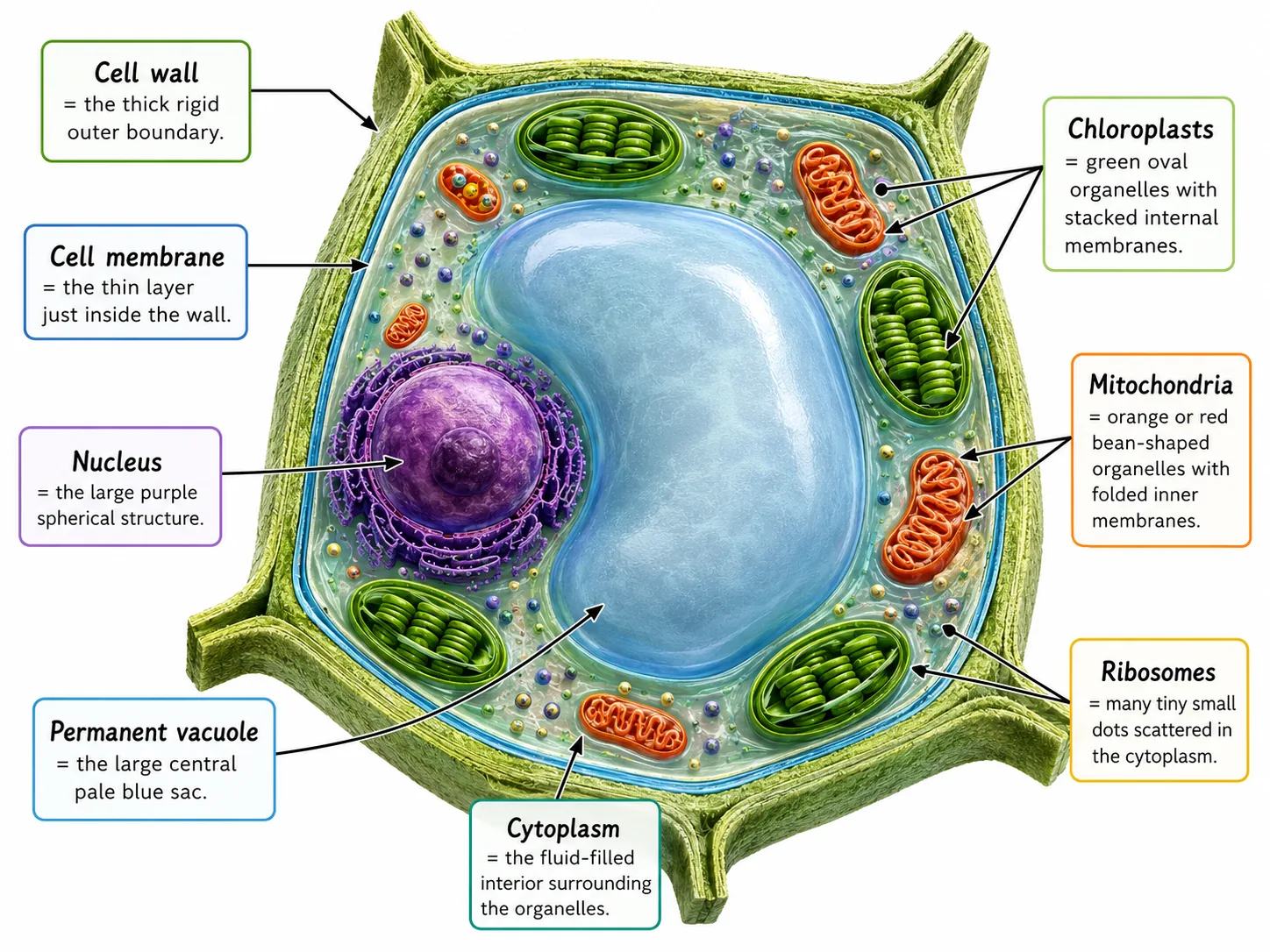
Plant cell labelled with descriptions
Typical plant cell (labelled)
Plant cell wall structure
Central vacuole and tonoplast
Chloroplast ultrastructure
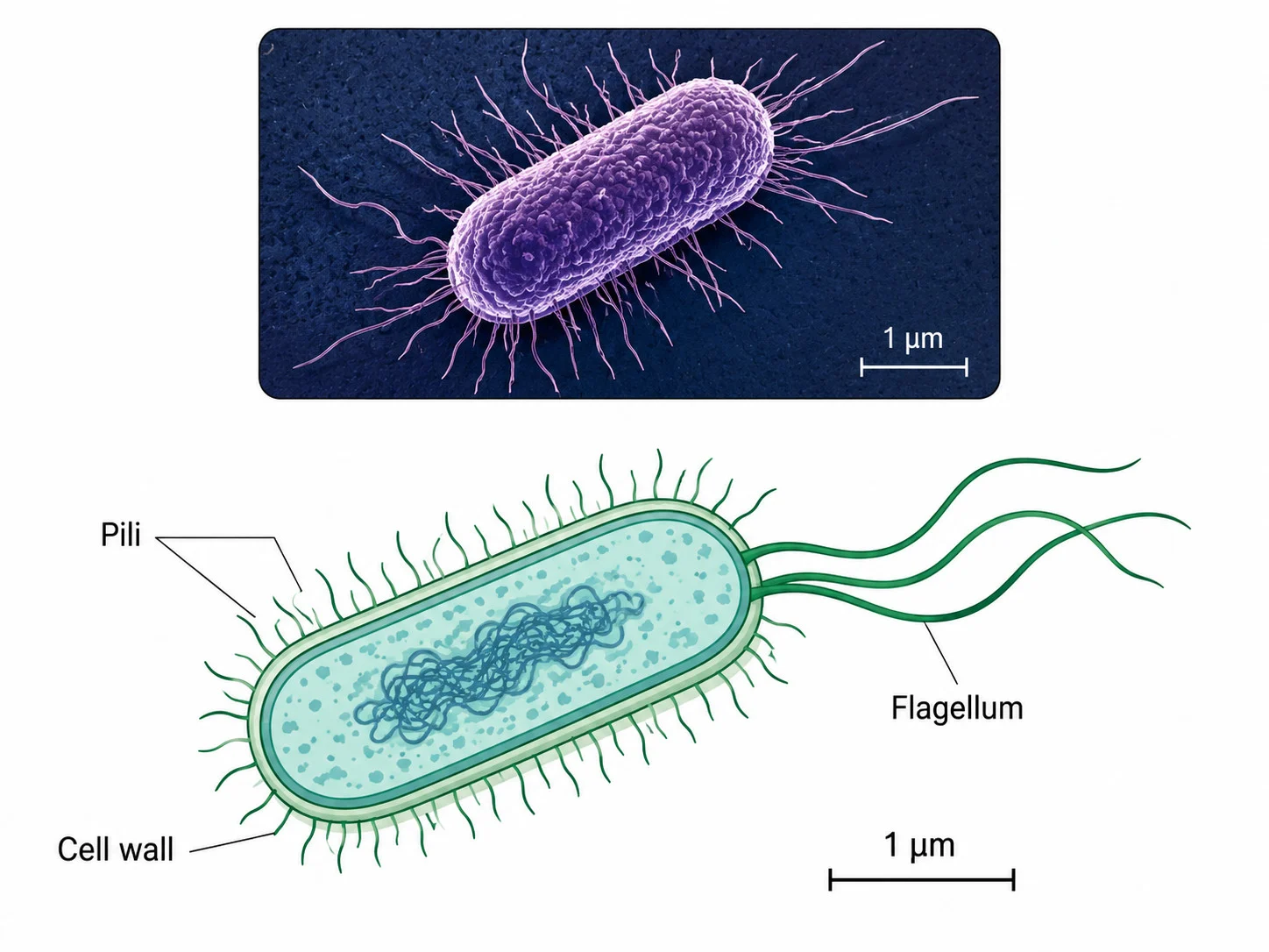
Bacterial ultrastructure
Plant cell structures in a micrograph-style view

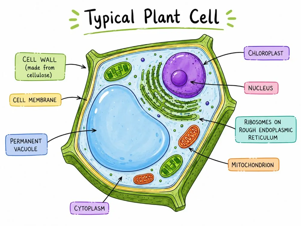
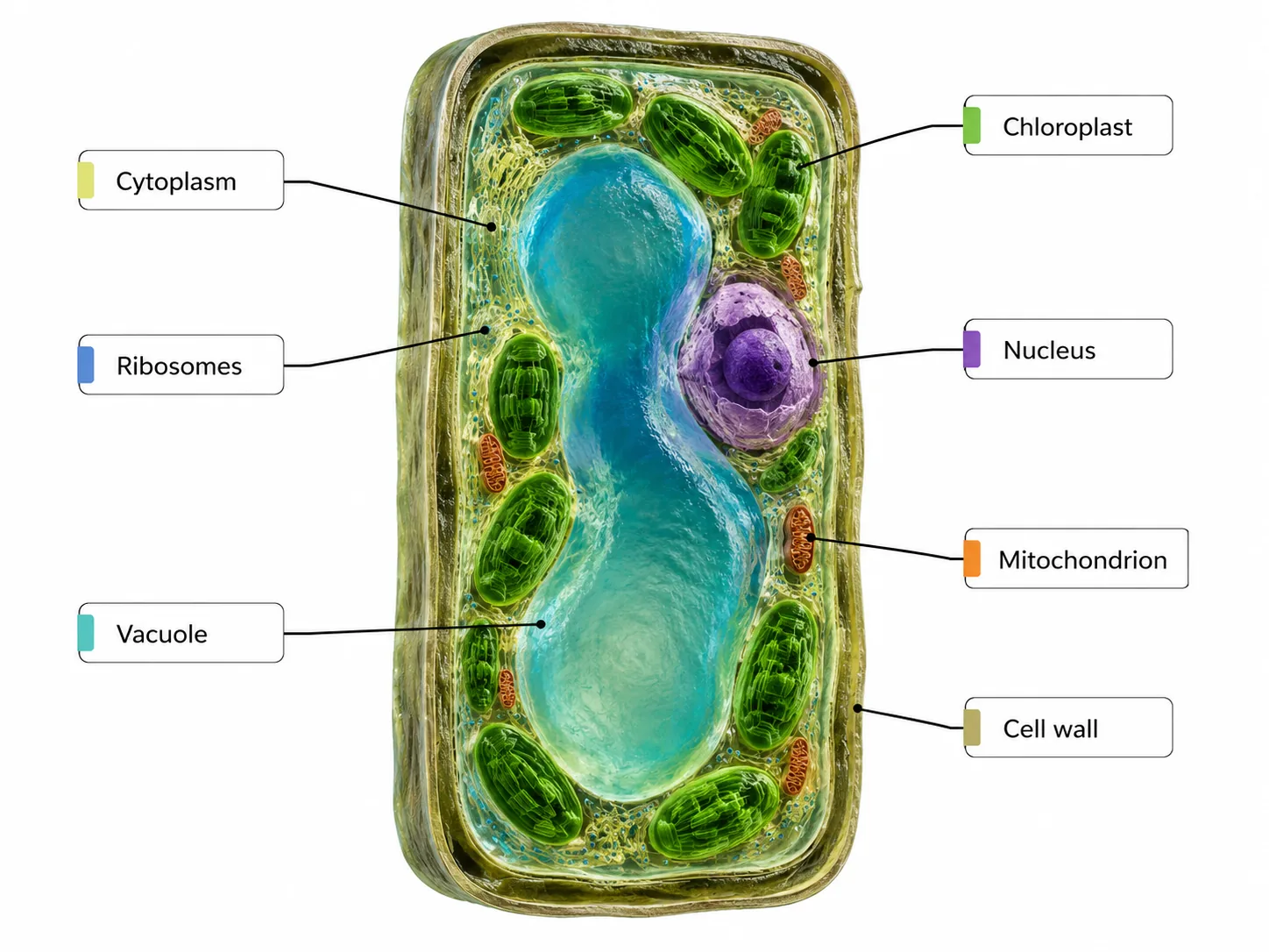
Plant cells have three additional features that animal cells lack:

Extra structures in plant cells
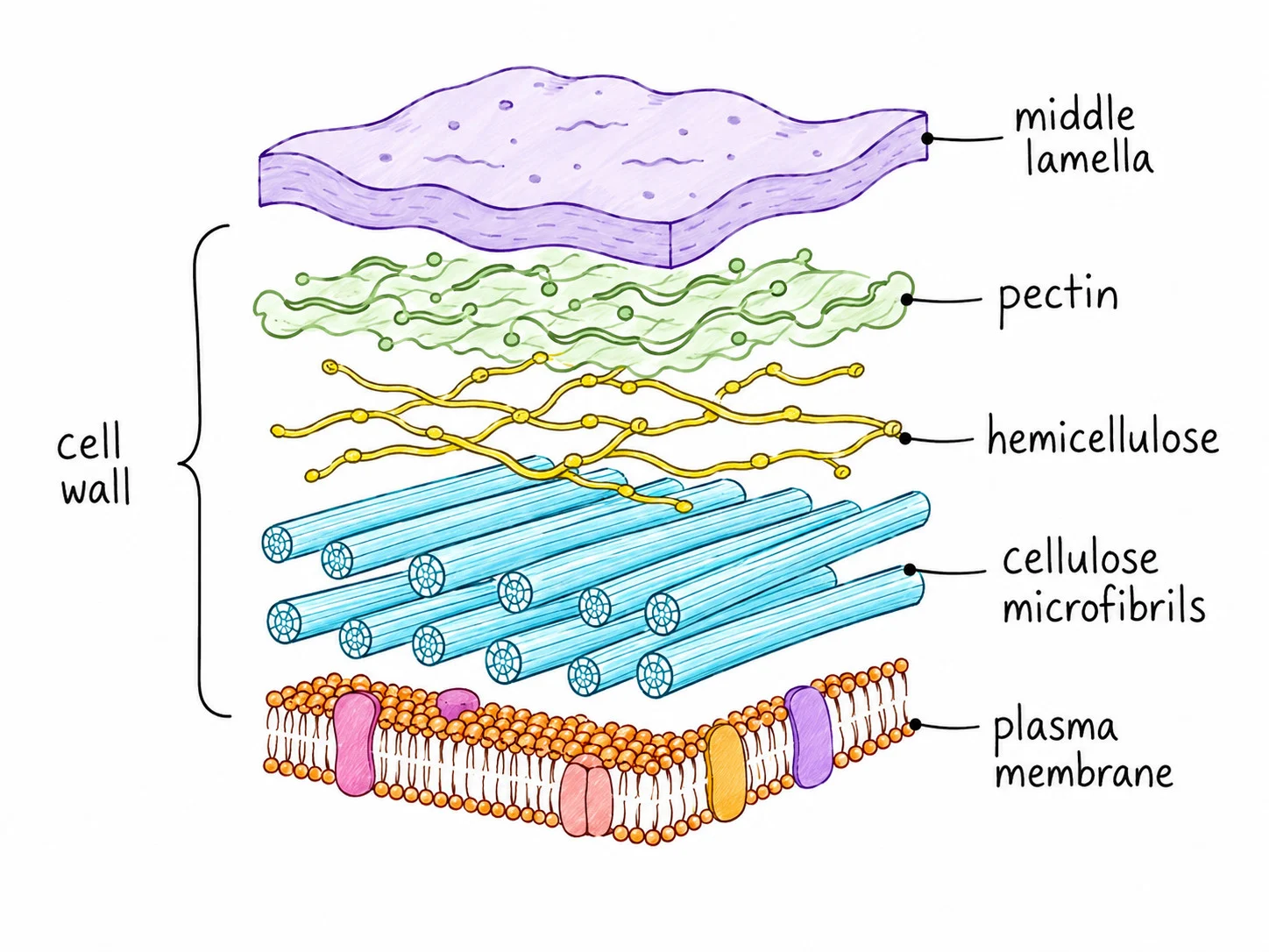
Cell wall
Cell wall a rigid layer made of cellulose outside the membrane. It strengthens the cell and supports the plant, and it stops the cell bursting when water enters.
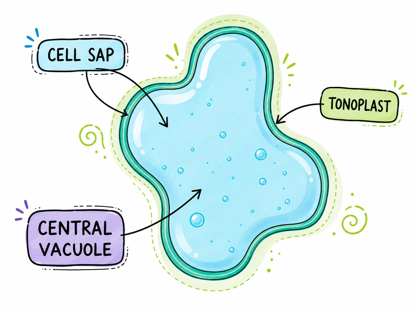
Permanent vacuole
Permanent vacuole a large space filled with cell sap (a solution of sugars and salts). It keeps the cell firm and stores substances.
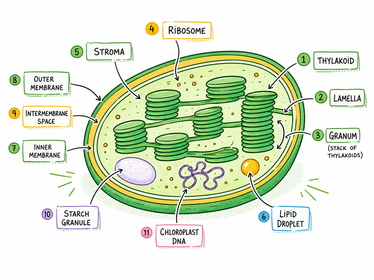
Chloroplasts
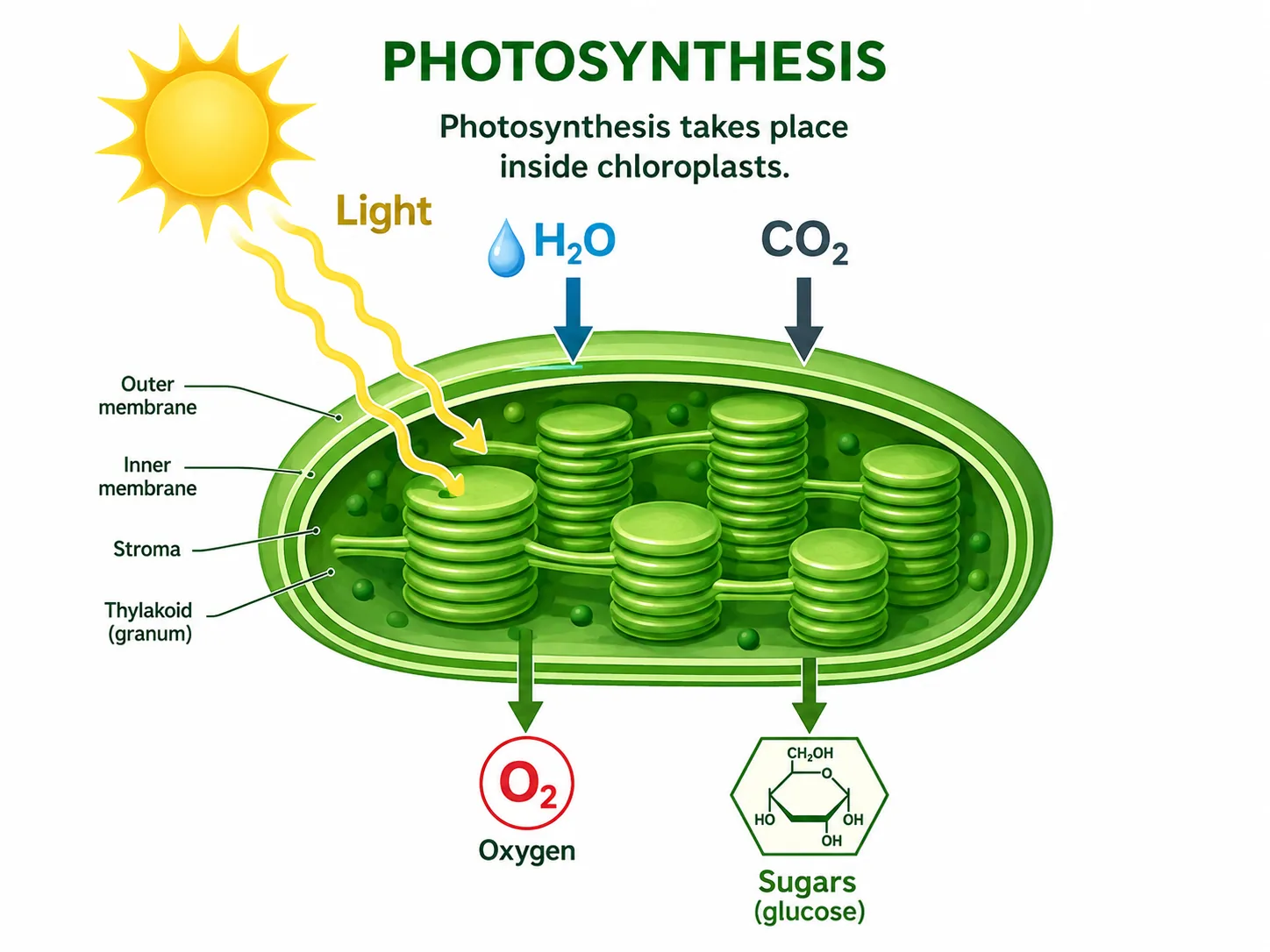
Chloroplasts contain the green pigment chlorophyll; they absorb light for photosynthesis. They are found in the green parts of a plant, such as leaf cells, but not in root cells.

Section 3 of 9Comparison table

StructureAnimal cellPlant cell
NucleusYesYes
Cell membraneYesYes
MitochondriaYesYes
RibosomesYesYes
Cellulose cell wallNoYes
Permanent vacuoleNoYes
ChloroplastsNoYes (green parts)

Section 4 of 9Linking structure to function

pasted 1782294312670 0
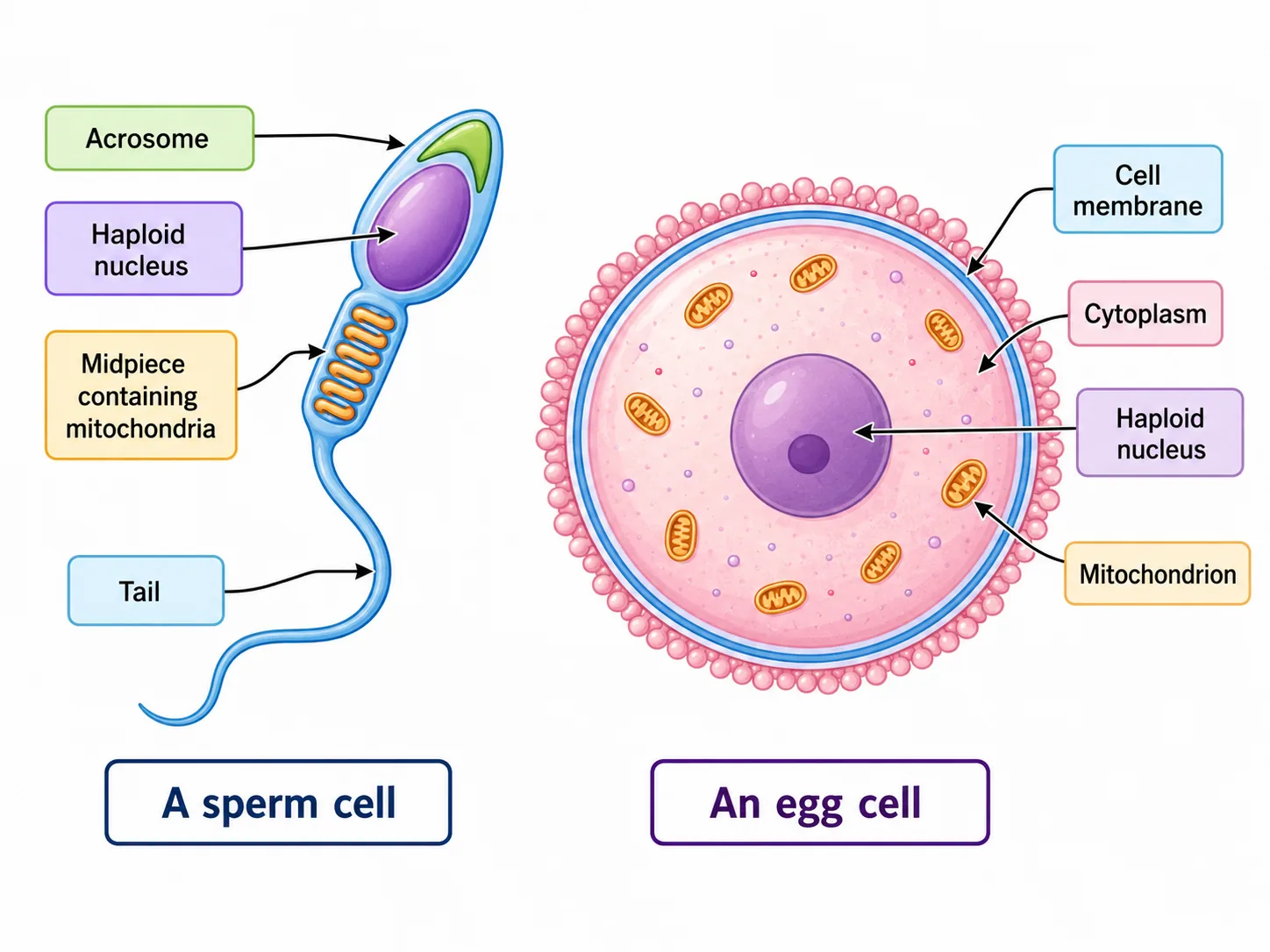
Sperm Cell — Structure
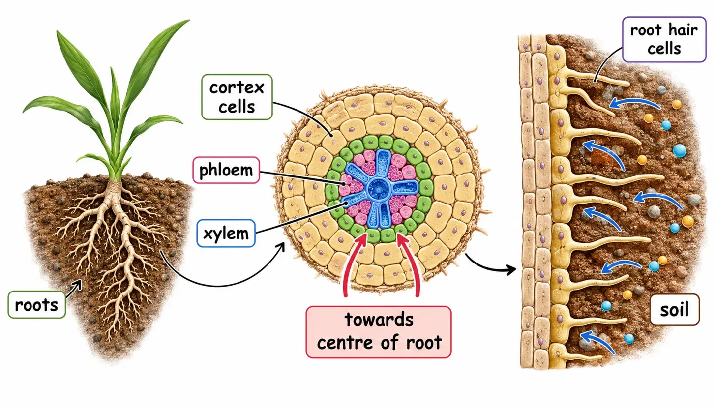
Root Structure and Root Hair Cells

Examination questions often give an unfamiliar cell and ask you to link a feature to a function.

1
The method is to name the structure and then state what it does and what that allows the cell to do.
2
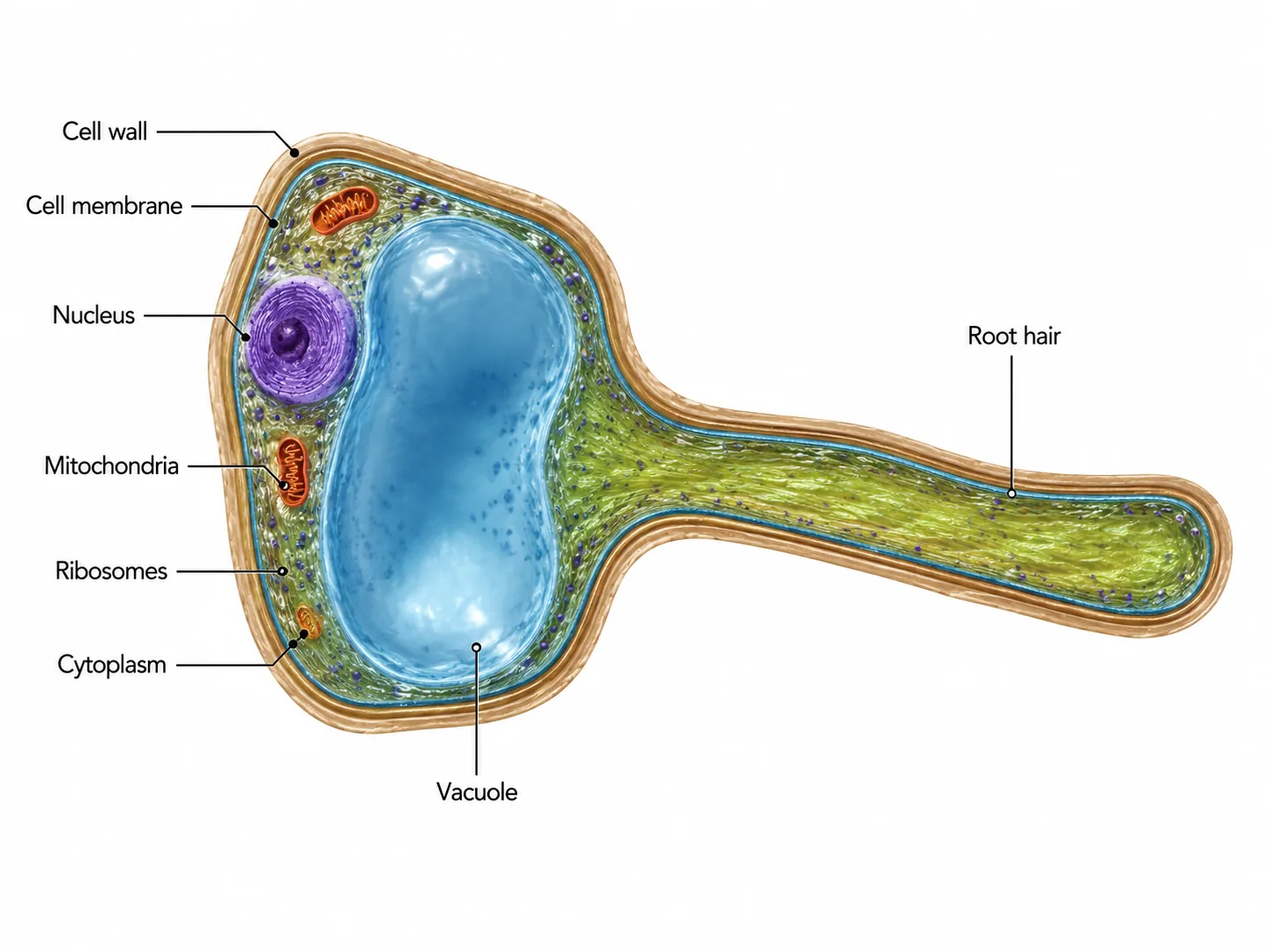
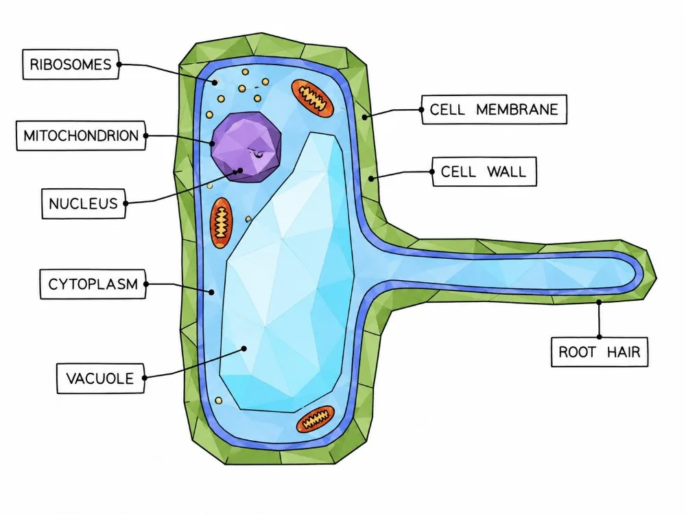
A root hair cell has no chloroplasts because it is underground and receives no light, so photosynthesis cannot occur there.
3
A leaf palisade cell is packed with chloroplasts so that it can absorb as much light as possible.
4
A sperm cell contains many mitochondria to supply the energy for swimming.
5
Notice that in each case the number or presence of a structure reflects the energy or process the cell must carry out.

The cell wall is a favourite exam trap. It is not the same as the cell membrane: the wall is fully permeable and rigid, while the membrane is thin and partially permeable and found in every cell.

Section 5 of 9Studying cells under the microscope

Light Microscope vs Electron Microscope
Plant Cell — Light vs Electron Microscope
Plant cell under light vs electron microscope
Plant cell under light microscope vs electron microscope

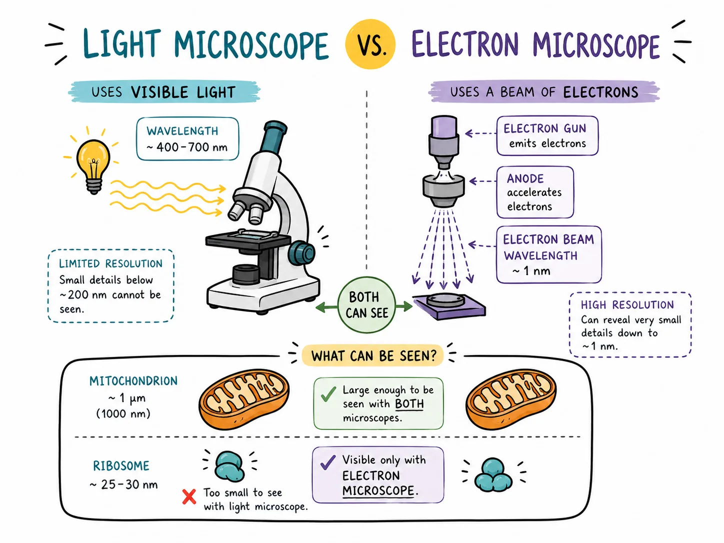
The nucleus, and in plants the cell wall, vacuole and chloroplasts, can be seen with a light microscope.

Studying cells under the microscope
Mitochondria and ribosomes are too small to see in detail, and need an electron microscope; this is covered in the microscopy lesson.
When drawing a cell, use a sharp pencil, label with ruled lines that do not cross, and draw the outline as a single continuous line.

Section 6 of 9Worked approach to unfamiliar cells
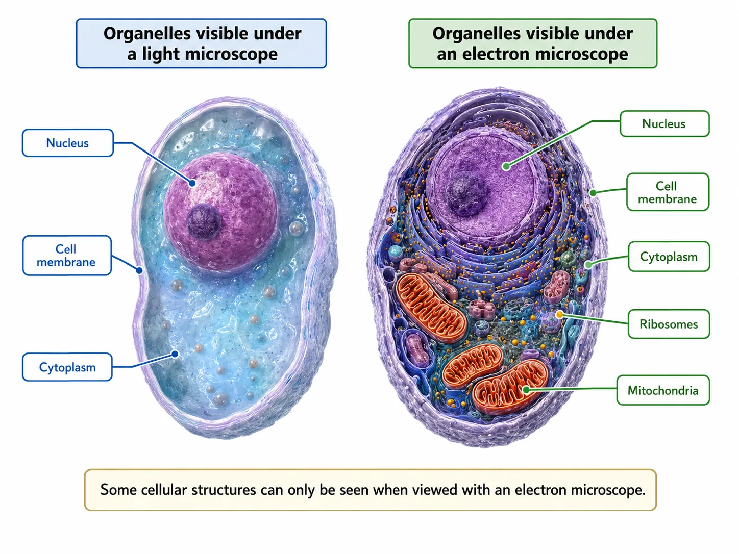
Organelles visible under light vs electron microscope

A reliable way to answer questions about a cell you have never seen is to ask three things in turn.

1
First, is it eukaryotic?
2
If a nucleus is visible, it is.
3
Second, is it plant or animal?
4
Look for a cell wall, a large vacuole or chloroplasts: if any is present the cell is a plant cell.
5
Third, what does its unusual feature tell you about its job?

Consider a cell from the lining of the small intestine.

Worked approach to unfamiliar cells
It has many mitochondria and a surface folded into tiny projections.
You would conclude that it carries out a process needing energy, such as absorption by active transport, and that the folds increase surface area.
Consider instead a cell from the middle of a leaf which has a large number of chloroplasts, a cell wall and a big vacuole; it is a plant cell specialised for photosynthesis.

Remember also that some structures can be linked to size. The nucleus is usually the largest organelle in an animal cell, about 10 micrometres across, while mitochondria are about 1 micrometre and ribosomes only around 25 nanometres. This is why a light microscope shows the nucleus clearly but cannot show ribosomes at all.

Finally, do not forget the cytoplasmalthough it looks featureless, it is where many enzyme-controlled reactions happen, and it holds the organelles in position.

Section 7 of 9Exam-style questions
Plant cell labelled (chloroplast, vacuole, cell wall)
Internal Structure of a Leaf and a Palisade Mesophyll Cell
Labelled root hair cell showing organelles
mitochondria chloroplast landscape
photosynthesis chloroplast
Aerobic Respiration
Aerobic Respiration
aerobic respiration
aerobic respiration
Root hair cell structure
  1. Name the structure in a plant cell that (a) contains chlorophyll, (b) is made of cellulose, (c) contains cell sap. [3]3 marks

  2. Muscle cells contain a large number of mitochondria. Explain why. [2]2 marks

  3. Explain why root hair cells do not contain chloroplasts, but leaf palisade cells do. [3]3 marks

Section 8 of 9Key terms

TermMeaning
Cell membraneThin partially permeable layer that controls what enters and leaves the cell.
MitochondriaOrganelles where aerobic respiration releases energy.
RibosomeSite of protein synthesis.
Cell wallRigid cellulose layer outside the plant cell membrane.
Permanent vacuoleSap-filled space that keeps a plant cell firm.
ChloroplastOrganelle containing chlorophyll, where photosynthesis occurs.
ChlorophyllGreen pigment that absorbs light energy.

Section 9 of 9Exam technique

1
Saying the cell wall controls what enters and leaves the cell. That is the job of the cell membrane.
2
Claiming plant cells do not have mitochondria. They do, because they respire as well as photosynthesise.
3
Writing that the nucleus 'makes' energy. The mitochondria release energy; the nucleus controls the cell.
4
Saying chloroplasts are found in all plant cells. Only the cells exposed to light have them.
Cambridge 9700 AS Biology Core Formula

Magnification Triangle & Unit Conversion

Formula: M = I / A  ⇔  A = I / M  ⇔  I = M × A

Units: 1 mm = 1,000 μm = 1,000,000 nm.
Rule: Convert Image size (ruler mm) to μm by ×1,000 before dividing by Actual size.