Section 1 of 9The need for greater resolving power

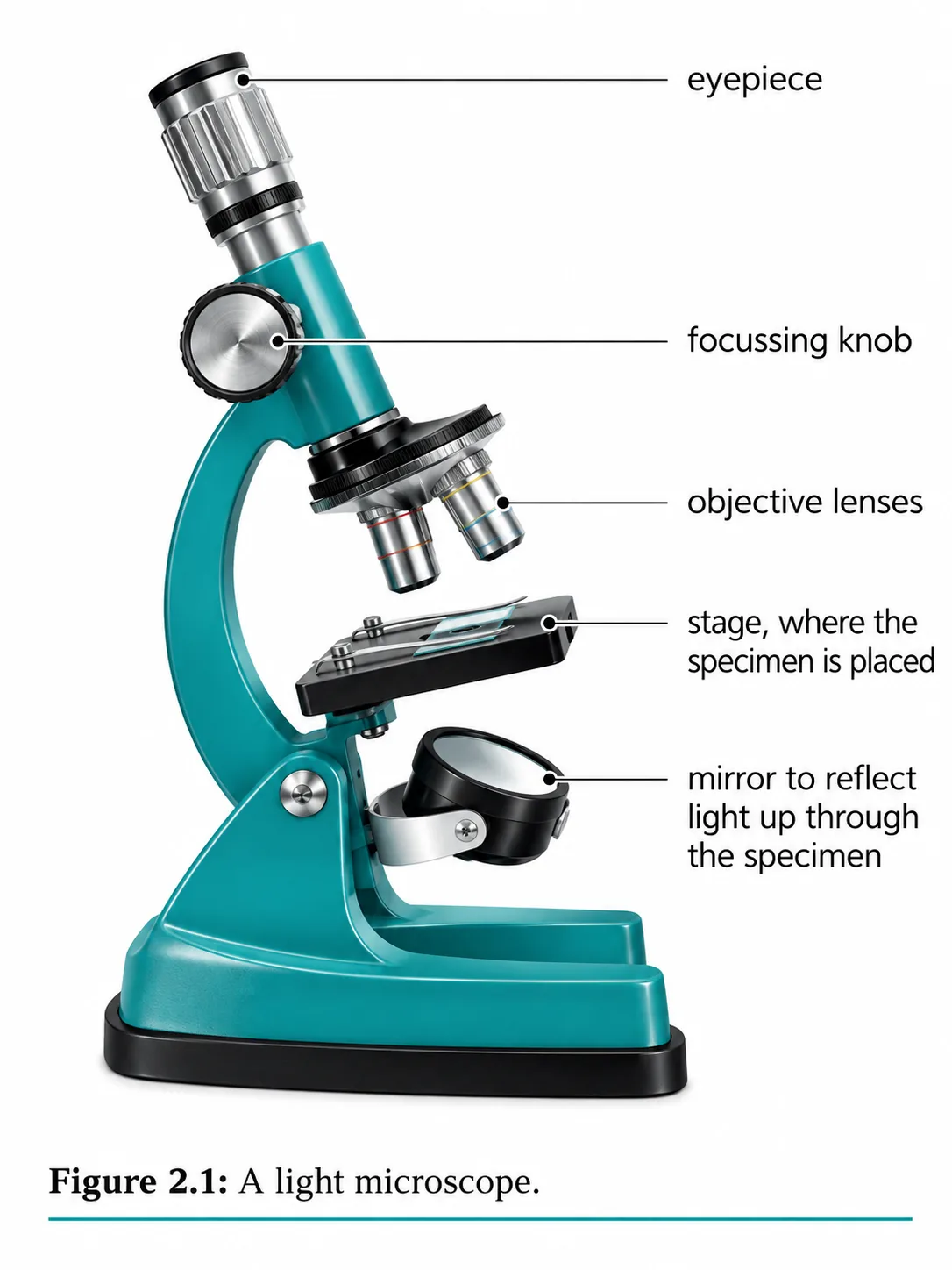

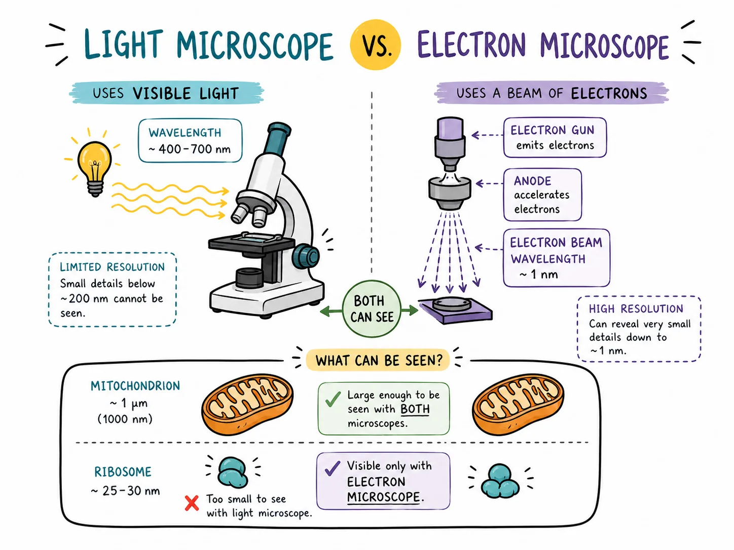
The light microscope is limited by the wavelength of visible light, so it cannot resolve objects smaller than about 0.2 micrometres.
- which allows far greater resolution, and magnifications of more than ×1 000 000 are possible.
Section 2 of 9Two types
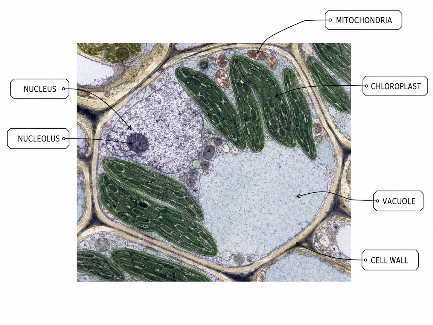
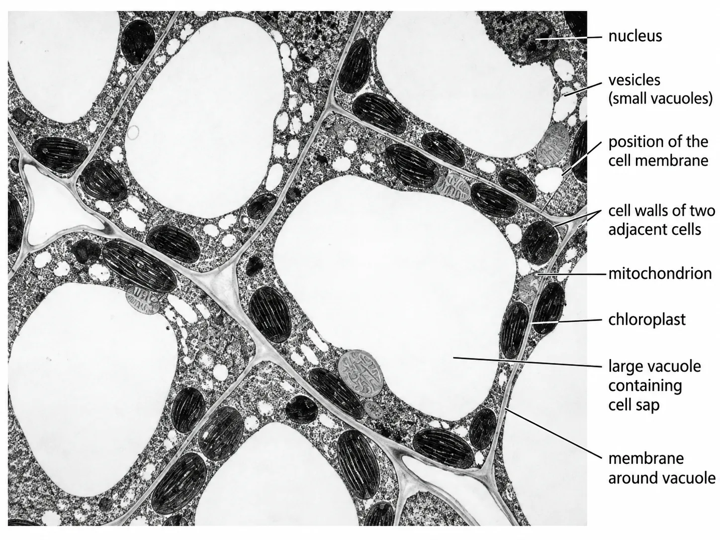


A transmission electron microscope (TEM) passes electrons through a very thin slice of specimen.
- giving a 2-D image of the internal structure of cells, such as the details of organelles.
- producing a black and white image (an electron micrograph) that may be coloured by computer afterwards.
Section 3 of 9Comparison with the light microscope


| Feature | Light microscope | Electron microscope |
|---|---|---|
| Radiation used | Visible light | Beam of electrons |
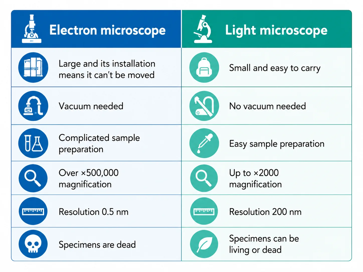
| Maximum magnification | About ×1500 | More than ×1 000 000 |
| Resolution | About 0.2 micrometres | About 0.1 nanometres (TEM) |
| Specimen | Can be living | Dead, in a vacuum |
| Image | Colour (with stains) | Black and white |
| Cost and size | Cheap, portable | Very expensive, large, needs training |
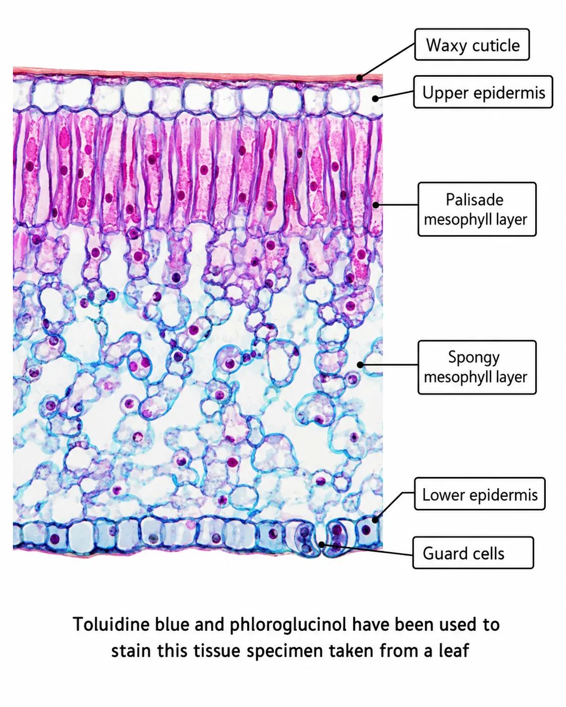
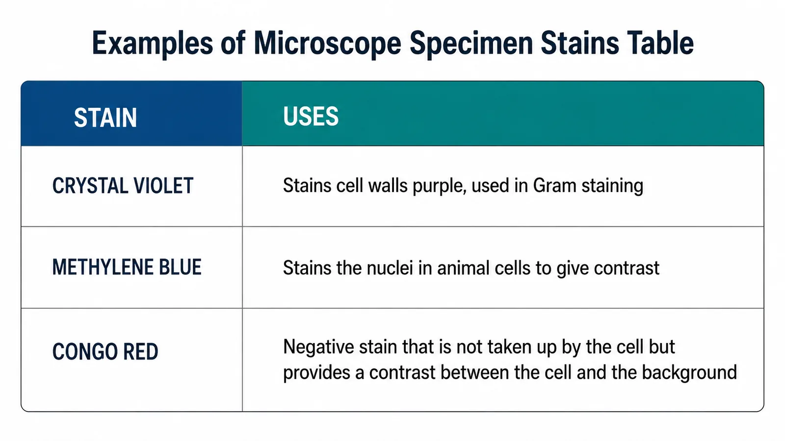
The specimen must be dead for an electron microscope because the electrons only travel through a vacuum, and the preparation, which includes fixing, dehydrating and staining with heavy metals, can create artefacts: structures in the image that were not present in the living cell.
Section 4 of 9What electron microscopy revealed
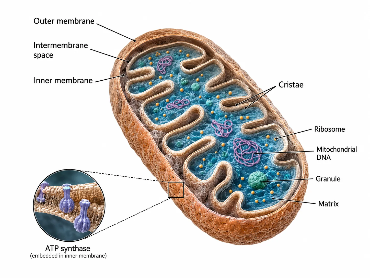

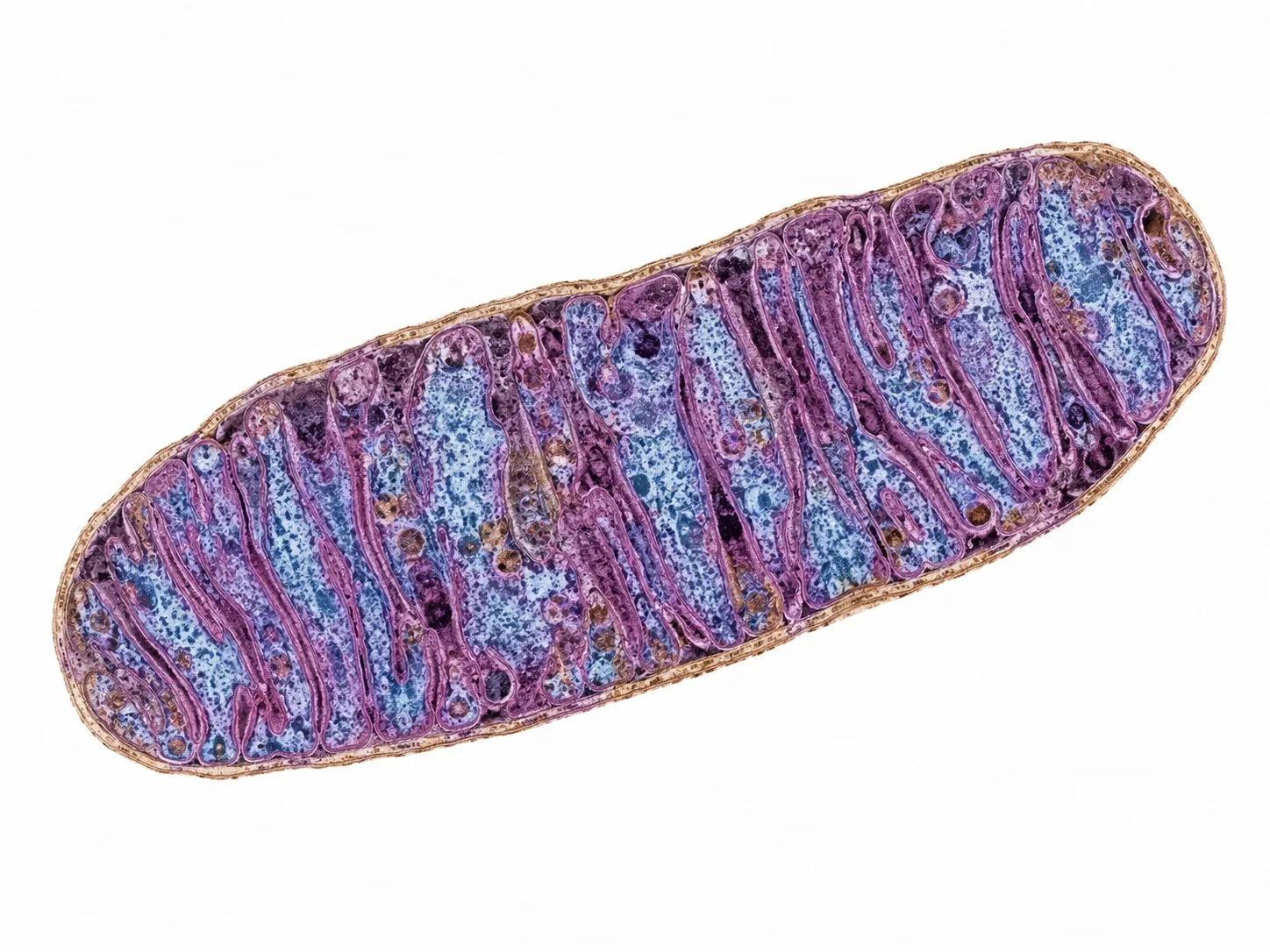
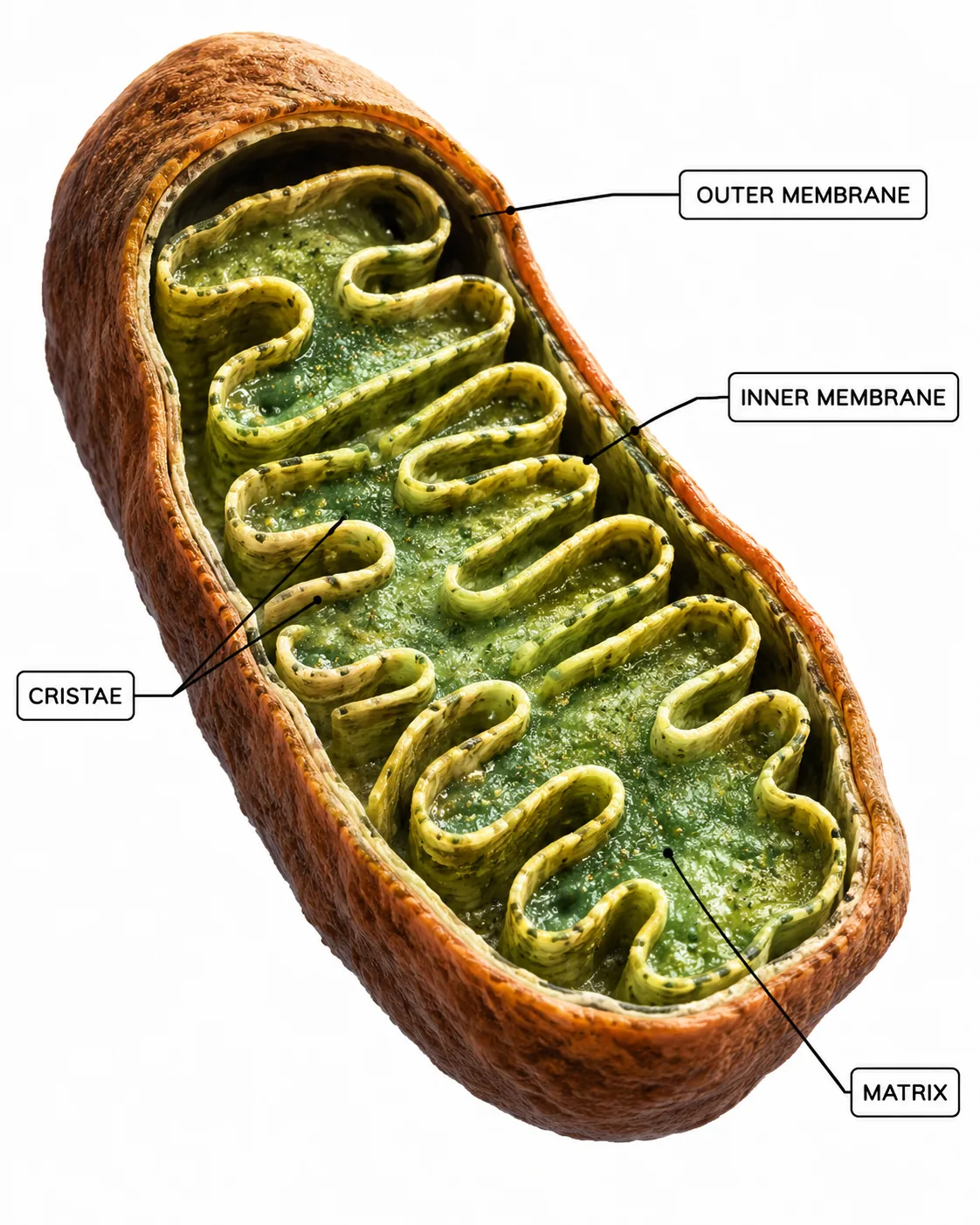

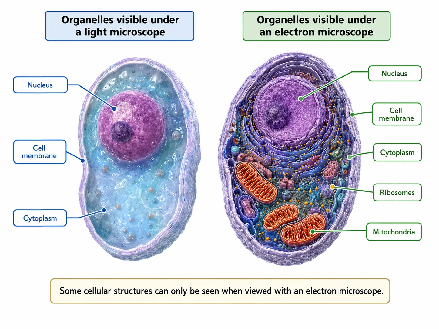


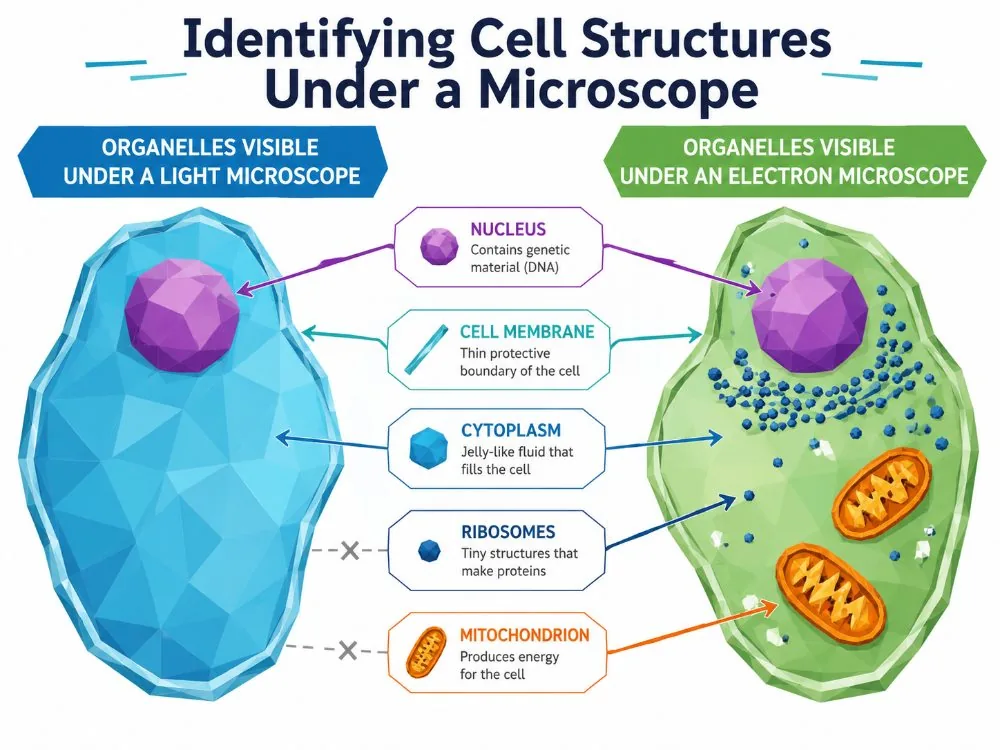
Electron microscopes have allowed biologists to see sub-cellular structures in detail.
- and the structure of bacteria and viruses.
Section 5 of 9Calculations with scale
For electron micrographs, the units are often micrometres or nanometres. 1 mm = 1000 µm and 1 µm = 1000 nm.
- and use it to calculate the magnification: magnification = measured length of scale bar / real length it represents.
Section 6 of 9Worked example: reading a micrograph


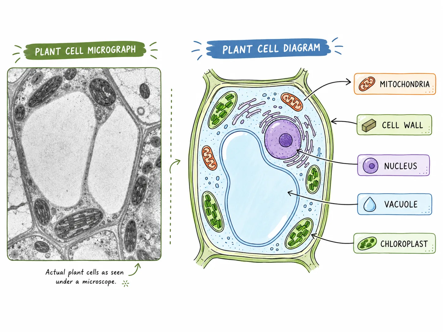

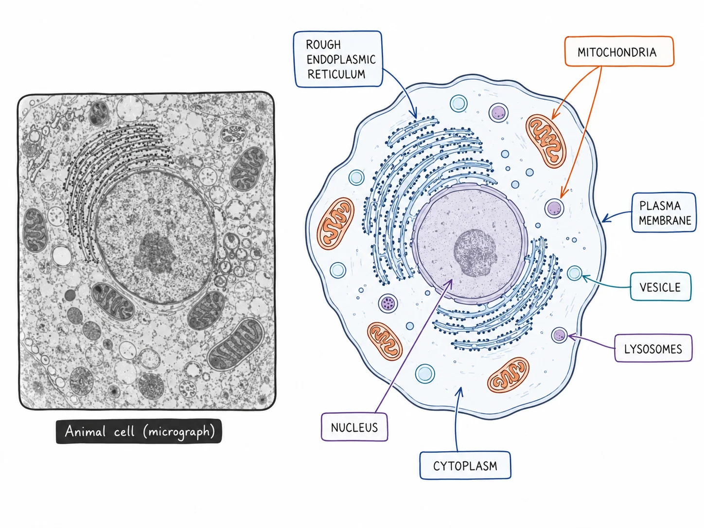
Suppose a TEM image shows a chloroplast and a scale bar of 20 mm that represents 2 micrometres.
- If the chloroplast in the image is 50 mm long, then its actual length is 50 / 10 000 = 0.005 mm = 5 micrometres.
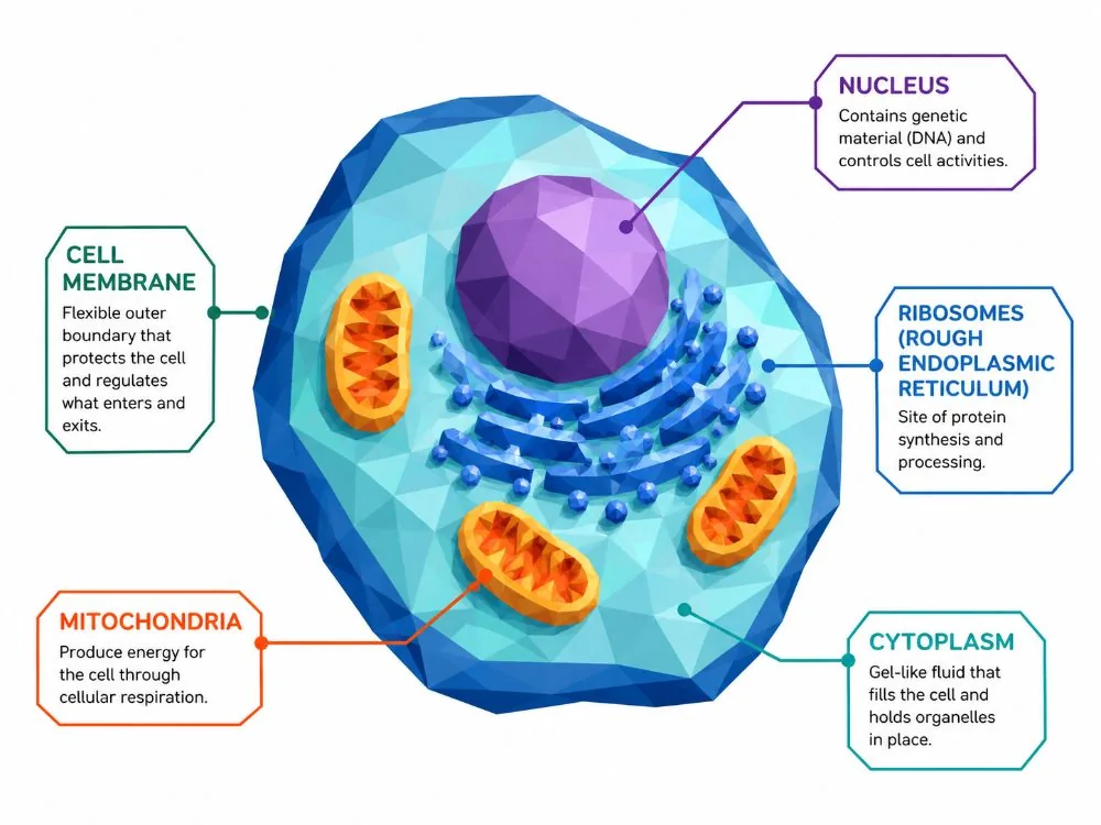
When describing what you can see on a micrograph, name the organelles by their features: a mitochondrion appears as a small oval with folded inner membranes; a nucleus is a large round structure with a double membrane and pores; ribosomes are tiny dark dots; chloroplasts have stacks of membranes; and a cell wall appears as a thick line outside the membrane in plant cells.
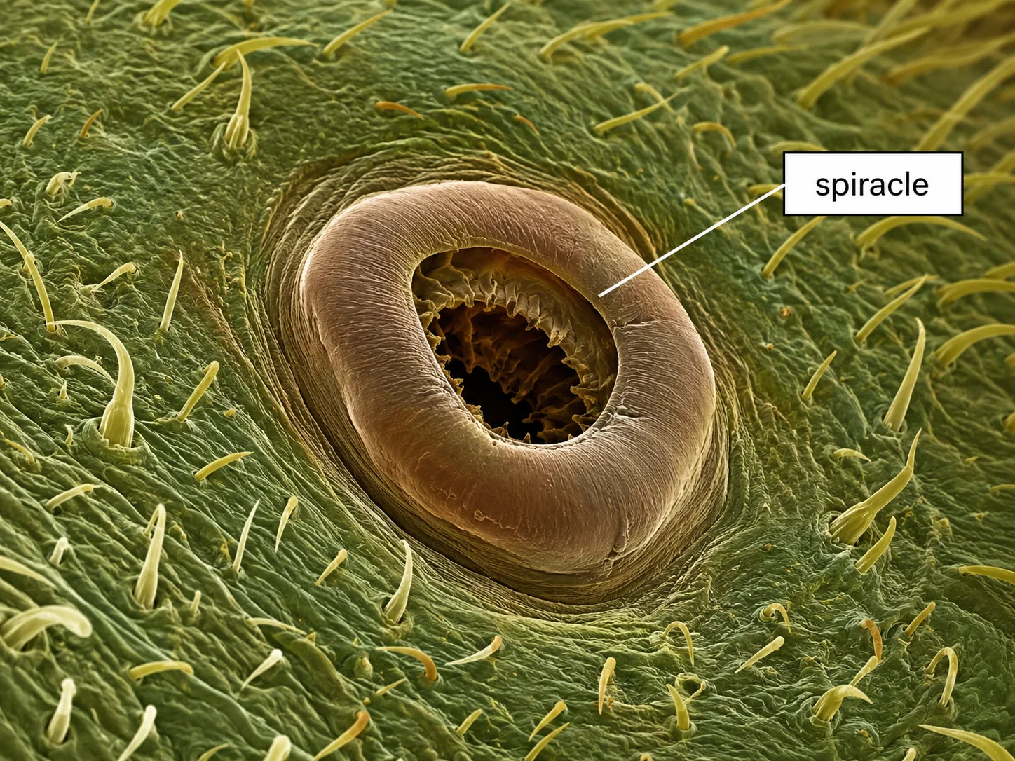
Applications also extend beyond cellsSEM images are used to study the structure of insects, pollen and fibres, while TEM is used to study viruses and the details of the cell membrane.
- which is why electron microscopy results are checked against other methods.
Section 7 of 9Exam-style questions



An electron micrograph shows a mitochondrion that measures 45 mm long. The magnification is ×30 000. Calculate the actual length in micrometres. [3]3 marks
1. Actual size = 45 / 30 000 [1].
2. = 0.0015 mm [1].
3. 0.0015 × 1000 = 1.5 µm [1].
Explain why electron microscopes have been able to show more detail of cell structures than light microscopes. [3]3 marks
Exam-style questions1. Electron microscopes use electron beams, which have a much shorter wavelength than light [1].2. This gives a much greater resolution, so very small structures such as ribosomes can be seen as separate [1].3. Higher magnification can be used without blurring, so internal details of organelles are seen [1].
Section 8 of 9Key terms
| Term | Meaning |
|---|---|
| Electron microscope | A microscope that uses a beam of electrons to give high magnification and resolution. |
| TEM | Transmission electron microscope, which shows thin sections in 2-D. |
| SEM | Scanning electron microscope, which shows the surface in 3-D. |
| Resolution | The ability to see two close points as separate. |
| Wavelength | The distance between two successive peaks of a wave. |
| Artefact | A feature on an image created by the preparation, not present in the living specimen. |
| Nanometre | One billionth of a metre (1 nm = 0.001 micrometre). |
Section 9 of 9Exam technique
Marked practiceQuizzes for this lesson
Magnification Triangle & Unit Conversion
Formula: M = I / A ⇔ A = I / M ⇔ I = M × A
Units: 1 mm = 1,000 μm = 1,000,000 nm.
Rule: Convert Image size (ruler mm) to μm by ×1,000 before dividing by Actual size.