Section 1 of 9Gas exchange in the human lungs


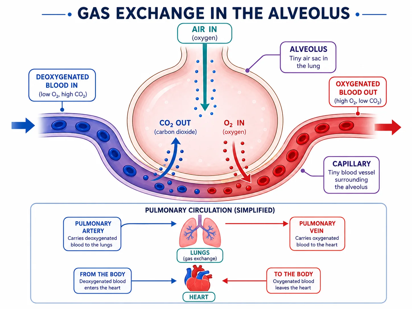

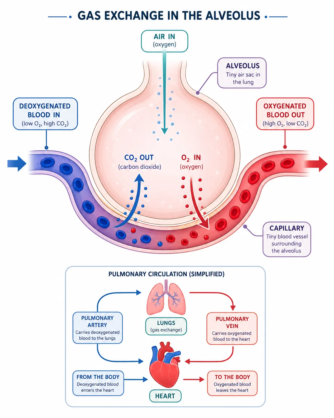

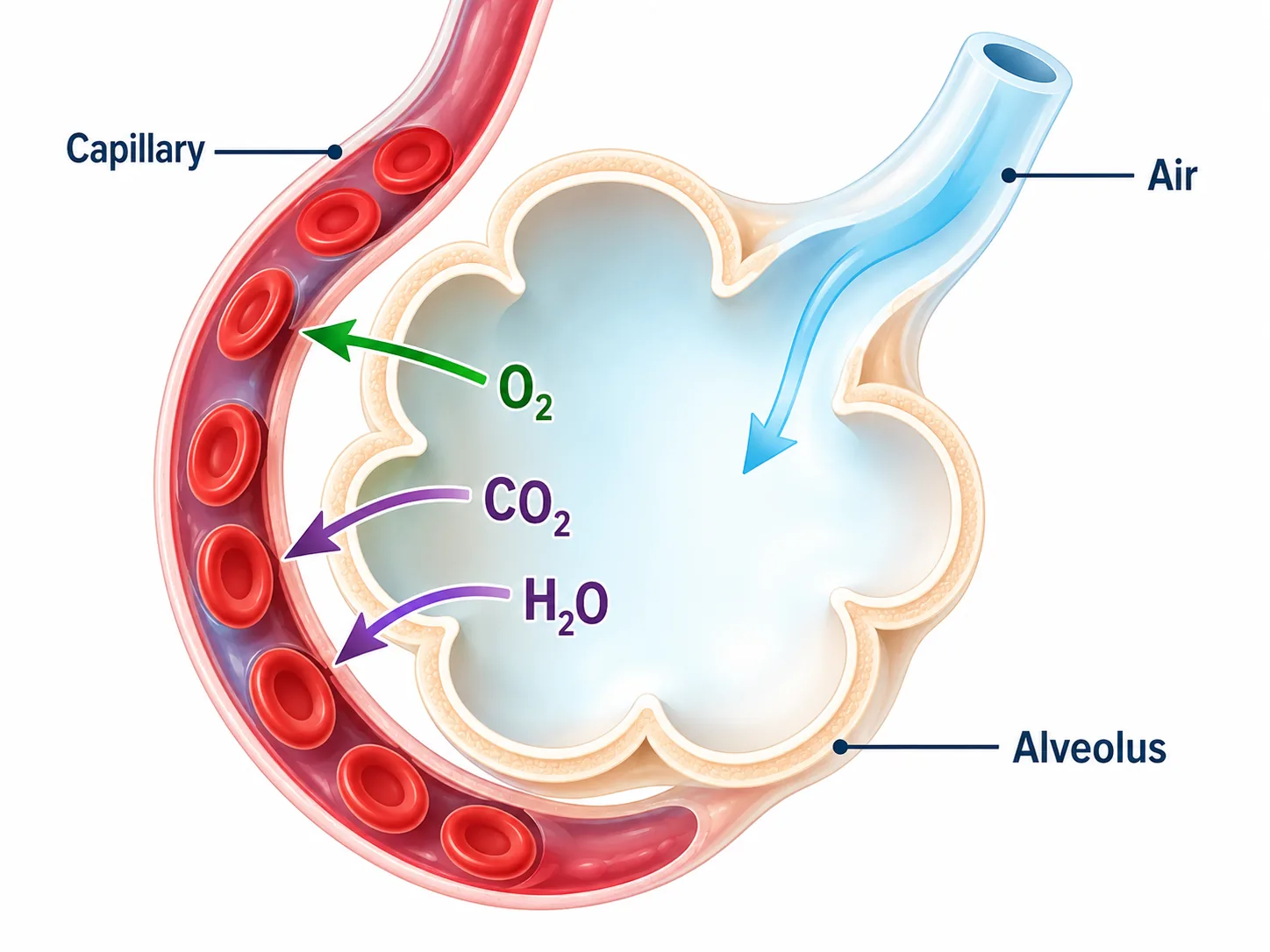


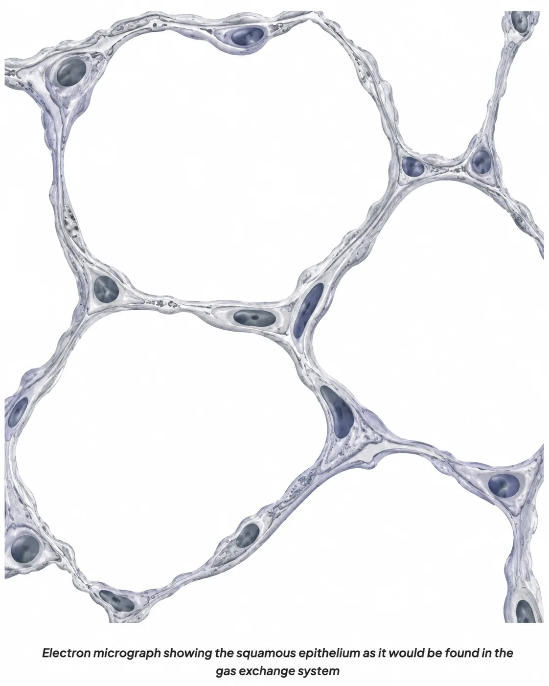

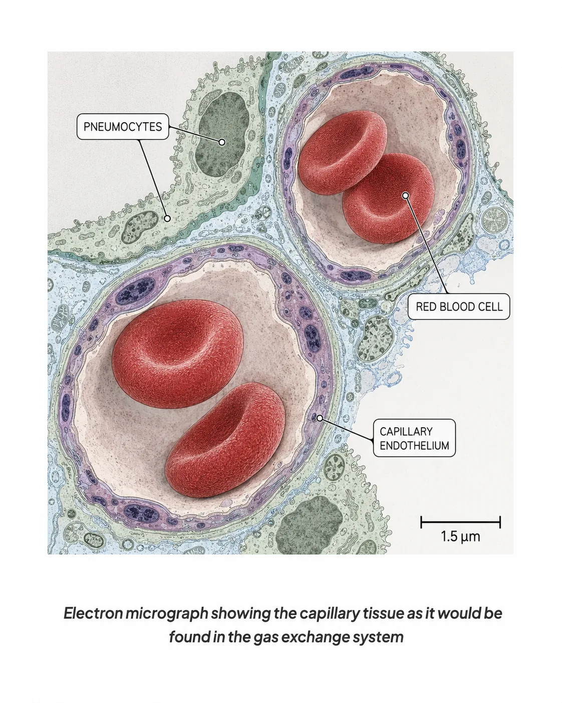




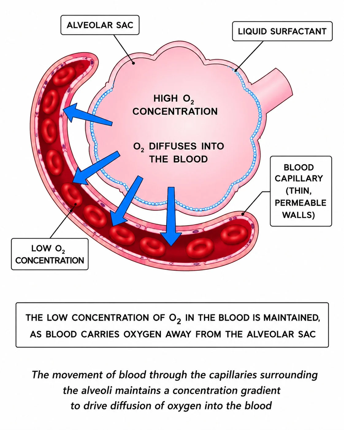
Air travels down the trachea, into the bronchi and then bronchioles to the alveoli.
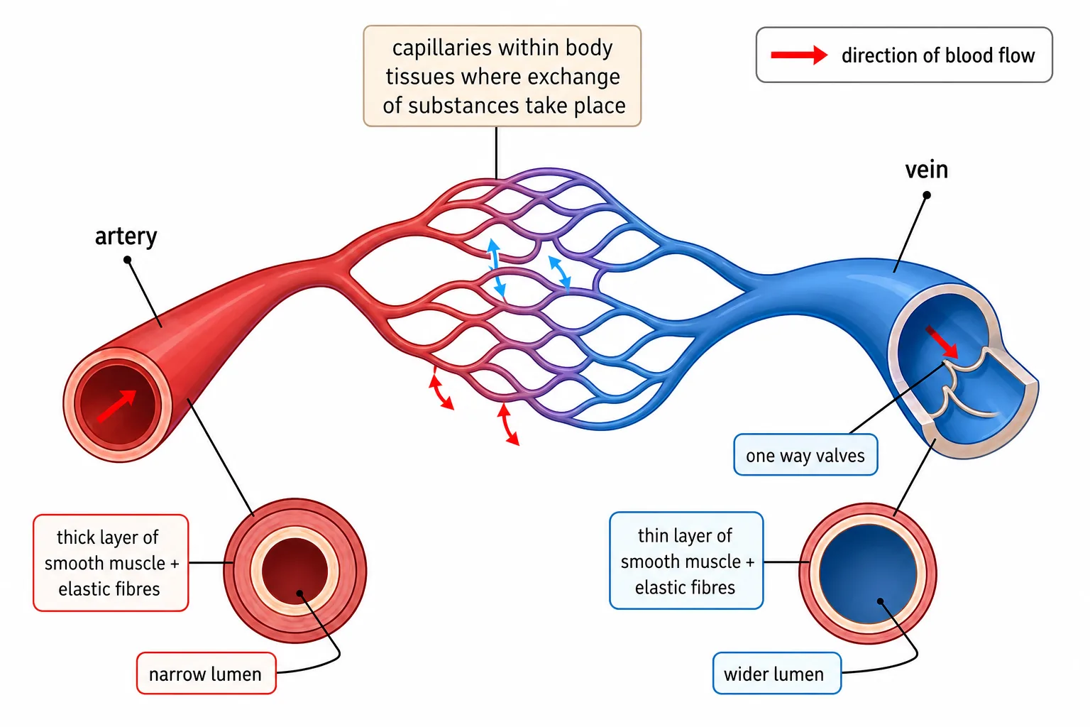
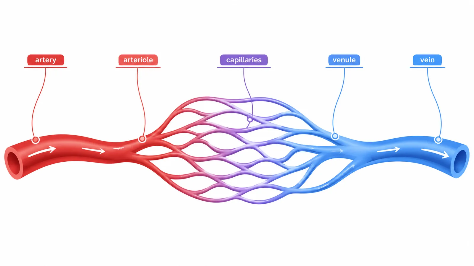
- and blood flow removes oxygen quickly,
- so the concentration gradient stays steep.
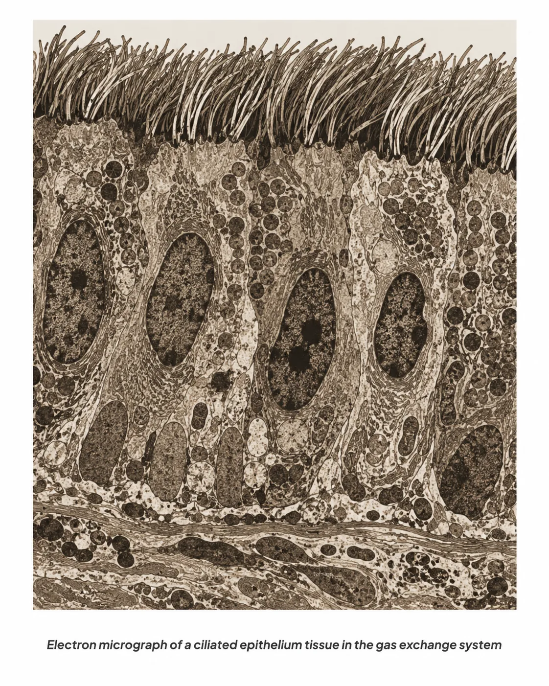
Adaptationshuge number of alveoli (large area), walls one cell thick, moist lining so gases dissolve, and a dense network of capillaries.
Section 2 of 9Absorption in the small intestine

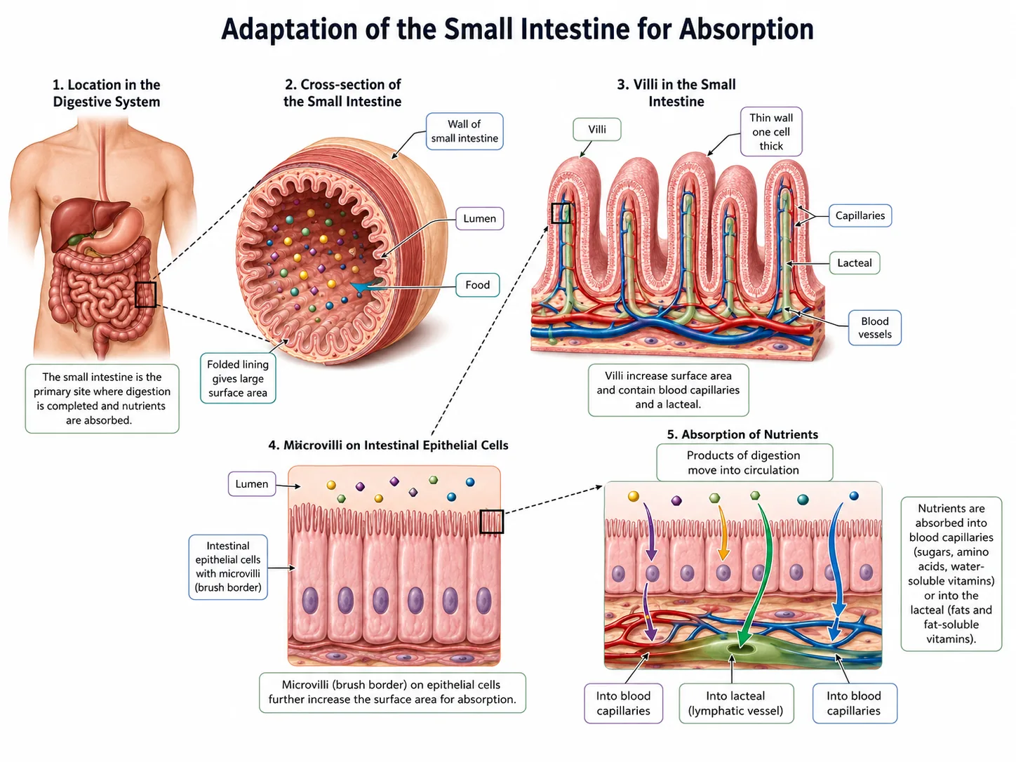


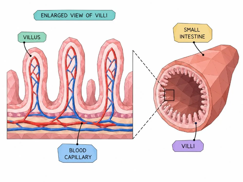
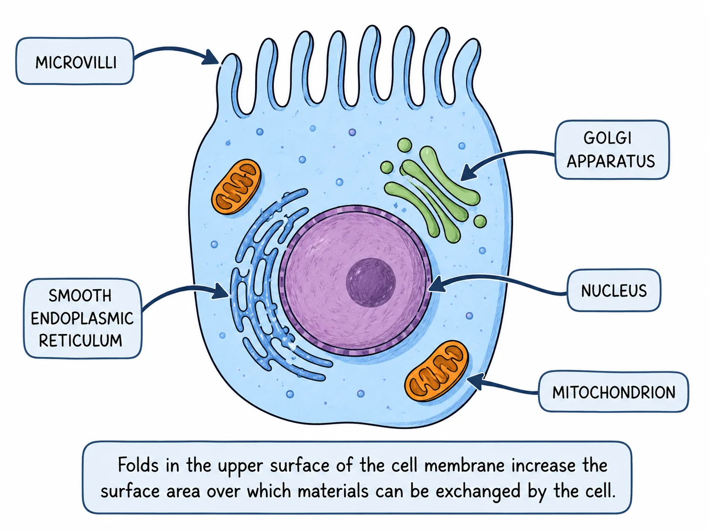

After digestion, soluble molecules such as glucose, amino acids, fatty acids and glycerol are absorbed into the blood across the wall of the small intestine.
Section 3 of 9Gills in fish


Fish take in oxygen dissolved in water.
- so blood always meets water with a higher oxygen concentration, and the gradient is maintained along the whole length.
Section 4 of 9Leaves and roots


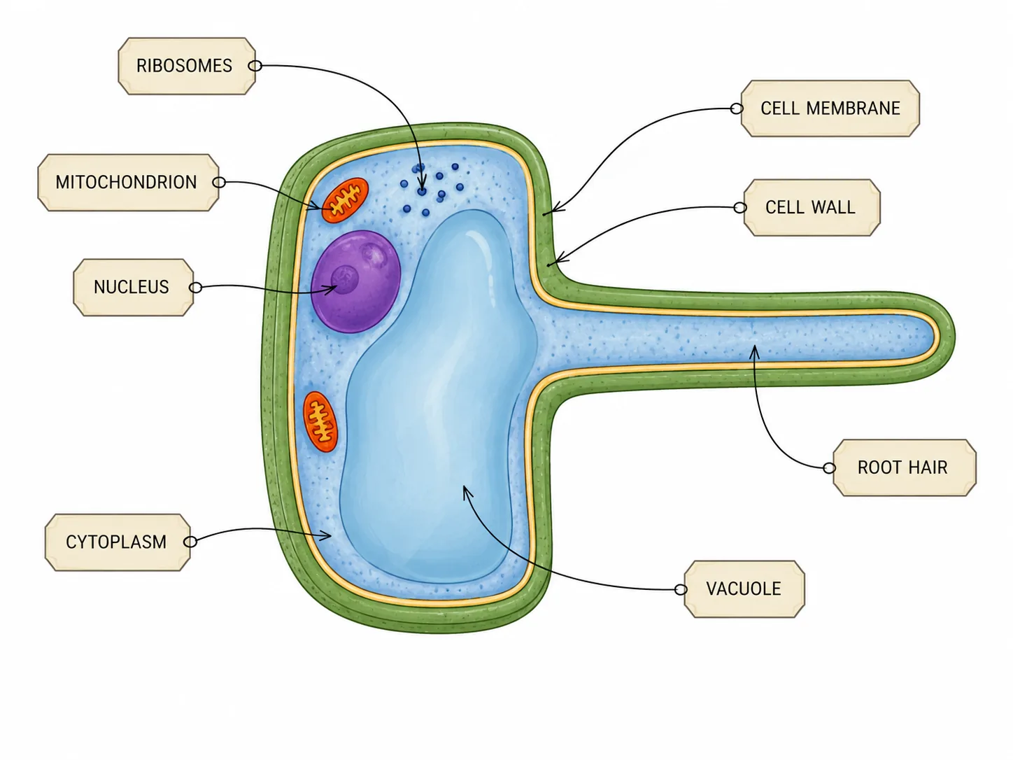

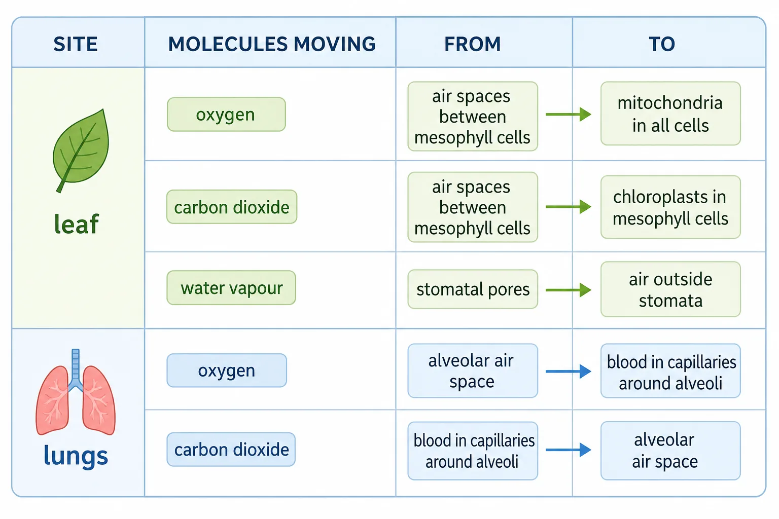
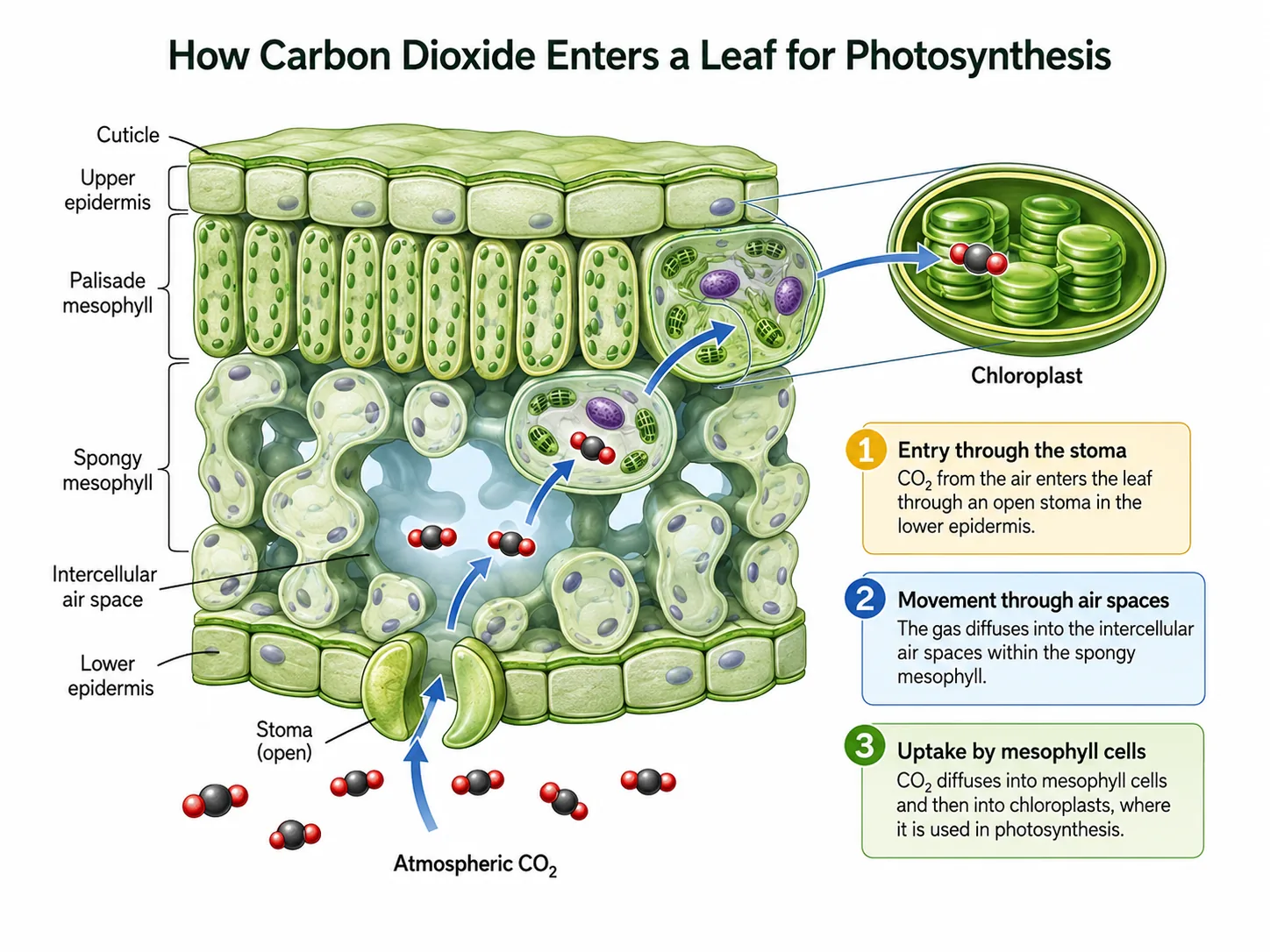
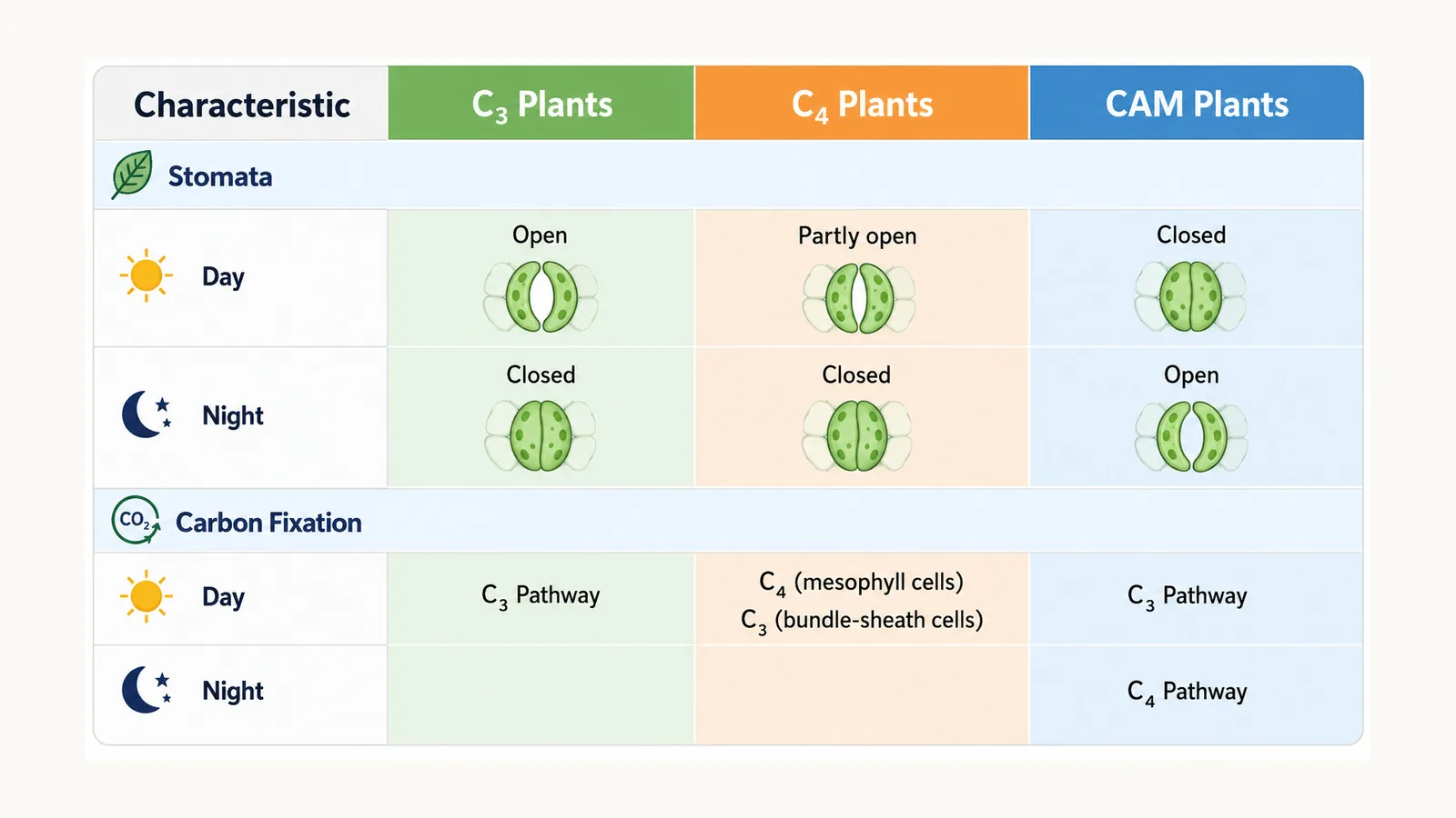
Leaves carbon dioxide diffuses in through the stomata and into the air spaces of the spongy mesophyll, where it dissolves in the film of water on cell walls and diffuses into the cells.
Roots
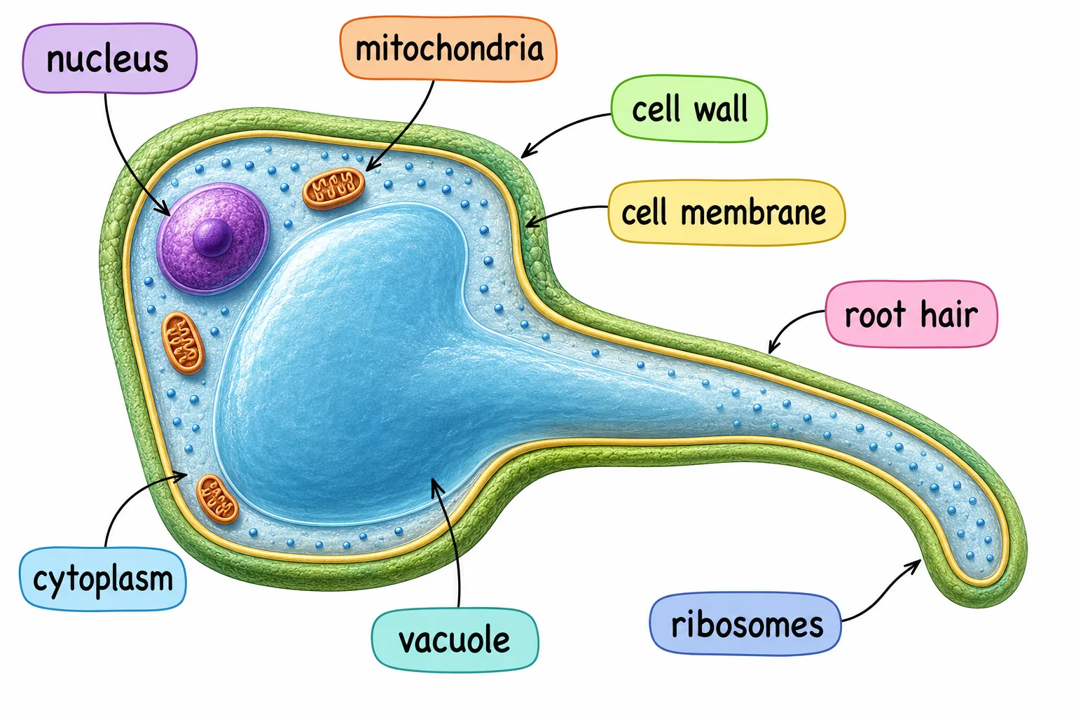
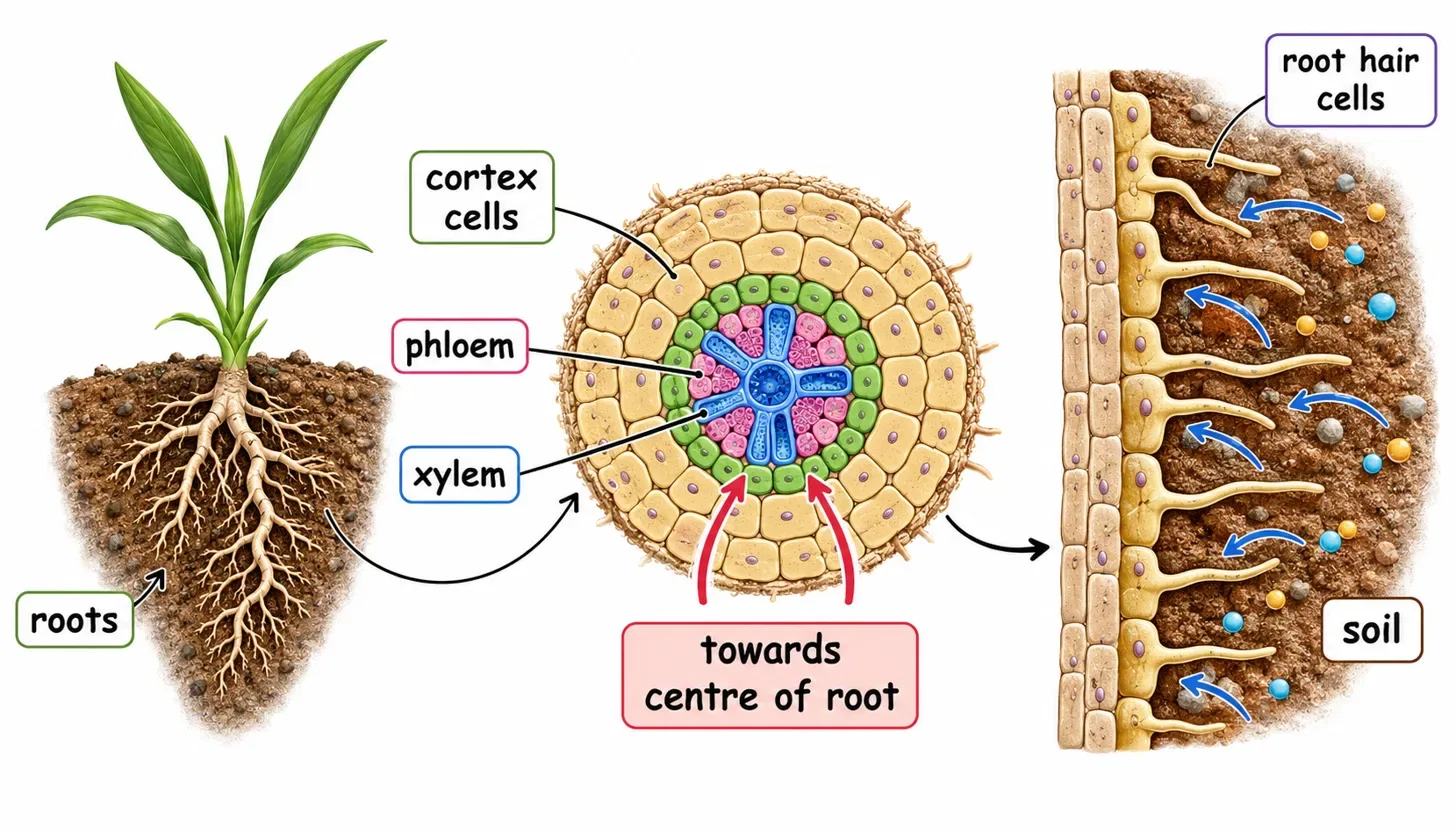
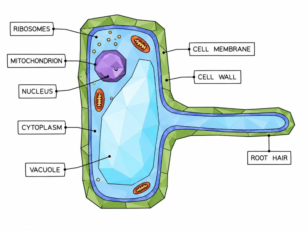
- root hair cells have long thin extensions that give a large surface area for absorbing water by osmosis and mineral ions by active transport.
- They are close to the xylem in the root.
Section 5 of 9Comparing surfaces
| Exchange surface | Substances | Key adaptations |
|---|---|---|
| Alveoli | O2 in, CO2 out | Many alveoli, thin walls, capillaries, ventilation |
| Villi | Digested food | Folding, microvilli, capillaries, lacteal |
| Gill lamellae | O2 and CO2 in water | Thin plates, counter-current blood flow |
| Leaf mesophyll | CO2, O2, water vapour | Air spaces, stomata, thin shape |
| Root hairs | Water, mineral ions | Extended hair, many mitochondria |
Section 6 of 9Describing and explaining a gradient
Questions on exchange surfaces use the same structure of answer: name the substance, state the direction of movement and the process, state the concentration difference, then explain how the adaptation maintains or exploits it.
- and the blood keeps flowing so that oxygenated blood is carried away and replaced by blood of lower oxygen concentration.
- such as a shorter diffusion distance or a steeper gradient.
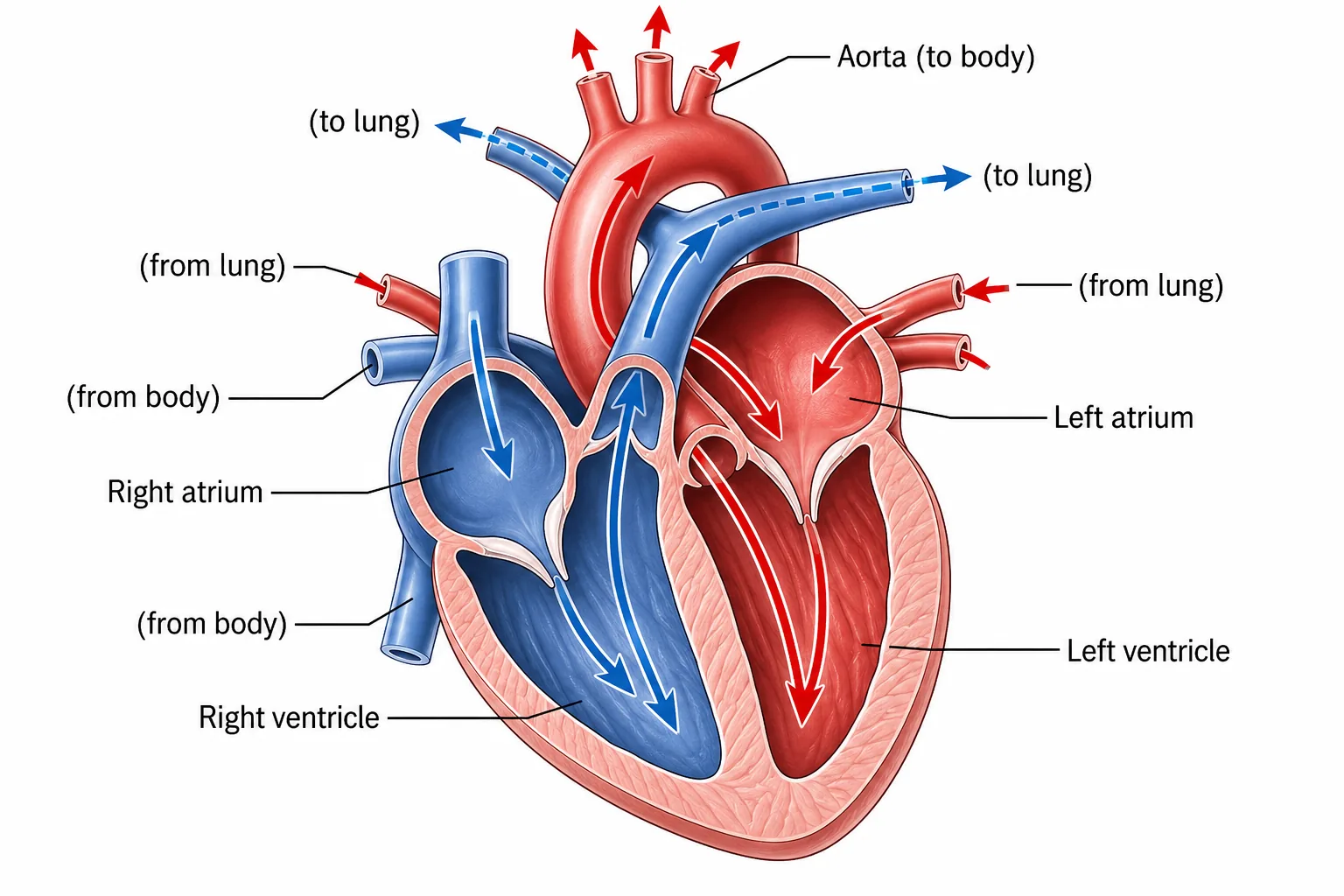
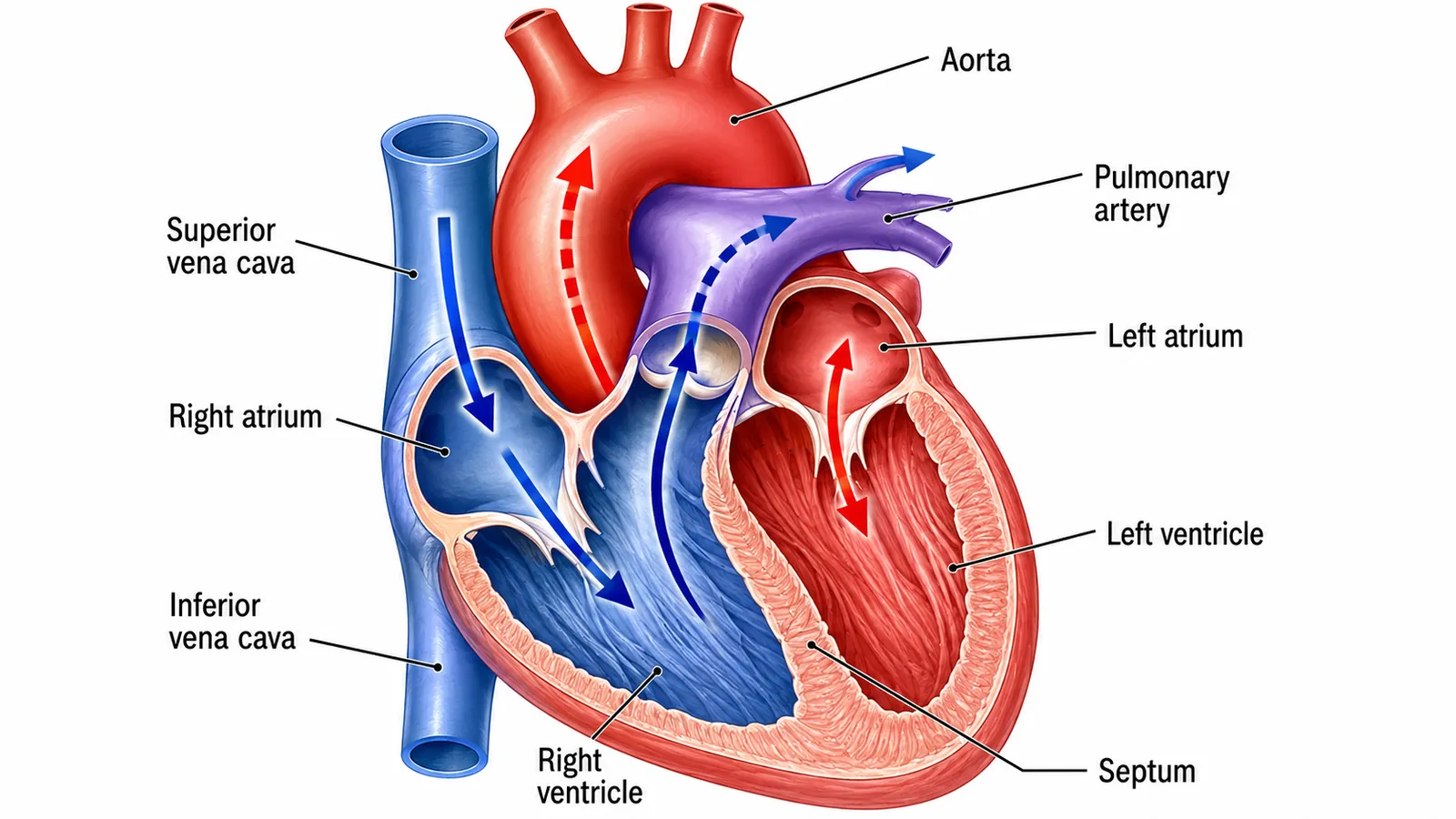
Exercise and disease change these conditions. During exercise, breathing becomes deeper and faster and the heart pumps faster, so the gradient is maintained even though the muscles use oxygen more quickly. In diseases such as emphysema the alveolar walls break down, reducing the surface area and making gas exchange less effective, so the patient feels breathless.
Remember that the same principles apply to every surface: a large area, a thin barrier, a steep gradient maintained by transport or movement of the medium, and in some cases active transport when diffusion alone cannot move the substance in the needed direction.
Section 7 of 9Exam-style questions






Describe how the structure of a villus helps absorption of glucose. [3]3 marks
The villus has a thin wall one cell thick, giving a short diffusion distance [1]. Microvilli and the villus folding give a large surface area [1]. A rich network of capillaries removes glucose and maintains the concentration gradient [1].
Explain why a fish dies quickly when taken out of water. [3]3 marks
Out of water the gill lamellae collapse and stick together [1], so the surface area for gas exchange is much reduced [1]; not enough oxygen can diffuse into the blood [1].
Explain how carbon dioxide enters a leaf for photosynthesis. [2]2 marks
It diffuses in through the open stomata from the air, where its concentration is higher [1], and then through the air spaces and into the mesophyll cells, which use it up and keep the gradient steep [1].
Section 8 of 9Key terms
| Term | Meaning |
|---|---|
| Ventilation | Movement of air in and out of the lungs. |
| Villi | Finger-like projections lining the small intestine. |
| Lacteal | A lymph vessel in a villus that absorbs fatty acids and glycerol. |
| Lamellae | Thin plates on gill filaments where gas exchange happens. |
| Counter-current flow | Water and blood flowing in opposite directions to keep a diffusion gradient along the whole surface. |
| Stomata | Pores in the leaf surface for gas exchange. |
| Root hair cell | Plant cell specialised to absorb water and mineral ions. |