Skip to content
Dabour HubLEARN · PRACTICE · EXPLORESign inStudent

2.2.3Scaling Up

The Heart

5 min read 9 parts Lesson 10 of 16 1 quiz

0%read

Section 1 of 9A double circulatory system

Heart structures and functions table
Double Circulation
Double Circulation
Double Circulation
Double Circulation
double circulation
The Heart
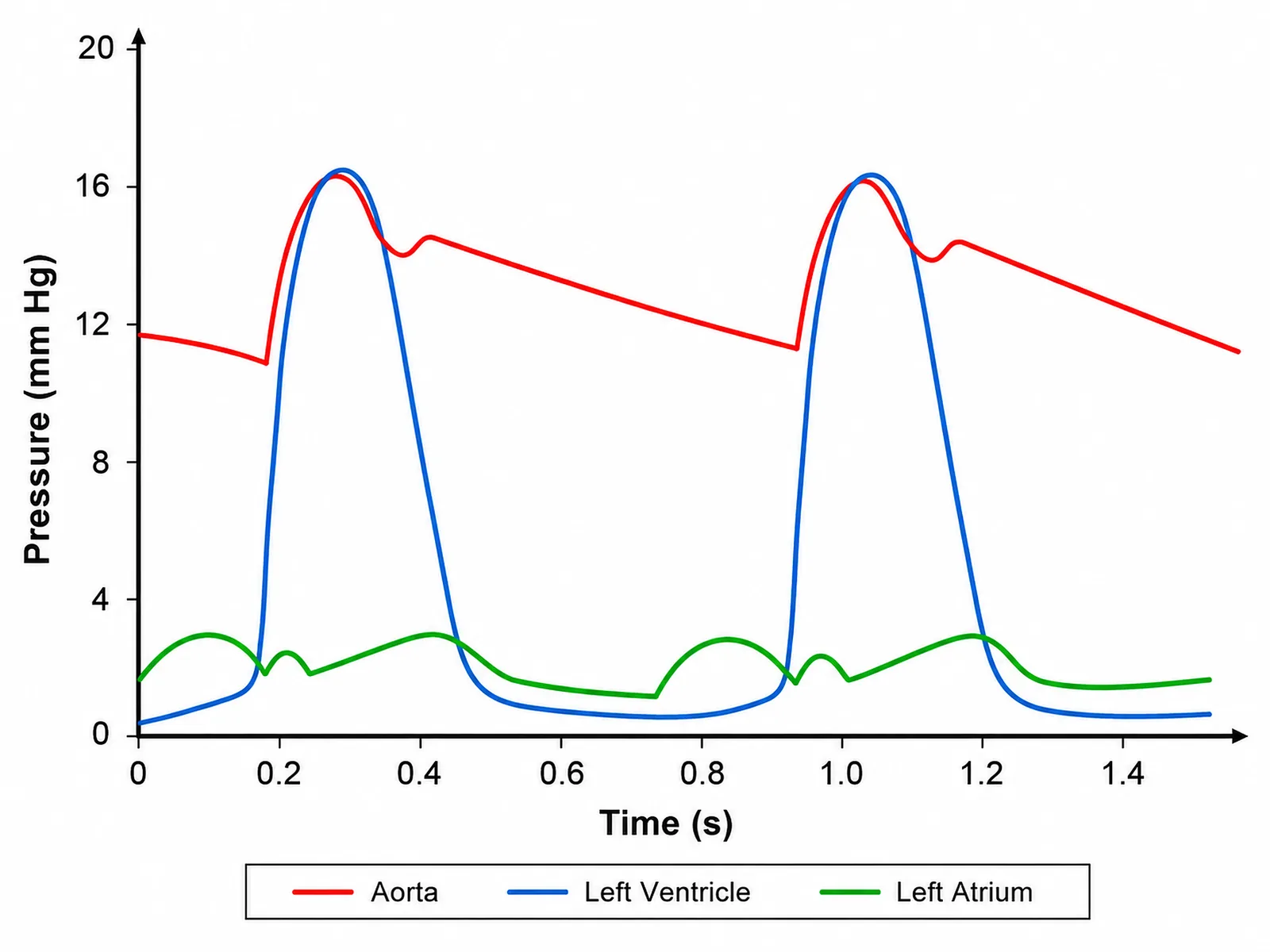
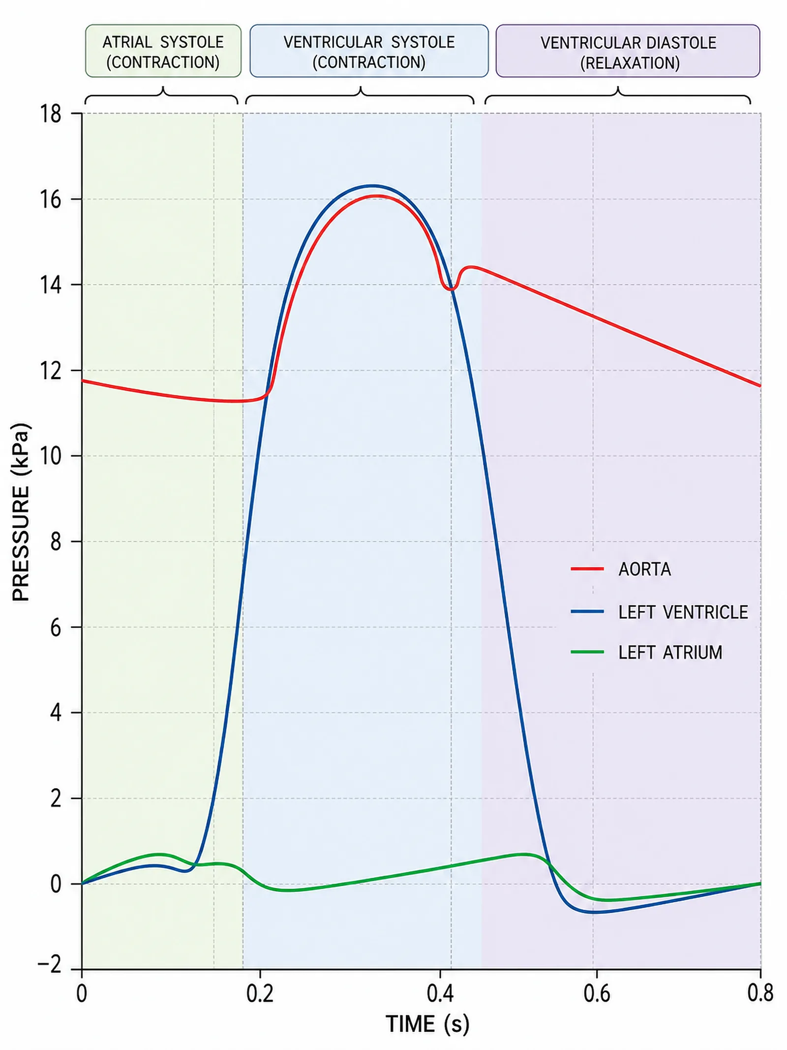
Pressure changes in the heart over time (graph)
The Heart
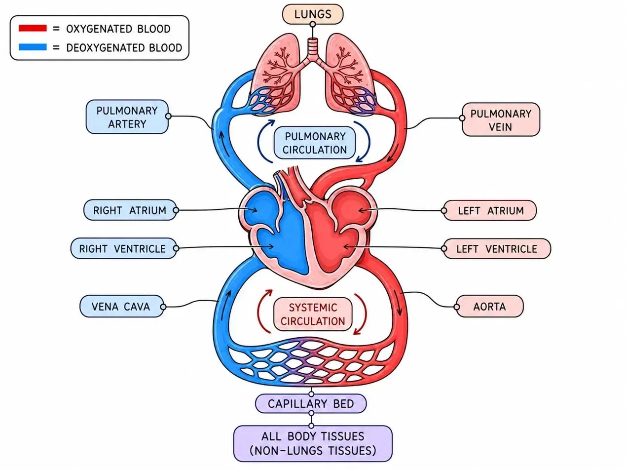
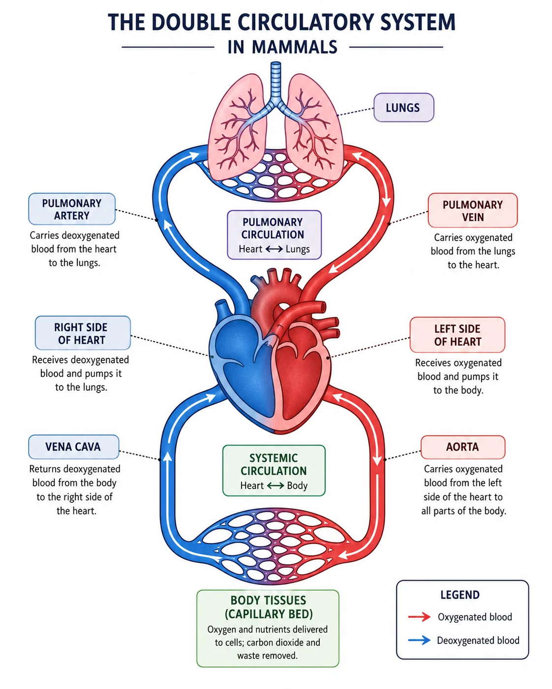
Double circulation in humans
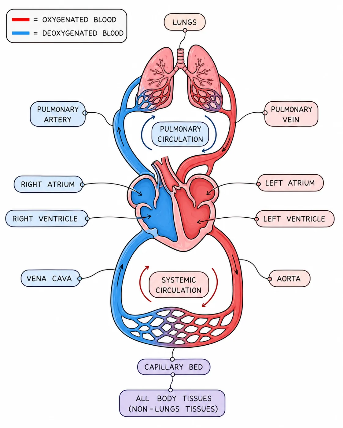
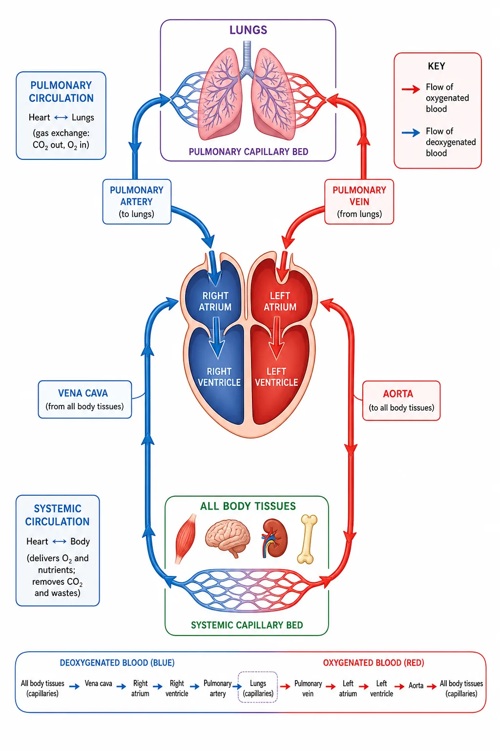
Double circulation diagram (heart, lungs, body)
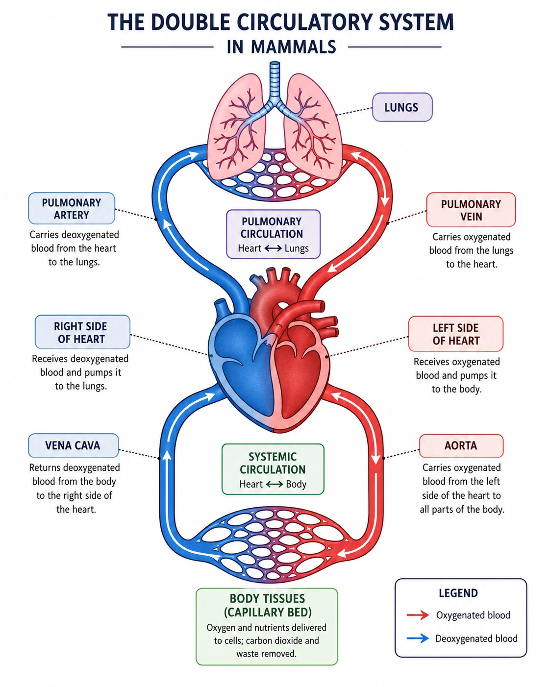
Double circulatory system in mammals

Humans have a double circulatory system: blood passes through the heart twice in each complete circuit of the body.

A double circulatory system
Pulmonary circulation
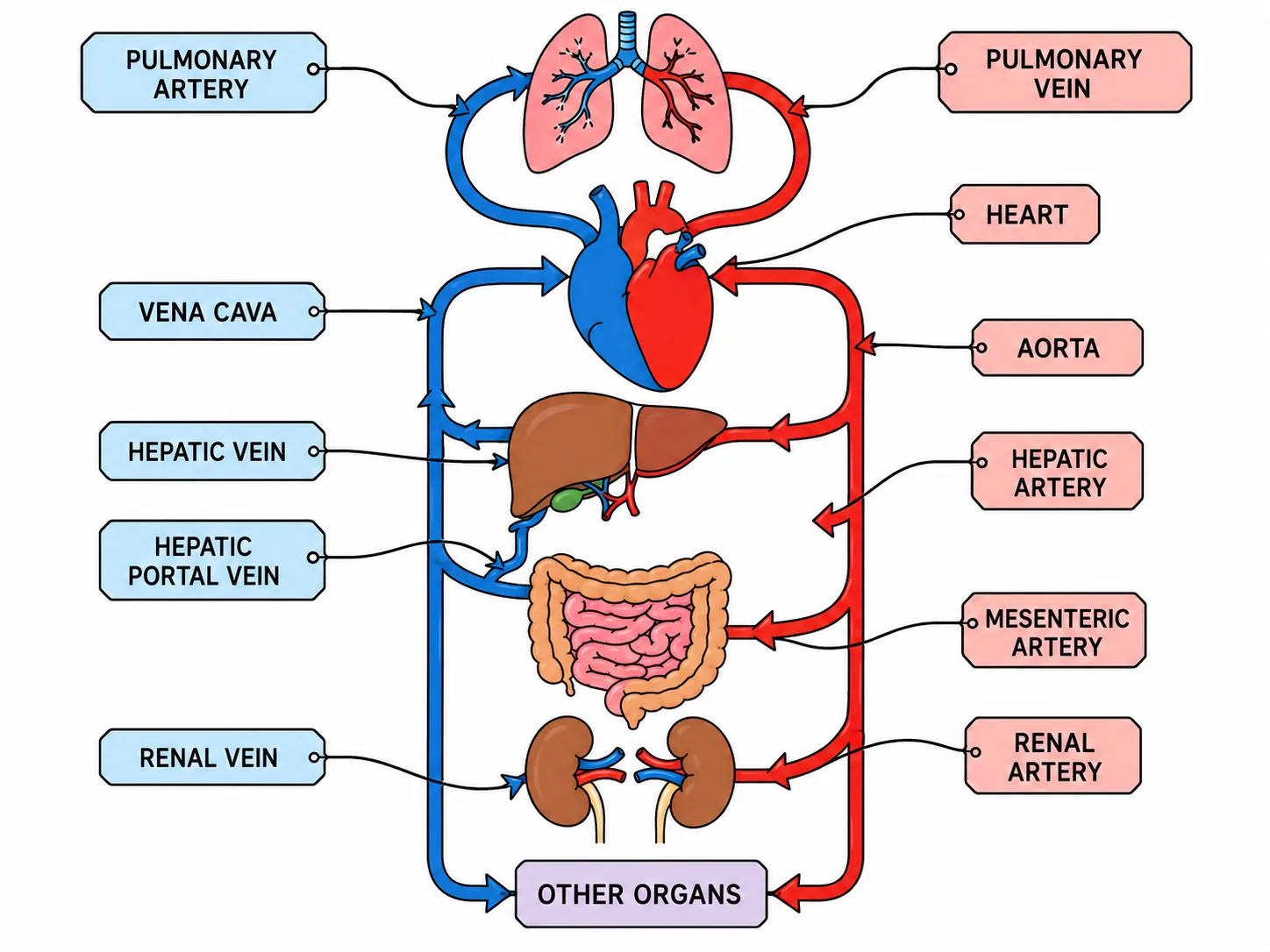
One circuit, the pulmonary circulation, carries deoxygenated blood from the heart to the lungs to pick up oxygen and returns it to the heart.
Systemic circulation
The other, the systemic circulation, carries oxygenated blood from the heart to the body organs and returns deoxygenated blood to the heart.
This arrangement is efficient: after passing through the lungs, the blood pressure drops,
  • so the blood is returned to the heart to be given a boost before travelling around the body.
This ensures that oxygenated blood is delivered to tissues quickly at high pressure.

Section 2 of 9Structure of the heart

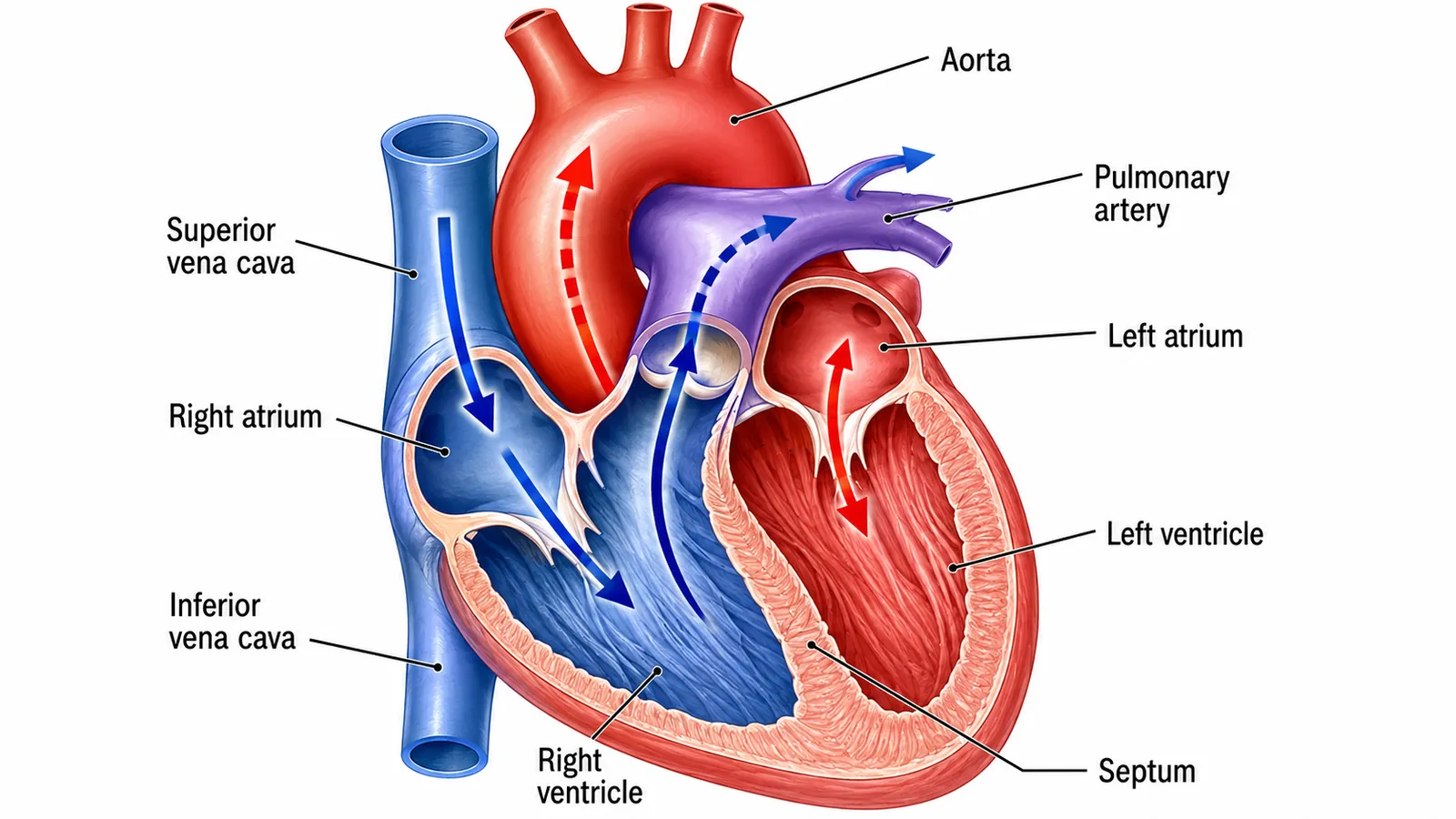
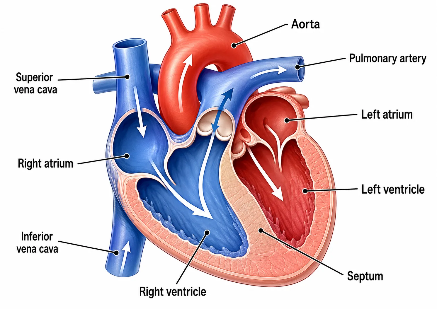
Heart structure (labelled chambers and vessels)
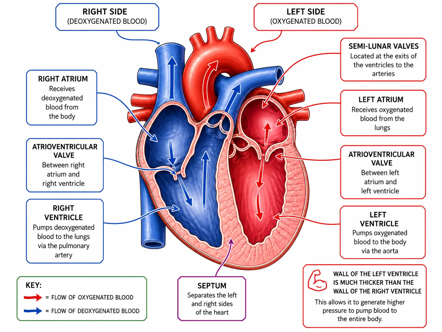
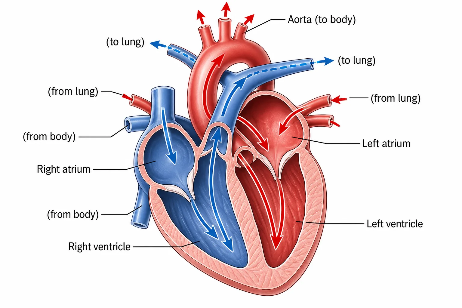
Heart diagram with oxygenated and deoxygenated flow
pasted 1782294538363 0
Labelled section of the human heart with blood flow arrows
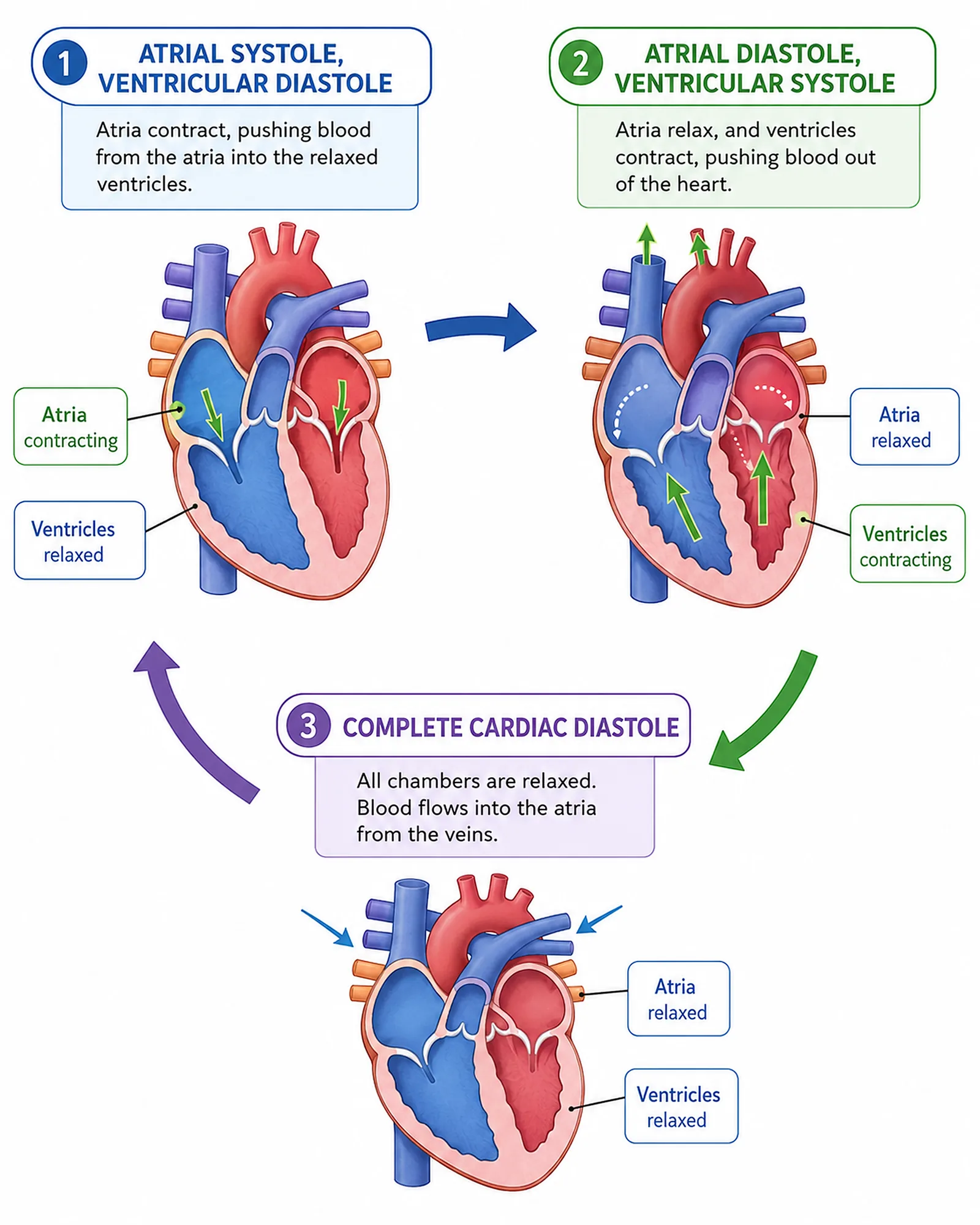
Cardiac cycle: atrial systole, ventricular systole, diastole
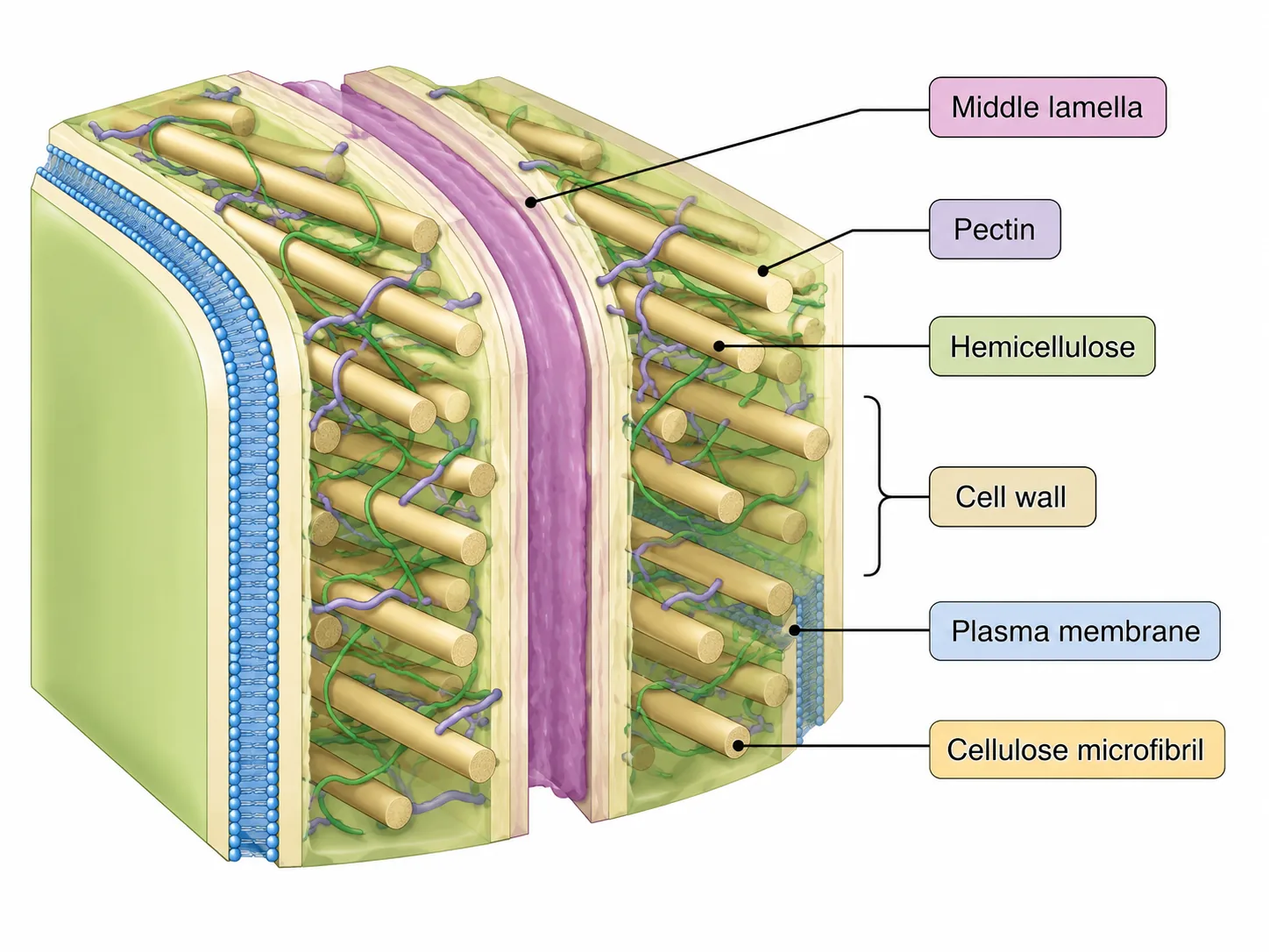
Plant Cell Wall Structure
plant cell wall structure
cardiac muscle
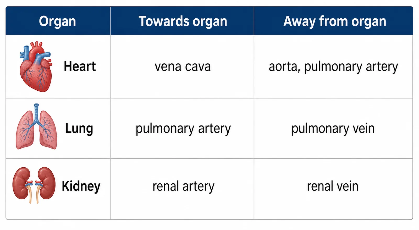
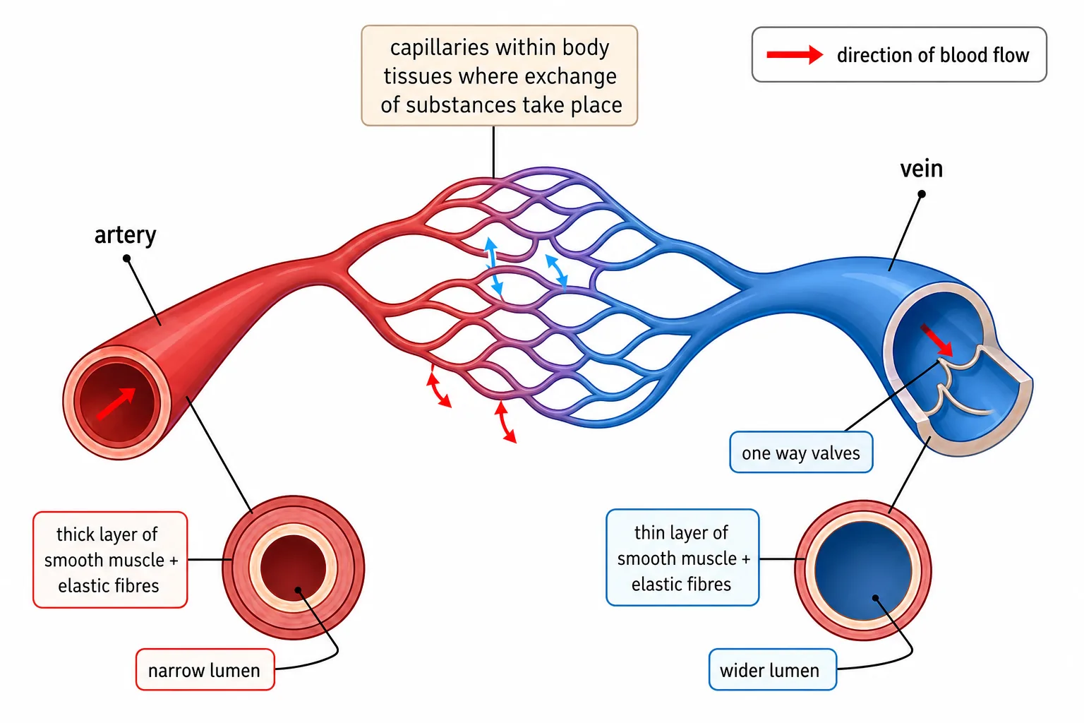
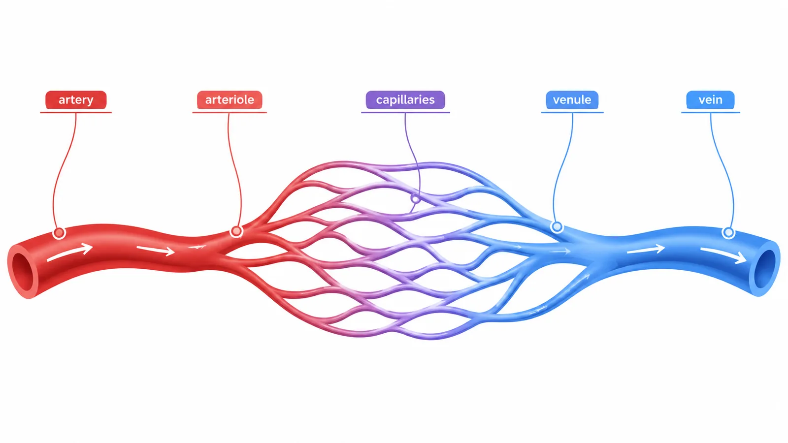
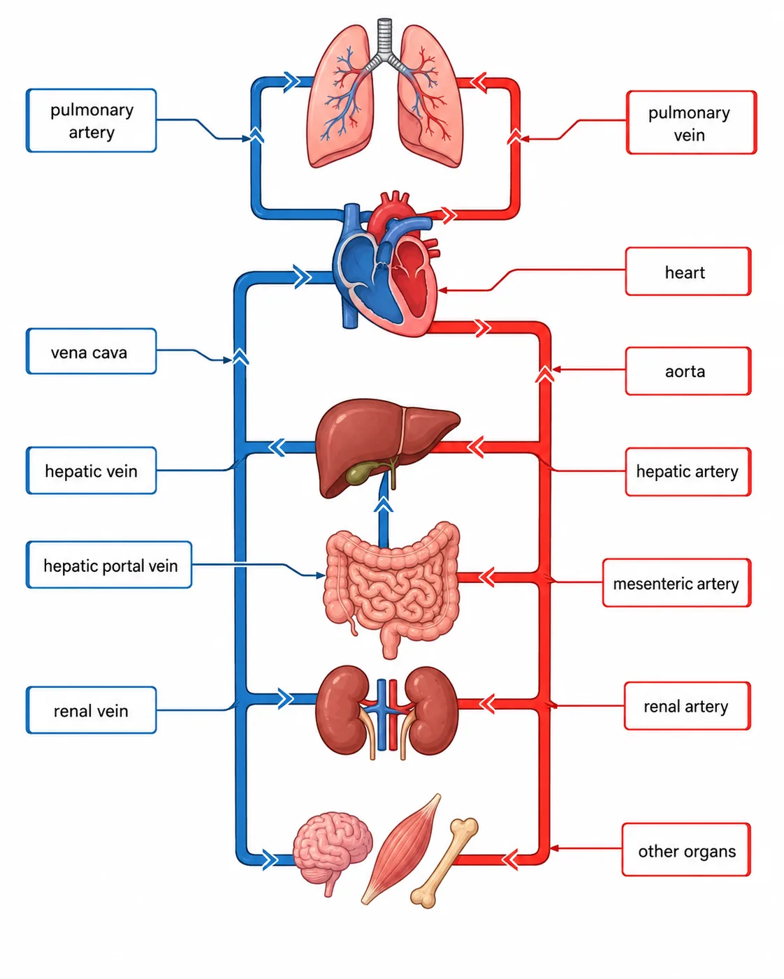
Blood vessels to and from heart, lung, kidney

The heart is a muscular organ made mostly of cardiac muscle, and it acts as a double pump.

Structure of the heart
Atria
It has four chambers: two thin-walled atria at the top that receive blood, and two thick-walled ventricles at the bottom that pump it out.
Valves
A wall of muscle, the septum, separates the right side from the left so the oxygenated and deoxygenated blood do not mix. Valves prevent backflow of blood.
The left ventricle has a thicker muscular wall than the right,
  • because it pumps blood at high pressure around the whole body,
  • while the right pumps only to the nearby lungs.
Blood vesselCarries bloodFrom / to
Vena cavaDeoxygenatedBody to right atrium
Pulmonary arteryDeoxygenatedRight ventricle to lungs
Pulmonary veinOxygenatedLungs to left atrium
AortaOxygenatedLeft ventricle to body
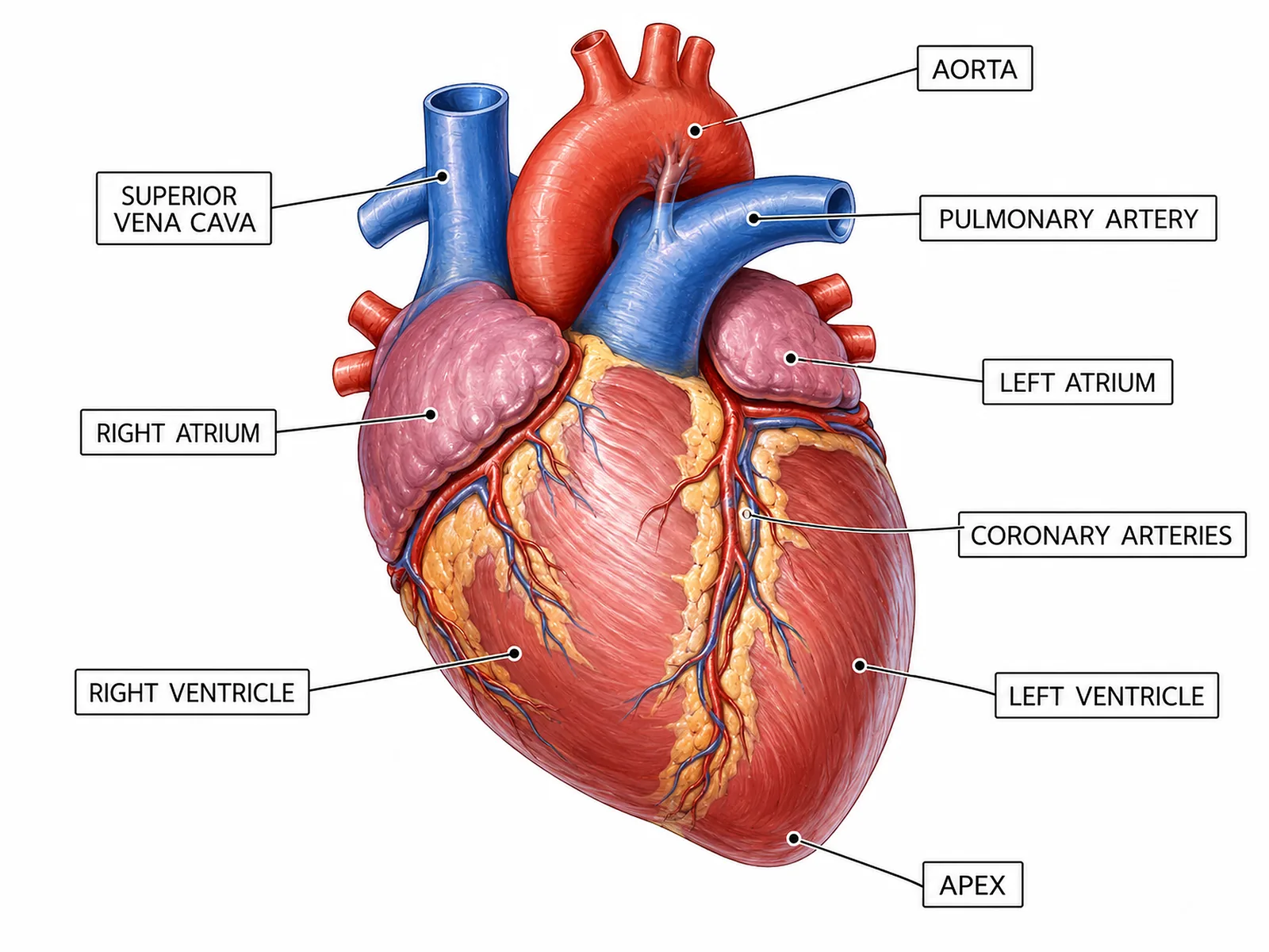
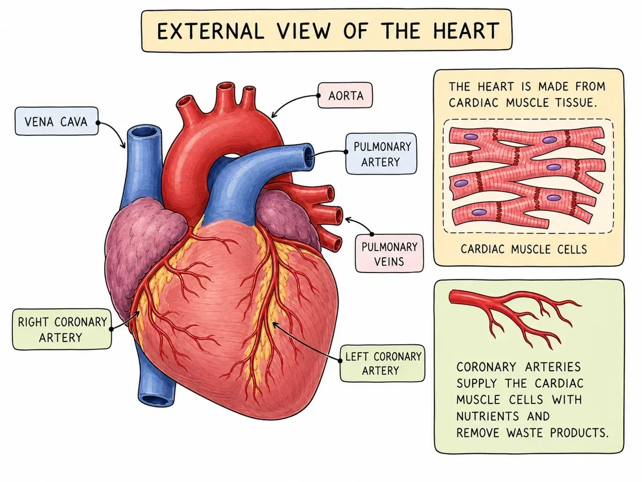
Coronary arteriesOxygenatedAorta to the heart muscle

Section 3 of 9The path of blood

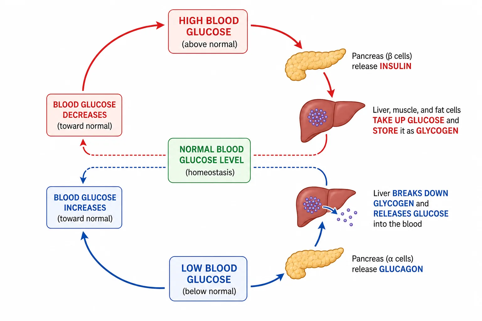
Control of Blood Glucose
Right atrium
Deoxygenated blood enters the right atrium via the vena cava.
Right ventricle
The atrium contracts, and blood passes through a valve to the right ventricle.
The ventricle contracts and pumps blood through the pulmonary artery to the lungs.
Left atrium
Oxygenated blood returns through the pulmonary vein to the left atrium.
Left ventricle
It passes through a valve to the left ventricle, which contracts and forces blood out of the aorta to the body.

The heart muscle itself needs its own supply of oxygen and glucose, delivered by the coronary arteries. If they become blocked, part of the heart muscle is starved of oxygen, leading to a heart attack.

Section 4 of 9Controlling heart rate and pacemakers

The resting heart rate is controlled by a group of cells in the right atrium wall called the pacemaker.

These cells produce electrical impulses that make the heart muscle contract regularly.
The rate adjusts with the body's needs: during exercise, nerves and the hormone adrenaline increase the rate so that more oxygen is supplied to the muscles.
Artificial pacemaker
If the natural pacemaker does not work properly, an artificial pacemaker, a small electrical device implanted under the skin, can be used to give a regular signal to the heart.

Section 5 of 9Heart problems and treatments

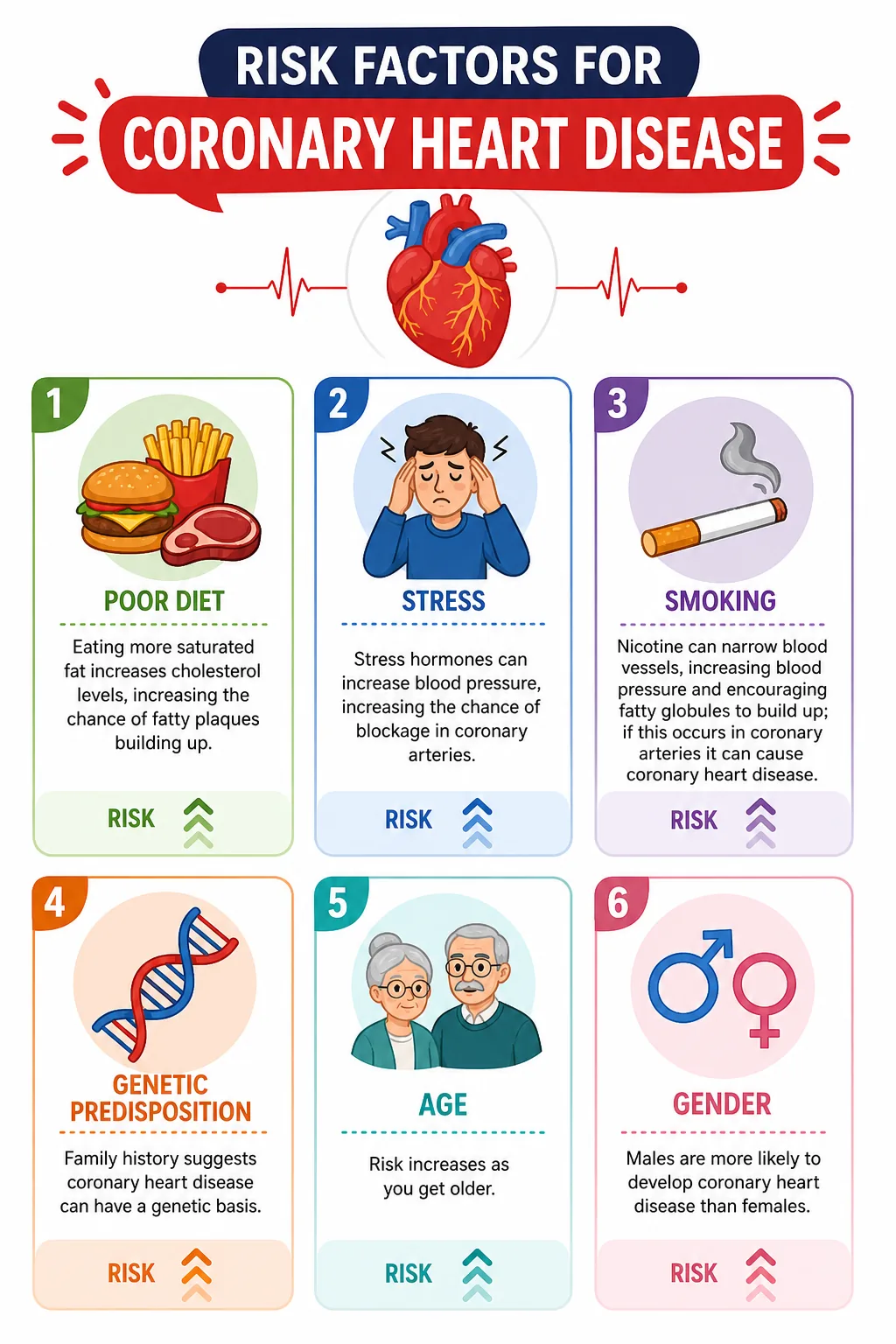
Risk Factors for Coronary Heart Disease
Risk factors for coronary heart disease
Coronary Heart Disease

In coronary heart disease, fatty deposits narrow the coronary arteries.

Statins
Treatments include statins to reduce blood cholesterol, stents to hold arteries open, and bypass surgery.
Faulty valves can be replaced with biological (animal) or mechanical valves.
Heart failure may be treated by a transplant from a donor, or an artificial heart as a temporary measure.
Each has drawbacks such as the risk of rejection and the need for immunosuppressant drugs, and a shortage of donors.
A healthy diet, no smoking and regular exercise reduce the risk.

Section 6 of 9Blood flow and the cardiac cycle

Blood Vessels
heart blood flow
heart blood flow
Graph of left ventricle volume over one cardiac cycle showing stroke volume
Blood Vessels
Blood Vessel Types
Blood Vessels

Every heartbeat follows a cycle.

1
First the atria contract, pushing blood down into the ventricles through the open atrioventricular valves.
2
Next the ventricles contract; the pressure shuts the atrioventricular valves,
  • making the 'lub' sound,
  • and forces blood out through the semilunar valves at the start of the aorta and pulmonary artery.
3
As the ventricles relax, the semilunar valves close, making the 'dub' sound, and the heart fills again.
4
The heart rate is the number of beats in one minute, and the stroke volume is the amount of blood pumped out of one ventricle each beat.

Cardiac output is stroke volume multiplied by heart rate. At rest, an adult's cardiac output is about 5 dm3 per minute, and during hard exercise it can rise to about 20 dm3 per minute. Fit people have a larger stroke volume, so a lower resting heart rate for the same output.

A frequent misconception is that blood on the right of the heart is mixed with that on the left.

Blood flow and the cardiac cycle
The septum prevents this,
  • and it is why the double circulation is so efficient: fully oxygenated blood goes to the body without being diluted by deoxygenated blood.
Make sure you name the correct valve or vessel in questions about where blood travels, and always follow the direction of flow.

Section 7 of 9Exam-style questions
Heart structure with left and right sides
heart anatomy
Pressure changes in aorta, left ventricle and left atrium (graph)
Blood vessels to organs: heart, liver, gut, kidneys
Blood vessels linking heart, lungs and organs
External View of the Heart
Heart external view with coronary arteries
External View of the Heart
  1. Explain why the wall of the left ventricle is thicker than that of the right ventricle. [2]2 marks

  2. Describe the pathway of blood from the vena cava to the aorta. [4]4 marks

  3. State the role of the pacemaker and suggest why a person might need an artificial one. [2]2 marks

Section 8 of 9Key terms

TermMeaning
AtriumUpper chamber of the heart that receives blood.
VentricleLower chamber of the heart that pumps blood out.
ValveStructure that stops the backflow of blood.
AortaMain artery carrying oxygenated blood to the body.
Vena cavaMain vein returning deoxygenated blood to the heart.
PacemakerGroup of cells that controls heart rate.
Coronary arteryArtery supplying blood to the heart muscle.
Double circulationSystem where blood passes through the heart twice per circuit.

Section 9 of 9Exam technique

1
Saying that all arteries carry oxygenated blood; the pulmonary artery carries deoxygenated blood.
2
Labelling the heart as it appears to you instead of from the patient's point of view (left is on your right).
3
Saying blood passes through the septum.
4
Forgetting the role of valves in preventing backflow.
\n