Section 1 of 9The circulatory system

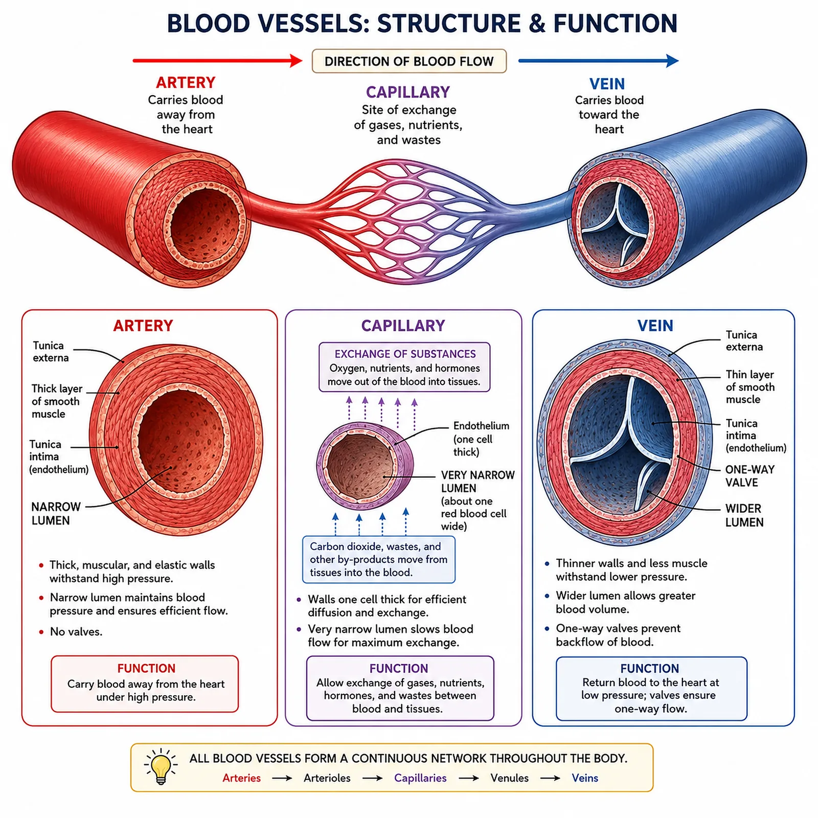
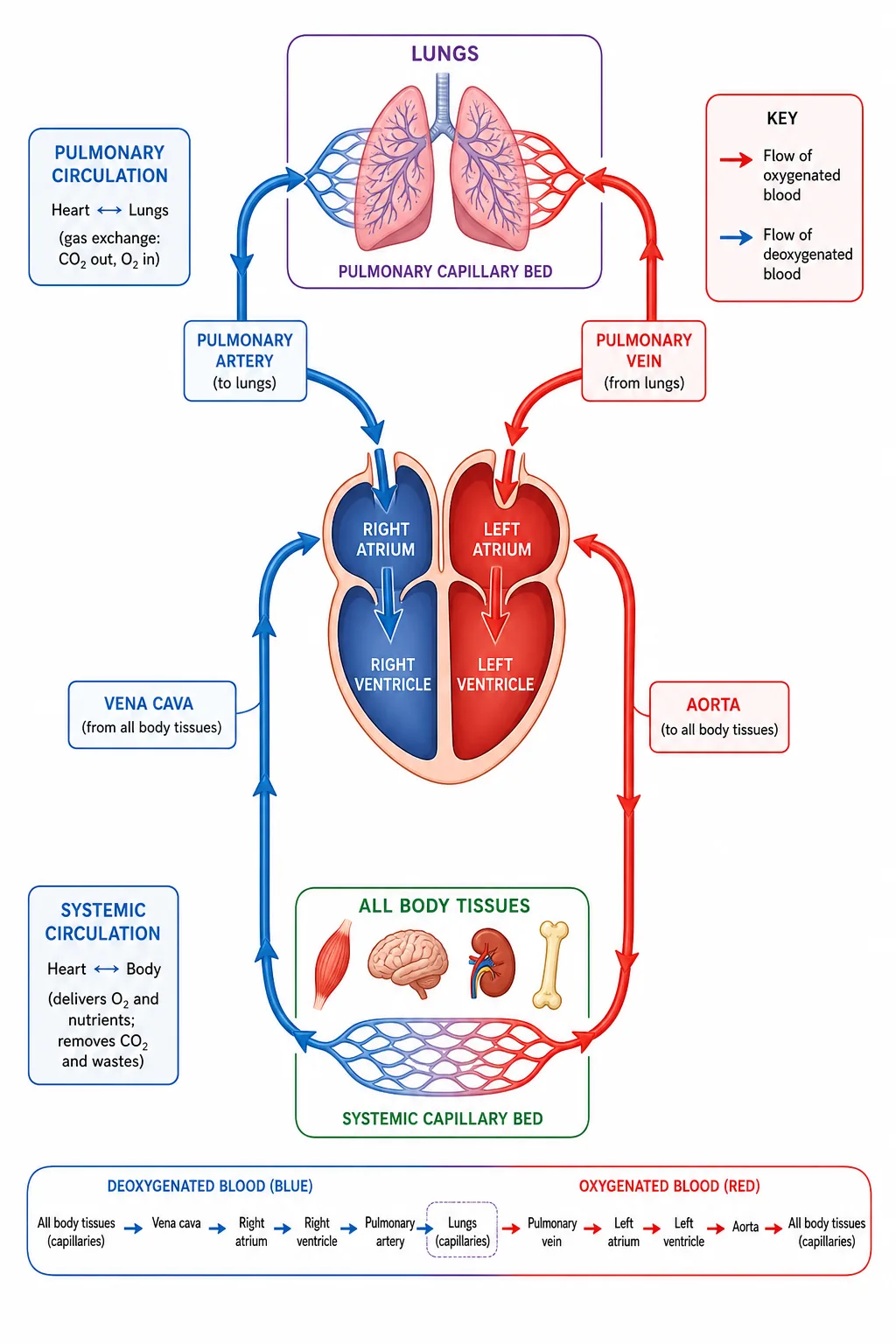


Humans have a double circulatory system.
- which allows it to be pumped at a higher pressure to the body after having been slowed in the lung capillaries.
Section 2 of 9Arteries
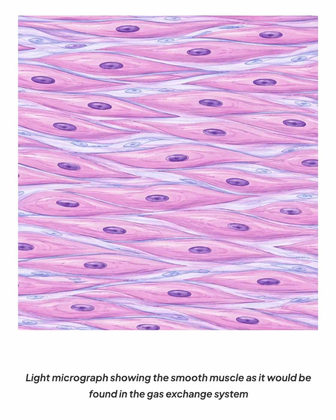
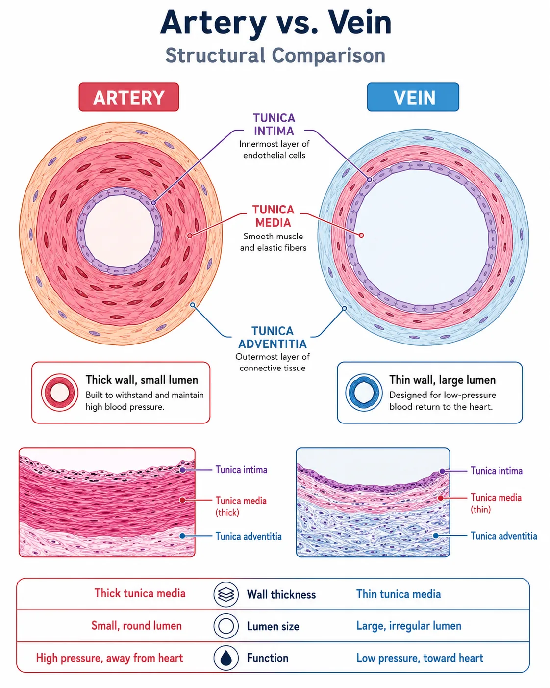
Arteries carry blood away from the heart, at high pressure.
- which evens out the flow, and the muscle can narrow or widen the vessel.
Section 3 of 9Veins and capillaries

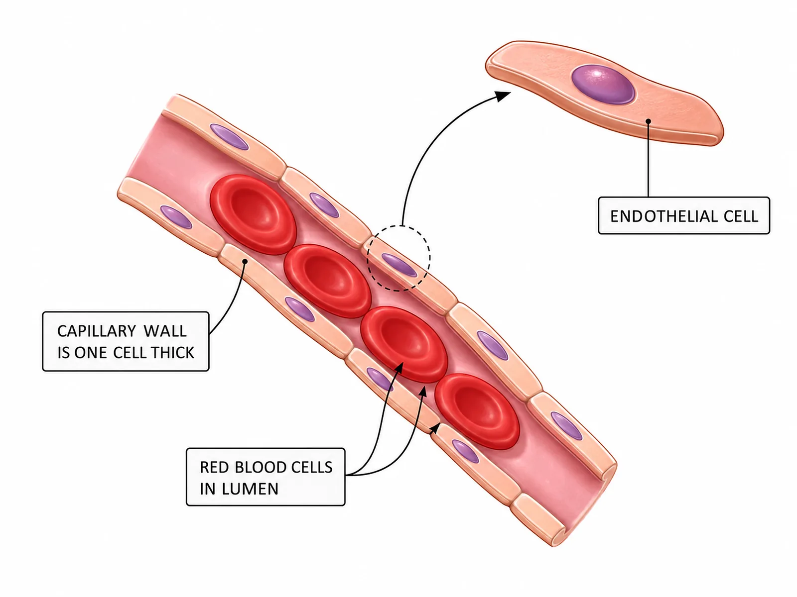

Veins carry blood back to the heart at low pressure. They have thinner walls with less muscle and elastic tissue, a wide lumen to reduce resistance to flow, and valves that stop the backflow of blood. Muscle movement around the veins squeezes the blood along. Veins generally carry deoxygenated blood except for the pulmonary vein.
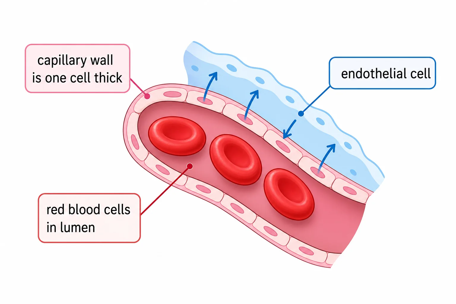
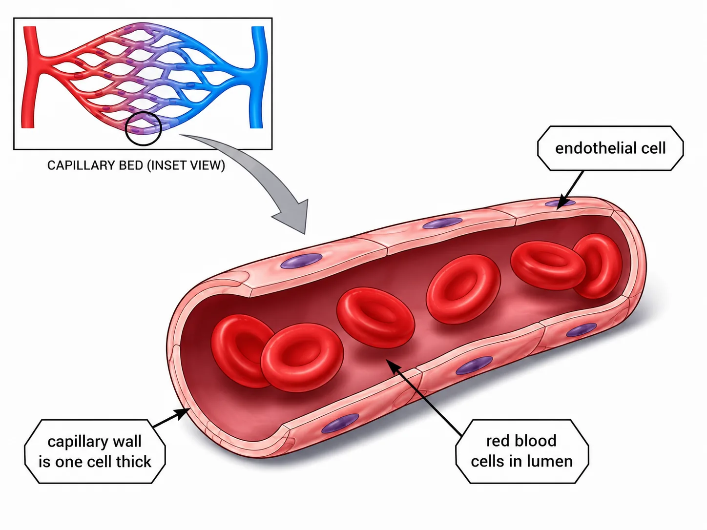
Capillaries are the smallest vessels, where exchange takes place between blood and the body cells. Their walls are one cell thick, giving a short diffusion distance. They are very narrow and form dense networks, giving a large total surface area, and they are leaky so some plasma can pass out to form tissue fluid.
Section 4 of 9Comparison table
| Feature | Artery | Vein | Capillary |
|---|---|---|---|
| Direction of flow | Away from heart | Towards heart | Between arteries and veins |
| Pressure | High | Low | Falling |
| Wall | Thick, elastic and muscular | Thin | One cell thick |
| Lumen | Narrow | Wide | Very narrow |
| Valves | No | Yes | No |
Section 5 of 9Interpreting data and links
Data questions often give blood pressure or velocity values for each vessel type.
Blood vessels link to heart disease
- fatty deposits narrow coronary arteries, reducing blood and oxygen supplied to the heart muscle, which can cause a heart attack.
- Stents hold arteries open and statins reduce cholesterol.
Section 6 of 9Blood vessels in health and disease
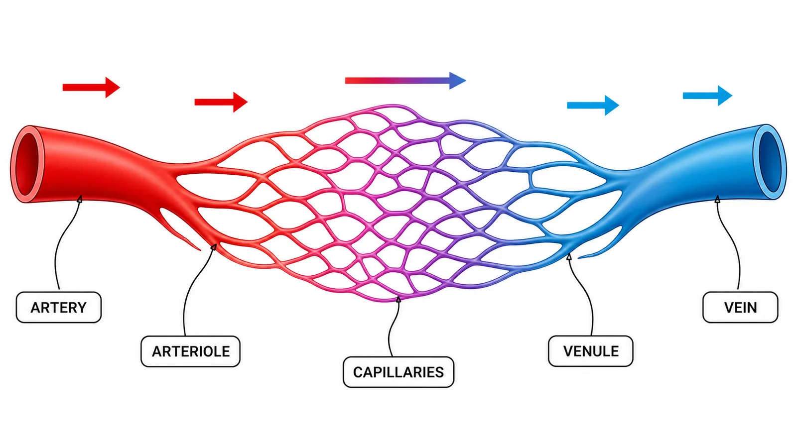
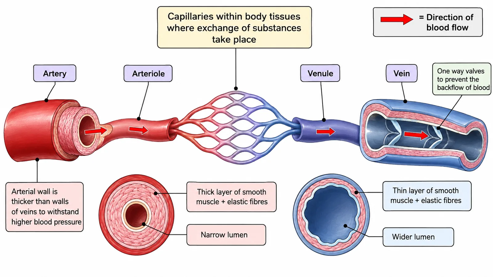
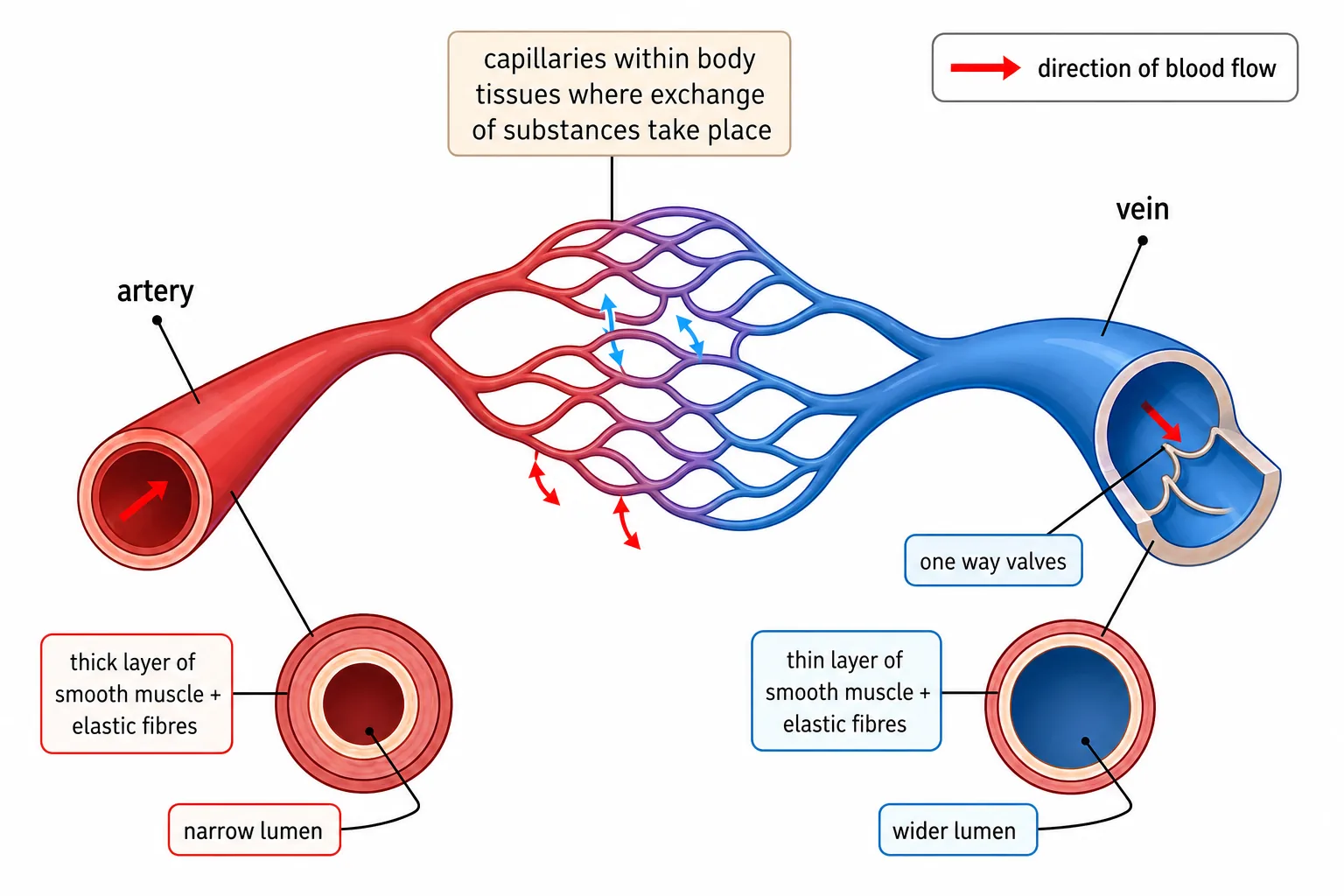


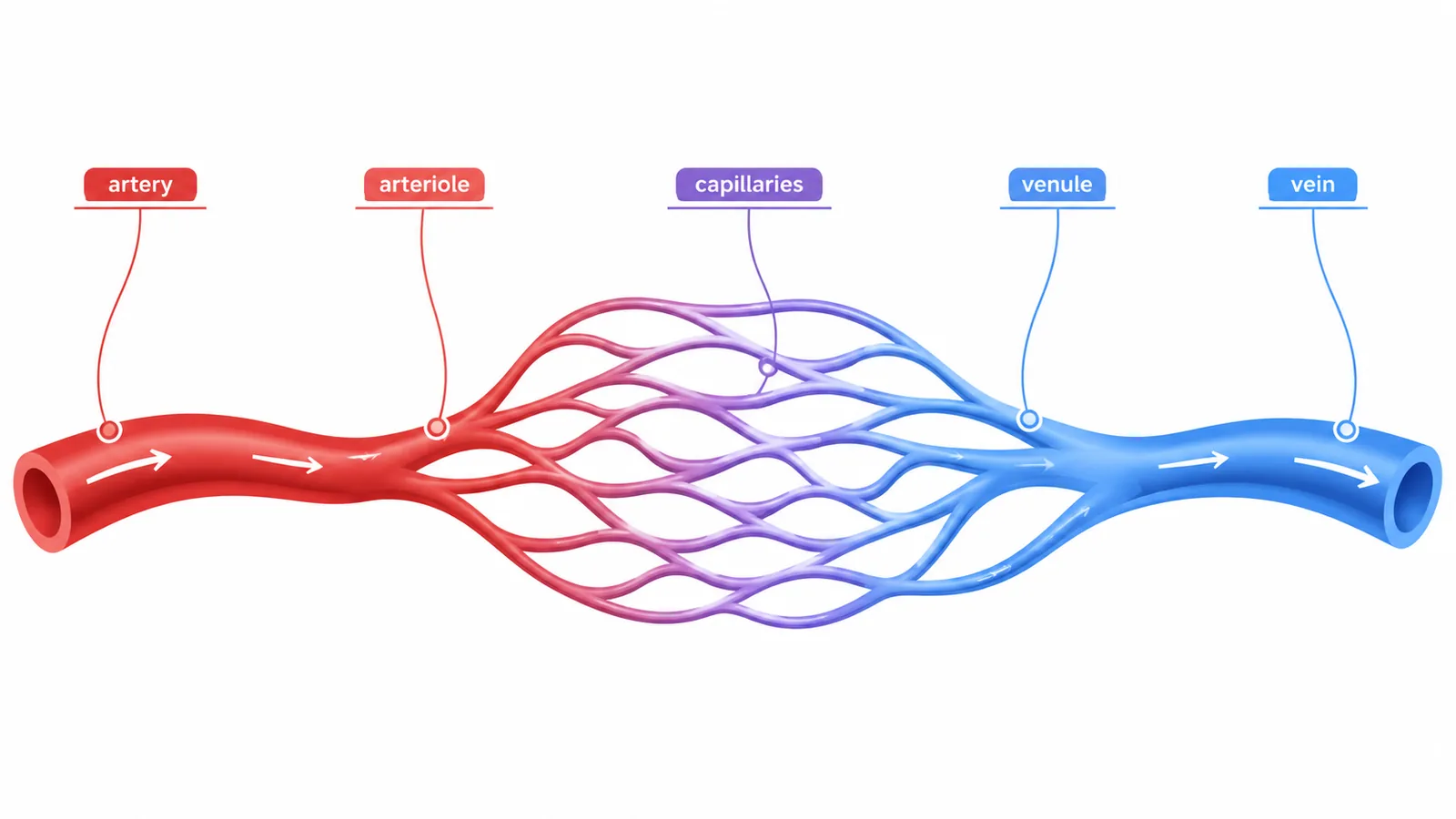

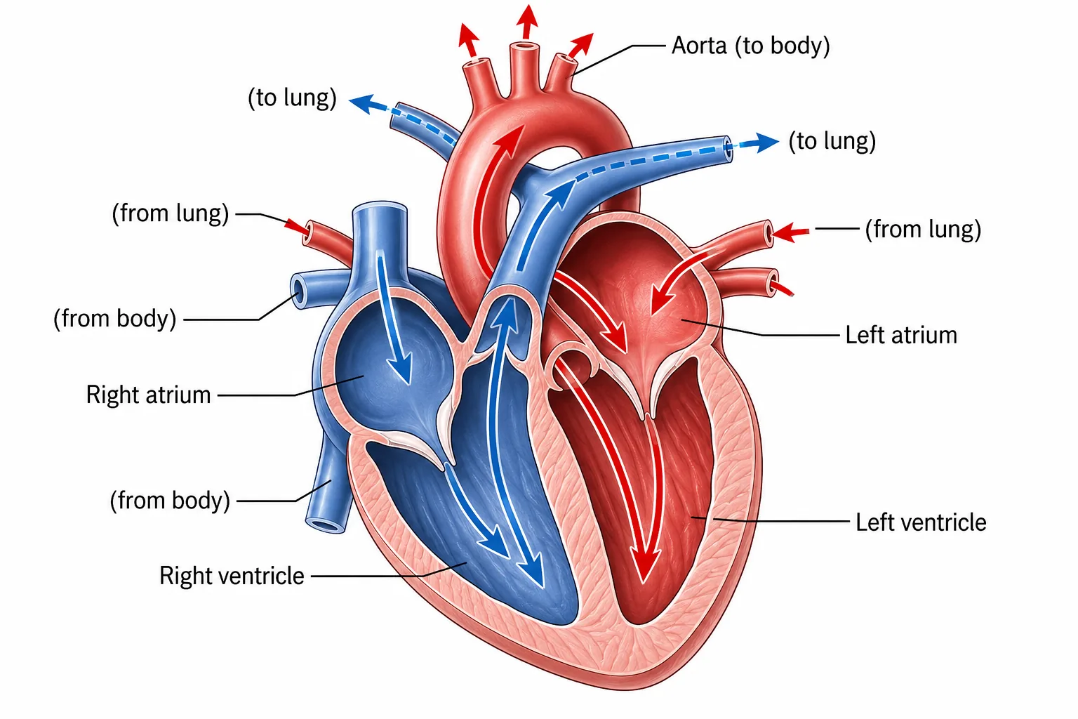
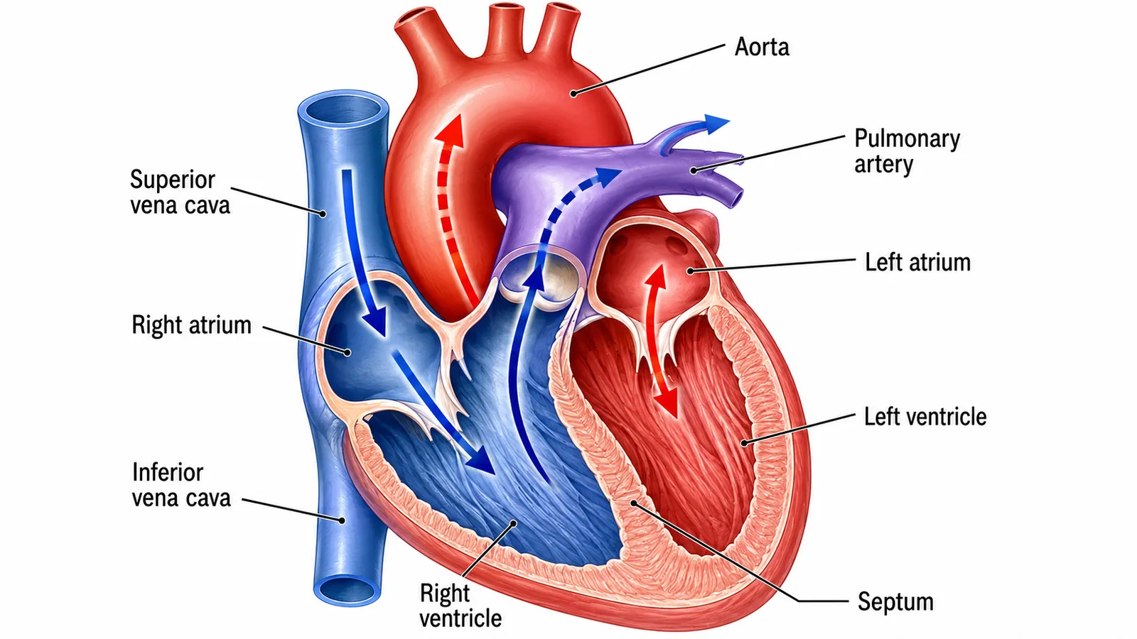


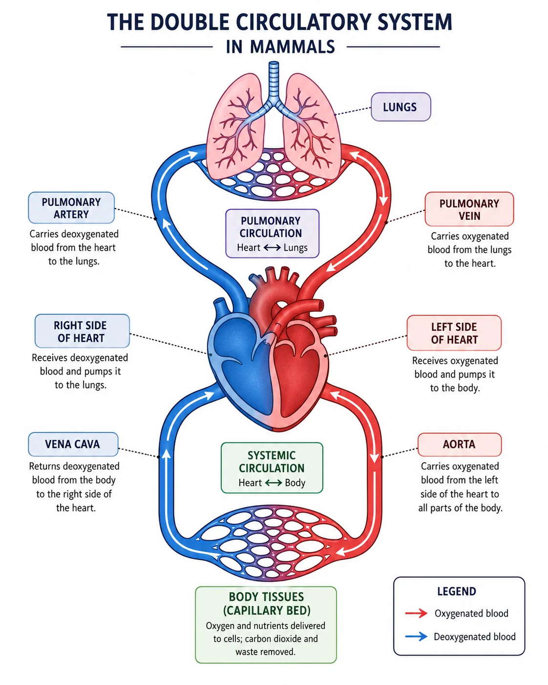



The structure of each vessel explains how it can go wrong.
- and bypass surgery, using a vein taken from the leg to carry blood around the blockage.
Varicose veins appear when the valves fail and blood pools in the veins.
- and the vein returns blood with a low resistance to flow.
In calculations of heart rate from a pulse, count the beats in a set time and scale up: 18 beats in 15 seconds is 72 beats per minute.
Section 7 of 9Exam-style questions
Explain how the structure of an artery is related to its function. [3]3 marks
Arteries carry blood at high pressure, so they have thick walls [1] containing elastic fibres that stretch and recoil to smooth blood flow [1] and muscle to help control the diameter of the vessel [1].
Give two ways a capillary is adapted for exchange. [2]2 marks
Wall is one cell thick, giving a short diffusion distance [1]; the network of many capillaries gives a large surface area [1].
The blood pressure in the aorta is 120 mmHg and in a vein is 10 mmHg. Calculate how many times greater the pressure in the aorta is and suggest why veins need valves. [3]3 marks
120 / 10 = 12 times [1]. Pressure in veins is too low to keep blood flowing back to the heart [1], so valves prevent backflow [1].
Section 8 of 9Key terms
| Term | Meaning |
|---|---|
| Artery | A vessel carrying blood away from the heart at high pressure. |
| Vein | A vessel carrying blood towards the heart, with valves. |
| Capillary | A very small vessel with walls one cell thick where exchange occurs. |
| Lumen | The space inside a blood vessel. |
| Valve | A flap that prevents the backflow of blood. |
| Double circulatory system | A system in which blood passes through the heart twice in one circuit of the body. |
| Aorta | The main artery leaving the left ventricle. |