Comprehensive Explanation: DNA Molecular Architecture, Replication and Genetic Engineering
Biology (General Secondary - 3rd Secondary) | Grade 12 (Thanaweya Amma) | 2026 / 2027 Official Curriculum | Chapter 2: Molecular Biology & Biotechnology
🎯 Targeted Learning Outcomes & Pedagogical Objectives:
📖 Core Terminology, Scientific Definitions & Glossaries:
⚖️ Governing Scientific Laws, Theorems & Mathematical Models:
📌 Exhaustive In-Depth Pedagogical Breakdown:
◄ 1. Historical Proofs of DNA as the Genetic Material
Before the mid-20th century, prevailing consensus favored proteins as genetic carriers due to their structural complexity (20 amino acids vs. only 4 nucleotides). Three seminal milestones refuted this hypothesis:
A. Griffith's Transformation Experiment (1928): Frederick Griffith worked with two strains of Streptococcus pneumoniae: virulent Smooth (S) strain with a protective polysaccharide capsule (causes fatal pneumonia in mice) and non-virulent Rough (R) strain without capsule (mouse survives). Heat-killed S strain alone did not kill mice. However, injecting mice with a mixture of heat-killed S strain and live R strain caused fatal septicemia, and living S bacteria were isolated from cardiac blood! Griffith deduced that a 'transforming principle' was transferred from dead S to live R bacteria, endowing them with capsules.
B. Avery, MacLeod, and McCarty Experiment (1944): Oswald Avery and colleagues isolated and biochemically purified the transforming principle. They treated the extract with proteases (protein destroying) and ribonucleases (RNA destroying); transformation still occurred! When treated with Deoxyribonuclease (DNAse), transformation was completely abolished! This provided direct chemical proof that DNA is the genetic material.
C. Hershey and Chase Bacteriophage T2 Experiment (1952): Alfred Hershey and Martha Chase labeled bacteriophage T2 proteins with radioactive sulfur-35 (³⁵S, present in methionine/cysteine but absent in DNA) and phage DNA with radioactive phosphorus-32 (³²P, present in DNA backbones but absent in proteins). After allowing phages to infect E. coli, blending sheared off ghost capsids, and centrifugation separated bacterial pellets from supernatant. Nearly 100% of ³²P entered the bacteria, whereas less than 3% of ³⁵S entered. The viral progeny emerging from lysed bacteria contained ³²P, concluding definitively that viral DNA, not protein, directs replication.
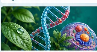
Figure (Figure 1 - Watson and Crick DNA Double Helix Model.png): Molecular spatial architecture of the DNA double helix showing antiparallel strands and hydrogen bonding.
◄ 2. Stereochemistry of the Watson-Crick DNA Double Helix
In 1953, James Watson and Francis Crick, incorporating Rosalind Franklin and Maurice Wilkins' critical X-ray diffraction fiber patterns ('Photo 51'), formulated the 3D model of the DNA double helix.
Backbone Geometry: The alternating sugar-phosphate chain forms the hydrophilic outer handrails of the helix. Adjacent deoxyribose units are linked by covalent 3'-5' phosphodiester bonds (formed between the 3'-OH group of one sugar and the 5'-phosphate group of the next). Because the two strands run in opposite polarities (one 5'->3', the other 3'->5'), they are termed antiparallel. This antiparallel geometry is geometrically required so that nitrogenous bases face each other at optimal spatial angles to establish hydrogen bonds.
Base Complementarity and Dimensions: The diameter of the double helix is uniform (2.0 nm) because a bulky, double-ringed Purine (Adenine or Guanine) always pairs with a smaller, single-ringed Pyrimidine (Thymine or Cytosine). Adenine binds Thymine via 2 hydrogen bonds (A = T), while Guanine binds Cytosine via 3 hydrogen bonds (G ≡ C). One complete helical turn spans 3.4 nm and contains exactly 10 base pairs (0.34 nm between successive base pairs). Chargaff's rules dictate that: %A = %T, %G = %C, and %Purines (%A + %G) = %Pyrimidines (%T + %C) = 50%.
Figure (Figure 2 - DNA Replication Fork and Okazaki Fragments.png): Replication fork mechanics illustrating leading strand continuous synthesis and lagging strand Okazaki fragments.
◄ 3. Enzymatic Mechanism of Semiconservative DNA Replication
Prior to mitotic or meiotic division (S phase of interphase), the genome must be duplicated with absolute fidelity via semiconservative replication, where each parental strand acts as a template for synthesizing an identical daughter strand.
Enzymatic Cascade: (1) DNA Helicase binds replication origins, hydrolyzing ATP to unwind the double helix and sever hydrogen bonds, forming the replication fork. (2) Single-Stranded Binding Proteins (SSBs) coat exposed single strands to prevent spontaneous re-annealing. Topoisomerase relieves torsional strain ahead of the fork.
(3) RNA Primase synthesizes a short RNA primer (~10-12 nucleotides) complementary to the template, providing the obligatory free 3'-OH group required by DNA Polymerase.
(4) DNA Polymerase reads template strands strictly in the 3' to 5' direction and synthesizes daughter strands exclusively in the 5' to 3' direction. On the Leading Strand (oriented 3'->5' towards the fork), synthesis is smooth and continuous. On the Lagging Strand (oriented 5'->3' into the fork), synthesis is discontinuous: multiple RNA primers are laid down, and polymerase synthesizes discrete fragments (100–200 nucleotides in eukaryotes) known as Okazaki Fragments.
(5) DNA Polymerase I removes RNA primers and fills the gaps with deoxyribonucleotides, while its proofreading exonuclease activity corrects mismatched bases. (6) DNA Ligase catalyzes phosphodiester bond formation between adjacent fragments, sealing the continuous strand.
Figure (Table 1 - Genetic Code Codons and Recombinant DNA Plasmids.png): Standard universal mRNA codon chart and recombinant plasmid cloning vector schematic.
◄ 4. Recombinant DNA Technology, Plasmids and Genetic Engineering
Genetic engineering allows isolating specific therapeutic genes (e.g., human insulin, human growth hormone, interferon) and cloning them in bacterial vectors for industrial production.
Restriction Endonucleases: Discovered in bacteria as defense systems against bacteriophages, these enzymes recognize palindromic sequences (e.g., EcoRI recognizes 5'-GAATTC-3' / 3'-CTTAAG-5') and cleave both strands between G and A, leaving complementary single-stranded overhanging sticky ends.
Cloning into Plasmids: A bacterial plasmid (carrying an antibiotic resistance marker, such as Ampicillin resistance) and target human cDNA are cleaved using the identical restriction endonuclease, generating mutually compatible sticky ends. DNA Ligase covalently seals the phosphodiester backbone, creating a Recombinant Plasmid.
Transformation and Expression: The recombinant vector is introduced into competent E. coli cells. Transformed bacteria are selected on ampicillin-containing nutrient agar. Cultured in large-scale industrial bioreactors, these recombinant colonies translate human mRNA into clinical-grade recombinant human insulin.
🧮 Exemplary Step-by-Step Worked Problems & Applied Exercises:
Worked Example 1: Quantitative Chargaff Calculations and Hydrogen Bond Determination
Problem Statement: A double-stranded DNA molecule consists of 1,500 base pairs (3,000 nucleotides). Biochemical analysis indicates that 450 nucleotides are Adenine. Calculate: (1) Total number of Thymine, Guanine, and Cytosine nucleotides; (2) Percentage of Guanine; (3) Total number of hydrogen bonds in this DNA segment; (4) Number of complete helical turns.
Detailed Solution & Derivation: Step 1: Chargaff's rule states A = T. Therefore, T = 450 nucleotides. Total A + T = 450 + 450 = 900 nucleotides. Step 2: Remaining nucleotides = Total - (A + T) = 3,000 - 900 = 2,100 nucleotides. Because G = C, Guanine = 2,100 / 2 = 1,050 nucleotides, and Cytosine = 1,050 nucleotides. Step 3: Percentage of Guanine = (1,050 / 3,000) × 100% = 35%. Step 4: Hydrogen bonds = (A × 2) + (G × 3) = (450 × 2) + (1,050 × 3) = 900 + 3,150 = 4,050 hydrogen bonds. Step 5: One complete helical turn in double-stranded DNA spans 10 base pairs. Total turns = 1,500 base pairs / 10 = 150 complete turns.
Worked Example 2: Restriction Enzyme Digestion & Gel Band Count
Problem Statement: A circular bacterial plasmid of 6,000 bp possesses three recognition sites for EcoRI. A linear human DNA fragment of 4,000 bp possesses two recognition sites for EcoRI. If both samples are digested completely with EcoRI and resolved on agarose gel electrophoresis, how many DNA fragments are generated from each?
Detailed Solution & Derivation: Step 1: For circular DNA, the number of fragments generated by restriction cleavage equals the number of recognition sites. Therefore, 3 cuts in a circular plasmid generate 3 DNA fragments. Step 2: For linear DNA, the number of fragments generated equals the number of recognition sites + 1. Therefore, 2 cuts in a linear DNA molecule generate 2 + 1 = 3 DNA fragments.
💡 Master Teacher Insights & Critical Exam Cautions:
🌍 Practical Real-World, Industrial & Societal Connections:
✔ Commercial Production of Recombinant Human Insulin: Humulin replaced bovine/porcine insulin in 1982, eliminating severe allergic reactions and antibodies in diabetic patients.
✔ Forensic DNA Profiling & PCR Amplification: Forensic laboratories amplify trace Short Tandem Repeats (STRs) from crime scenes using Polymerase Chain Reaction (PCR) and Taq polymerase to identify suspects with 99.9999% statistical certainty.
✔ Genetically Modified Crops (Bt Crops): Splicing the insecticidal endotoxin gene from Bacillus thuringiensis into Egyptian cotton plants confers innate resistance against bollworms, reducing chemical pesticide spraying by over 70%.