Structures and Functions in Living Organisms
Transport systems — Edexcel Linear - OL Revision Notes & Topical Questions
15 min read 19 parts Lesson 14 of 25 1 quiz
Transpiration Practical, Blood Components and Immunity — EDEXCEL LINEAR - OL notes: Investigate transpiration with a potometer, describe the components of blood and explain how white blood cells and antibodies give immunity.A potometer measures water uptake, which is a good estim…
- phagocytes
- lymphocytes
- memory cells
- Worked example 1
- Worked example 2
- Worked example 3
Section 1 of 19Practical: investigating transpiration with a potometer
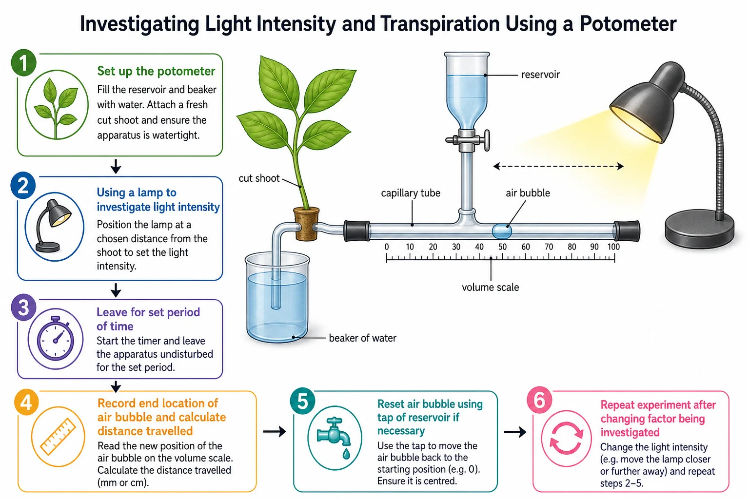
A potometer measures water uptake, which is a good estimate of the transpiration rate. There are two types: a simple one with a capillary tube and air bubble, and a reservoir type that lets the bubble be reset.
| Lamp distance (cm) | Bubble distance (mm) | Rate (mm/min) |
|---|---|---|
| 10 | 32 | 1.07 |
Using CORMS for light intensity: Change the intensity of the light; Observe/measure the distance moved by the bubble; Repeat; Measure the time; Same plant, temperature, humidity and air movement.
Section 2 of 19Components of blood
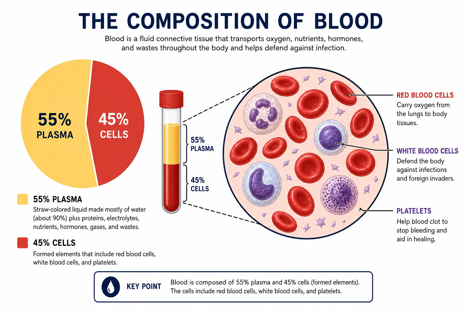
Blood consists of red blood cells, white blood cells, platelets and plasma. Over half the volume is plasma; most of the rest is red blood cells, with a small fraction of white cells and platelets.
| Component | Structure | Function |
|---|---|---|
| Red blood cells | Biconcave discs, no nucleus, contain haemoglobin | Carry oxygen |
| White blood cells | Large cells with a large nucleus; types differ | Defence against pathogens |
| Platelets | Cell fragments | Blood clotting |
| Plasma | Clear, straw-coloured aqueous solution | Transports dissolved substances |
Plasma transports
- digested food and mineral ions (absorbed from the small intestine to cells);
- carbon dioxide;
- urea (a waste product carried to the kidneys);
- hormones (from endocrine glands to target organs);
- and heat energy from respiration, moved to cooler parts of the body or the skin where it can be lost.
Section 3 of 19White blood cells and immunity
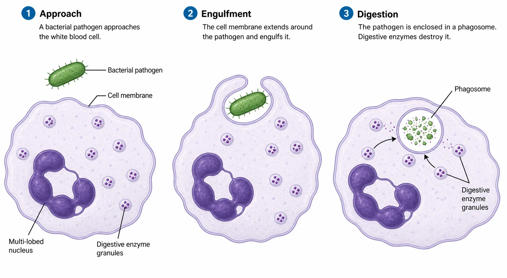
White blood cells are part of the immune system and defend against pathogens.
An organism has immunity when its immune system can respond quickly if the pathogen re-enters, because antibodies are already present or memory cells can make antibodies rapidly. A person infected with a pathogen they are immune to does not suffer the disease or its symptoms.
Section 4 of 19Transpiration and the factors the potometer can test
Transpiration is the loss of water vapour from the leaves, mainly through the stomata.
- but it also includes a small amount of water used in photosynthesis and for support.
Section 5 of 19Worked examples
A bubble moves 32 mm in 30 minutes. Calculate the rate in mm/min. [2]2 marks
32 ÷ 30 = 1.07 mm/min.
State what a potometer actually measures and why this estimates transpiration. [2]2 marks
It measures water uptake; most water taken up is lost by transpiration, so the uptake is a good estimate of the rate.
Name three substances transported by plasma. [3]3 marks
Digested food (glucose), urea, hormones (also carbon dioxide, heat).
Explain why antibodies are described as specific. [2]2 marks
Each antibody has a shape that fits only one type of antigen, so it works only against one pathogen.
Explain why a second infection by the same pathogen causes no symptoms in an immune person. [3]3 marks
Memory cells remain and rapidly make large amounts of the right antibody, destroying the pathogen before it can cause disease.
Section 6 of 19Vaccination
Vaccines help the body develop immunity to infectious diseases and have drastically reduced, or even eradicated, some diseases. A vaccine contains a harmless version of a pathogen (weakened or dead pathogens, or parts of them), and scientists make sure of this by killing or weakening the pathogen or using only its antigens.
The individual is then immunethe response is so rapid that the pathogen cannot cause disease.
Section 7 of 19Platelets and blood clotting
Platelets are fragments of cells involved in clotting and forming scabs.
- which form a mesh across the wound trapping blood cells and forming a clot; this dries to a scab.
Section 8 of 19Structure and function of the heart
The heart is a double pump.
- whereas the right pumps only to the nearby lungs.
Remember arteries carry blood away from the heart. Both atria contract together and both ventricles contract together. A diagram is drawn as if the heart were in the chest, so the left side of the diagram is the person’s right.
Heart rate is the number of beats per minute (bpm). Exercise and adrenaline raise it. The resting rate is controlled by a group of cells in the right atrium called the pacemaker, which coordinates contraction and regulates the heart rate.
Section 9 of 19Coronary heart disease and its risk factors
The coronary arteries supply the cardiac muscle with oxygen and glucose. In coronary heart disease (CHD), layers of fatty material (plaque, mainly cholesterol) build up inside them, narrowing the lumen and reducing blood flow. Partial blockage causes severe chest pain (angina); complete blockage means cells cannot respire aerobically, leading to a heart attack.
| Risk factor | Effect |
|---|---|
| High blood pressure | Increases the force on artery walls and damages vessels |
| High-fat diet | Speeds up the build-up of fatty plaques |
| Smoking | Chemicals increase plaque build-up and blood pressure; carbon monoxide reduces the oxygen-carrying capacity of red blood cells |
| Genetics, stress, inactivity | Increase the likelihood of CHD |
Section 10 of 19Putting it together: exam focus
Questions on this topic often ask you to follow blood from one place to another, so rehearse the route vena cava, right atrium, tricuspid valve, right ventricle, semilunar valve, pulmonary artery, lungs, pulmonary vein, left atrium, bicuspid valve, left ventricle, aorta. Heart valves ensure one-way flow; the muscle wall of the heart is cardiac muscle supplied by coronary arteries.
Section 11 of 19Worked examples
Explain why the left ventricle wall is thicker than the right. [2]2 marks
It pumps blood at high pressure around the whole body, while the right ventricle only pumps to the lungs.
Explain how vaccination gives long-term immunity. [4]4 marks
A harmless form of the pathogen triggers antibody production and memory cells. If infected later, memory cells produce antibodies sooner, faster and in greater quantity, so the pathogen cannot cause disease.
Describe the role of platelets in clotting. [3]3 marks
They release chemicals that convert fibrinogen to insoluble fibrin, forming a mesh that traps cells and seals the wound.
State where the pacemaker is and what it does. [2]2 marks
In the right atrium; it coordinates the contraction of the heart muscle and regulates heart rate.
Explain how smoking increases the risk of CHD. [3]3 marks
Chemicals increase plaque build-up and blood pressure, and carbon monoxide reduces oxygen carrying by red blood cells.
Section 12 of 19Three types of blood vessel
The three main types of blood vessel are arteries, veins and capillaries. Smaller vessels that branch from arteries are arterioles (small arteries) and those that join into veins are venules (small veins). Each vessel is adapted to its function.
| Feature | Artery | Capillary | Vein |
|---|---|---|---|
| Direction of blood | Away from the heart | Between arteries and veins, through tissues | Towards the heart |
| Pressure | High | Falling | Low |
| Wall | Thick, muscular and elastic | One cell thick | Thin, less muscle |
| Lumen | Narrow | Very narrow | Wide |
| Valves | No | No | Yes |
Section 13 of 19Why each vessel is adapted as it is
Arteries carry blood at high pressure, so their walls are thick, muscular and elastic to withstand the pressure and recoil to push blood along. Veins carry blood at low pressure; a large lumen reduces resistance to flow and valves prevent the backflow of blood. Capillaries carry blood at low pressure within tissues and have walls one cell thick (a short diffusion distance), so substances easily diffuse in and out. Their walls are “leaky”, allowing blood plasma to leak out and form tissue fluid surrounding the cells.
Section 14 of 19General structure of the circulatory system
The circulatory system is a closed network of blood vessels connected to the heart, with blood flowing in one direction.
- so blood passes through the heart twice on each complete circuit.
- and this blood is usually oxygenated;
- the pulmonary artery is the exception, carrying deoxygenated blood away from the heart.
| Organ | Vessel towards organ | Vessel away from organ |
|---|---|---|
| Heart | Vena cava, pulmonary vein | Aorta, pulmonary artery |
| Lung | Pulmonary artery | Pulmonary vein |
| Liver | Hepatic artery, hepatic portal vein | Hepatic vein |
| Kidney | Renal artery | Renal vein |
Section 15 of 19Exchange at the capillaries and the role of tissue fluid
Blood pressure forces plasma through the thin capillary walls, forming tissue fluid that bathes the cells.
Section 16 of 19Pulse and pressure in different vessels
Blood leaves the heart in surges, so the pressure in arteries rises and falls with each beat; this surge can be felt as the pulse in an artery near the surface, for example at the wrist or neck.
- so veins need valves and rely on contraction of surrounding skeletal muscles squeezing the veins to help push the blood towards the heart.
In the exam, link each feature to a function: thick elastic wall to high pressure, thin wall to short diffusion distance, valves to preventing backflow, wide lumen to low resistance.
Section 17 of 19Worked examples
Explain two ways in which the structure of a vein differs from that of an artery. [4]4 marks
A vein has a larger lumen and thin walls, and it has valves to stop backflow; an artery has a thick, elastic, muscular wall and a narrow lumen with no valves.
Explain why capillary walls are one cell thick. [2]2 marks
It gives a short diffusion distance so oxygen, nutrients and carbon dioxide can pass quickly between blood and cells.
Name the vessel taking blood from the heart to the lungs and say what type of blood it carries. [2]2 marks
The pulmonary artery; deoxygenated blood.
Why is the circulation described as a double circulation? [2]2 marks
Blood passes through the heart twice in one complete circuit: once to the lungs and once to the body.
State the function of the valves in veins. [1]1 mark
They prevent the backflow of blood.
Section 18 of 19Key terms
| Term | Meaning |
|---|---|
| Potometer | Apparatus that measures water uptake by a shoot. |
| Plasma | The liquid part of blood. |
| Antigen | A marker on a cell or pathogen that triggers an immune response. |
| Antibody | A protein made by lymphocytes that binds a specific antigen. |
| Phagocyte | A white blood cell that engulfs and digests pathogens. |
| Memory cell | A long-lived lymphocyte that gives a faster response on re-infection. |
| Immunity | The ability to respond quickly to a pathogen and avoid disease. |
| Term | Meaning |
|---|---|
| Vaccine | A harmless form of a pathogen that causes immunity without disease. |
| Fibrin | Insoluble protein fibres that form a blood clot. |
| Pacemaker | Group of cells in the right atrium that controls heart rate. |
| Coronary artery | Artery supplying the heart muscle with oxygen and glucose. |
| Angina | Severe chest pain caused by partial blockage of coronary arteries. |
| Plaque | Fatty deposit, mainly cholesterol, inside arteries. |
| Term | Meaning |
|---|---|
| Artery | A vessel carrying blood away from the heart at high pressure. |
| Vein | A vessel carrying blood back to the heart at low pressure. |
| Capillary | A tiny vessel with walls one cell thick where exchange occurs. |
| Tissue fluid | Plasma that leaks out of capillaries and surrounds cells. |
| Double circulation | A system in which blood passes through the heart twice per circuit. |
| Arteriole | A small artery. |
Section 19 of 19Exam technique
Marked practiceQuizzes for this lesson
Last updated:
\n