1.1Cell Structure and Molecular Biology
Chemistry of life — EST 2 Biology Revision Notes & Topical Questions
20 min read 30 parts Lesson 1 of 11 2 quizzes
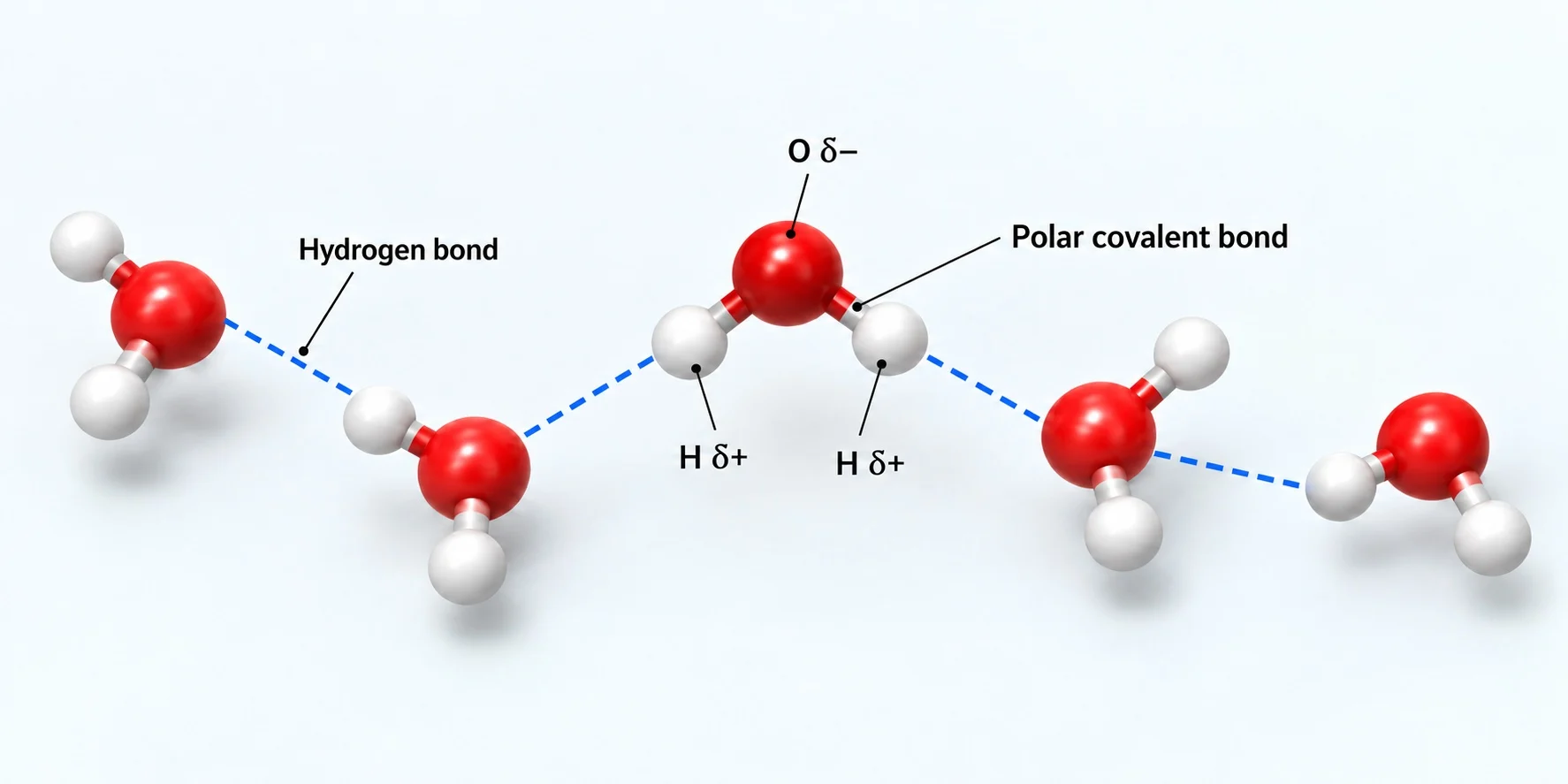
SAT Biology chemistry of life: connect water, pH and the four major biological macromolecules to cell structure and function. Water is a polar molecule: oxygen carries a partial negative charge and the hydrogen atoms carry partial positive charges.
- hydrogen bonds
- Dehydration synthesis
- Hydrolysis
Section 1 of 30Water and hydrogen bonding
Section 2 of 30Cohesion, adhesion and solvent action
Section 3 of 30pH and buffers
Section 4 of 30Build and break polymers
Section 5 of 30Protein structure and enzymes
Section 6 of 30Nucleotides and the 5′ and 3′ ends
DNA and RNA are polynucleotides: many nucleotide monomers linked in a chain. Each nucleotide has three components: a pentose (five-carbon) sugar, a phosphate group and a nitrogenous base. DNA bases are adenine, guanine, cytosine and thymine; RNA bases are adenine, guanine, cytosine and uracil.
A polynucleotide has two distinct ends.
- forming strong covalent bonds between the nucleotides.
Section 7 of 30DNA versus RNA
DNA is an antiparallel double helix: the two strands of nucleotides run in opposite 5′ → 3′ orientations. Hydrogen bonds join complementary bases between the strands. RNA is usually single-stranded, but it can fold, and the folds are held together by base pairing, with adenine pairing with uracil (not thymine) and cytosine pairing with guanine as in DNA.
| Feature | DNA | RNA |
|---|---|---|
| Pentose sugar | Deoxyribose | Ribose |
| Nitrogenous bases | A, T, C, G | A, U, C, G |
| Base pairing | A–T, C–G | A–U, C–G |
| Strands | Double helix | Usually single-stranded |
Section 8 of 30Amino acids and peptide bonds
Proteins are made of linear chains of monomers called amino acids.
Amino acids are joined by peptide bonds, which are covalent bonds formed at the carboxyl end of the growing peptide chain: a hydroxyl group (–OH) is lost from the carboxyl group of one amino acid and a hydrogen atom is lost from the amino group of the neighbouring amino acid.
Section 9 of 30Primary and secondary structure
DNA determines the primary structure of a protein by instructing the cell to add particular amino acids in a specific sequence.
- and a β-pleated sheet forms when the chain folds so that two parts lie parallel and hydrogen bonds form between the folded layers.
Remember that the four common macromolecule classes are carbohydrates, lipids, proteins and nucleic acids, and that nucleic acids and proteins are information-carrying polymers: the order of bases in DNA codes for the order of amino acids in a protein, which determines its shape and function.
Section 10 of 30Worked examples
State two differences between DNA and RNA. [2]2 marks
DNA has deoxyribose and thymine and is a double helix; RNA has ribose and uracil and is usually single-stranded.
Explain what antiparallel means in DNA. [2]2 marks
The two strands run in opposite 5′ → 3′ directions.
Describe how a peptide bond forms. [3]3 marks
–OH is lost from the carboxyl group of one amino acid and H from the amino group of the next; a covalent bond forms between C and N; water is released.
Why does a change in one amino acid in the sequence sometimes alter a protein's function? [2]2 marks
It can change the folding of the chain and so the shape of the protein.
Section 11 of 30Tertiary and quaternary structure of proteins
The tertiary structure is the overall three-dimensional folding of one polypeptide, held by interactions between R groups: hydrogen bonds, disulphide bonds between cysteine amino acids, ionic bonds between charged R groups and weak hydrophobic interactions between non-polar R groups. The quaternary structure is the arrangement of two or more polypeptide subunits working together; haemoglobin, for example, has four subunits that carry oxygen.
Section 12 of 30Carbohydrates
Carbohydrates are biological molecules made of monosaccharide monomers, such as glucose. Two monosaccharides join by dehydration synthesis to form a disaccharide, and many join to form a polysaccharide by covalent glycosidic bonds. Complex carbohydrates can be linear or branched.
| Polysaccharide | Found in | Structure and function |
|---|---|---|
| Cellulose | Plant cell walls | Long chains of β-glucose joined by 1,4 glycosidic bonds; alternate glucose molecules are rotated through 180°. Many hydrogen bonds between chains give great strength |
| Starch | Plants (storage) | α-glucose; amylose is unbranched and amylopectin is a branched form |
| Glycogen | Animals and fungi (storage) | Always branched, so large quantities can be stored in a small space |
Storage polysaccharides do not dissolve in the cytoplasm, so they have no effect on the water potential of cells.
Section 13 of 30Lipids
Lipids are energy-dense molecules that provide energy storage and support cell function.
- which affects structure and function.
Phospholipids have a hydrophilic head and hydrophobic tails and form bilayers in plasma and cell membranes. Steroids are a class of lipids; steroid hormones support growth and development, energy metabolism and homeostasis.
Section 14 of 30Polymers, hydrolysis and dehydration synthesis
A polymer is a large molecule made of many repeated monomers; the term macromolecule is often used for biological polymers.
| Monomer | Polymer |
|---|---|
| Monosaccharide (glucose) | Polysaccharide (starch, glycogen, cellulose) |
| Amino acid | Polypeptide / protein |
| Nucleotide | Polynucleotide (DNA, RNA) |
Dehydration synthesis (a condensation reaction) joins two smaller molecules through covalent bonding and removes a molecule of water. Hydrolysis (hydro = water, lysis = splitting) breaks covalent bonds by adding water, splitting large molecules into smaller ones.
Section 15 of 30Linking structure to function
Each molecule's properties arise from its bonding.
Section 16 of 30Identifying molecules in diagrams
Recognise carbohydrates by their rings of carbon, hydrogen and oxygen in the ratio near 1 : 2 : 1; amino acids by the amine and carboxyl groups; triglycerides by glycerol joined to three fatty acid chains; and nucleotides by a sugar bonded to a phosphate and a base. Check for double bonds in the fatty acid tails to decide if a lipid is saturated or unsaturated.
Finish by checking that you can state, for every molecule type, its monomer, its bond, the reaction that forms it and the reaction that breaks it down.
Section 17 of 30Worked examples
Name the bonds that hold the tertiary structure of a protein. [3]3 marks
Hydrogen bonds, disulphide bonds (between cysteines), ionic bonds, and hydrophobic interactions between R groups.
Explain why cellulose is strong. [2]2 marks
Long β-glucose chains with alternate molecules rotated 180° form many hydrogen bonds between chains.
Give two reasons why glycogen is a good storage molecule. [2]2 marks
It is branched, so a lot can be stored in a small space, and insoluble, so it does not affect water potential.
Define hydrolysis. [1]1 mark
Breaking of covalent bonds by the addition of water.
Section 18 of 30Nucleotides and polynucleotide polarity
DNA and RNA are polynucleotides: many nucleotide monomers linked in a chain. Each nucleotide has three components: a pentose (five-carbon) sugar, a phosphate group and a nitrogenous base. DNA bases are adenine, guanine, cytosine and thymine; RNA bases are adenine, guanine, cytosine and uracil.
A polynucleotide has two distinct ends.
- forming strong covalent bonds between the nucleotides.
Section 19 of 30DNA and RNA compared
DNA is an antiparallel double helix: the two strands of nucleotides run in opposite 5′ → 3′ orientations. Hydrogen bonds join complementary bases between the strands. RNA is usually single-stranded, but it can fold, and the folds are held together by base pairing, with adenine pairing with uracil (not thymine) and cytosine pairing with guanine as in DNA.
| Feature | DNA | RNA |
|---|---|---|
| Pentose sugar | Deoxyribose | Ribose |
| Nitrogenous bases | A, T, C, G | A, U, C, G |
| Base pairing | A–T, C–G | A–U, C–G |
| Strands | Double helix | Usually single-stranded |
Section 20 of 30Amino acids and peptide bonds
Proteins are made of linear chains of monomers called amino acids.
Amino acids are joined by peptide bonds, which are covalent bonds formed at the carboxyl end of the growing peptide chain: a hydroxyl group (–OH) is lost from the carboxyl group of one amino acid and a hydrogen atom is lost from the amino group of the neighbouring amino acid.
Section 21 of 30How DNA determines primary structure
DNA determines the primary structure of a protein by instructing the cell to add particular amino acids in a specific sequence.
- and a β-pleated sheet forms when the chain folds so that two parts lie parallel and hydrogen bonds form between the folded layers.
Practise by writing a complementary strand
- given the DNA sequence 5′-ATGC-3′, the complementary strand is 3′-TACG-5′ and the corresponding RNA is 5′-AUGC-3′ when transcribed from the complementary template.
- This links base pairing rules, antiparallel orientation and the difference between DNA and RNA bases.
Check that you can state the five key contrasts: sugar (deoxyribose or ribose), bases (T or U), strands (two or one), pairing (A–T or A–U) and ends (5′ phosphate and 3′ hydroxyl).
Section 22 of 30Worked examples
Name the three components of a nucleotide. [3]3 marks
Pentose sugar, phosphate group, nitrogenous base.
How does the sequence of DNA affect a protein? [3]3 marks
DNA specifies the order of amino acids; this primary structure determines folding and therefore function.
Identify the peptide bond in a diagram. [1]1 mark
Where nitrogen is bonded to a carbon that has a double bond to oxygen.
State which bases pair in RNA folds. [2]2 marks
A with U, and C with G.
Section 23 of 30Secondary, tertiary and quaternary structure
DNA determines the primary structure of a protein by instructing the cell to add particular amino acids in a specific sequence.
- and a β-pleated sheet forms when the chain folds so that two parts lie parallel and hydrogen bonds form between the folded layers.
The tertiary structure is the overall three-dimensional folding of one polypeptide, held by interactions between R groups: hydrogen bonds, disulphide bonds between cysteine amino acids, ionic bonds between charged R groups and weak hydrophobic interactions between non-polar R groups. The quaternary structure is the arrangement of two or more polypeptide subunits working together; haemoglobin, for example, has four subunits that carry oxygen.
Section 24 of 30Carbohydrate polymers
Carbohydrates are biological molecules made of monosaccharide monomers, such as glucose. Two monosaccharides join by dehydration synthesis to form a disaccharide, and many join to form a polysaccharide by covalent glycosidic bonds. Complex carbohydrates can be linear or branched.
| Polysaccharide | Found in | Structure and function |
|---|---|---|
| Cellulose | Plant cell walls | Long chains of β-glucose joined by 1,4 glycosidic bonds; alternate glucose molecules are rotated through 180°. Many hydrogen bonds between chains give great strength |
| Starch | Plants (storage) | α-glucose; amylose is unbranched and amylopectin is a branched form |
| Glycogen | Animals and fungi (storage) | Always branched, so large quantities can be stored in a small space |
Storage polysaccharides do not dissolve in the cytoplasm, so they have no effect on the water potential of cells.
Section 25 of 30Lipids and membranes
Lipids are energy-dense molecules that provide energy storage and support cell function.
- which affects structure and function.
Phospholipids have a hydrophilic head and hydrophobic tails and form bilayers in plasma and cell membranes. Steroids are a class of lipids; steroid hormones support growth and development, energy metabolism and homeostasis.
Section 26 of 30Linking structure to function in macromolecules
Each class of biological macromolecule is built from monomers joined by dehydration synthesis and broken by hydrolysis.
- and ionic and disulphide bonds lock the tertiary shape.
Section 27 of 30Exam technique
When asked to explain why a molecule suits its function, state the feature, the bond or property responsible, and the benefit.
Practise identifying unfamiliar diagramsfind the amine and carboxyl groups for amino acids, ring structures for sugars, long chains attached to glycerol for triglycerides.
Section 28 of 30Worked examples
Explain why a high temperature can make an enzyme stop working. [3]3 marks
Hydrogen bonds and other weak bonds break, so the tertiary structure unfolds and the active site shape is lost.
Compare starch and cellulose. [3]3 marks
Starch: α-glucose, storage, branched or unbranched. Cellulose: β-glucose, structural, 1,4 bonds with rotated alternate units.
Why are lipids a good energy store? [2]2 marks
They are energy-dense, so more energy is stored per gram.
Describe how a phospholipid is arranged in a membrane. [2]2 marks
It forms a bilayer with hydrophilic heads outwards and hydrophobic tails inwards.
Section 29 of 30Key terms
| Term | Meaning |
|---|---|
| Nucleotide | A monomer of nucleic acids made of sugar, phosphate and a base. |
| Antiparallel | Running in opposite directions. |
| Amino acid | The monomer of proteins. |
| Primary structure | The sequence of amino acids in a polypeptide. |
| R group | The variable side chain of an amino acid. |
| α-helix | A secondary structure with hydrogen bonds between every fourth peptide bond. |
| Term | Meaning |
|---|---|
| Polymer | A large molecule made of many repeating monomers. |
| Dehydration synthesis | A reaction that joins monomers and releases water. |
| Hydrolysis | A reaction that splits a molecule by adding water. |
| Peptide bond | The covalent bond between amino acids. |
| Quaternary structure | Arrangement of multiple polypeptide subunits. |
| Glycogen | Branched storage polysaccharide in animals and fungi. |
| Triglyceride | A lipid made of glycerol and three fatty acids. |
| Term | Meaning |
|---|---|
| Polymer | A large molecule made of many repeating monomers. |
| Dehydration synthesis | A reaction that joins monomers and releases water. |
| Hydrolysis | A reaction that splits a molecule by adding water. |
| Peptide bond | The covalent bond between amino acids. |
| Polynucleotide | A chain of many nucleotides. |
| Uracil | The base in RNA that pairs with adenine. |
| Deoxyribose | The sugar in DNA. |
| Term | Meaning |
|---|---|
| β-pleated sheet | A secondary structure where chain regions lie parallel, bonded by hydrogen bonds. |
| Tertiary structure | 3D folding of a polypeptide held by R group interactions. |
| Amylopectin | A branched form of starch. |
| Cellulose | A structural polysaccharide of β-glucose. |
| Steroid | A class of lipids including some hormones. |
Section 30 of 30Exam technique
Marked practiceQuizzes for this lesson
Last updated:
\n