A1.2Theme A — Unity and diversity
Nucleic acids — IB Biology HL Revision Notes & Topical Questions
10 min read 12 parts Lesson 6 of 14 2 quizzes
Curriculum Equation Key قوانين الرقم الهيدروجيني والاتزان الأيوني - pH & Acid-Base Equilibrium FormulasFast Exam Solution
pH = -log[H⁺] | pOH = -log[OH⁻] | pH + pOH = 14 | [H⁺][OH⁻] = 10⁻¹⁴
Word Equation & Conditions: الرقم الهيدروجيني = -لوغاريتم تركيز أيون الهيدروجين | pH + pOH = 14
Location & Specifications: الاتزان الأيوني، كيمياء 3 ثانوي، التحصيلي، و AP Chemistry.
Lesson summary
A1.2 Nucleic acids — IB BIOLOGY HL notes: Describe the structure of DNA and RNA nucleotides and polynucleotides, and explain how DNA stores and passes on the genetic code.DNA — it carries the genetic code in all living organisms, which is why the code is universal.DNA is mainly i…
- RNA viruses
- Drawing nucleotides
- Condensation
- Phosphodiester bond
- Sugar-phosphate backbone
- Sequence variety
Part 1 of 9DNA and RNA as genetic material
DNA
- DNA it carries the genetic code in all living organisms, which is why the code is universal.
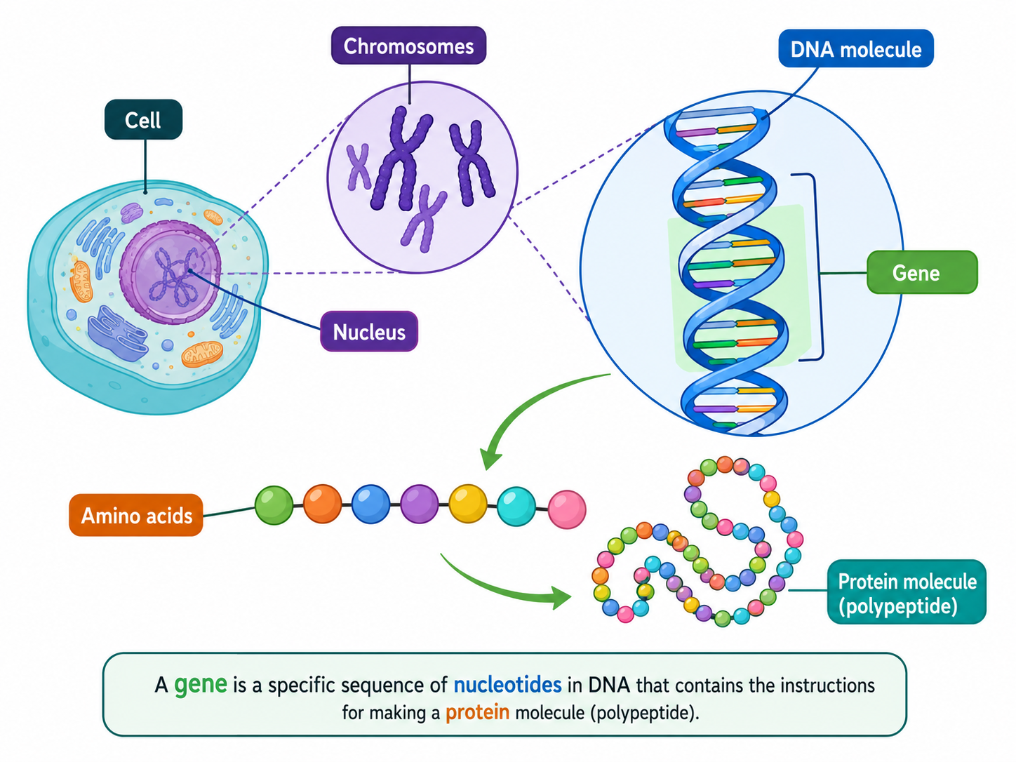
- DNA is mainly in the nucleus as chromosomes, and also in chloroplasts and mitochondria of eukaryotic cells.
RNA
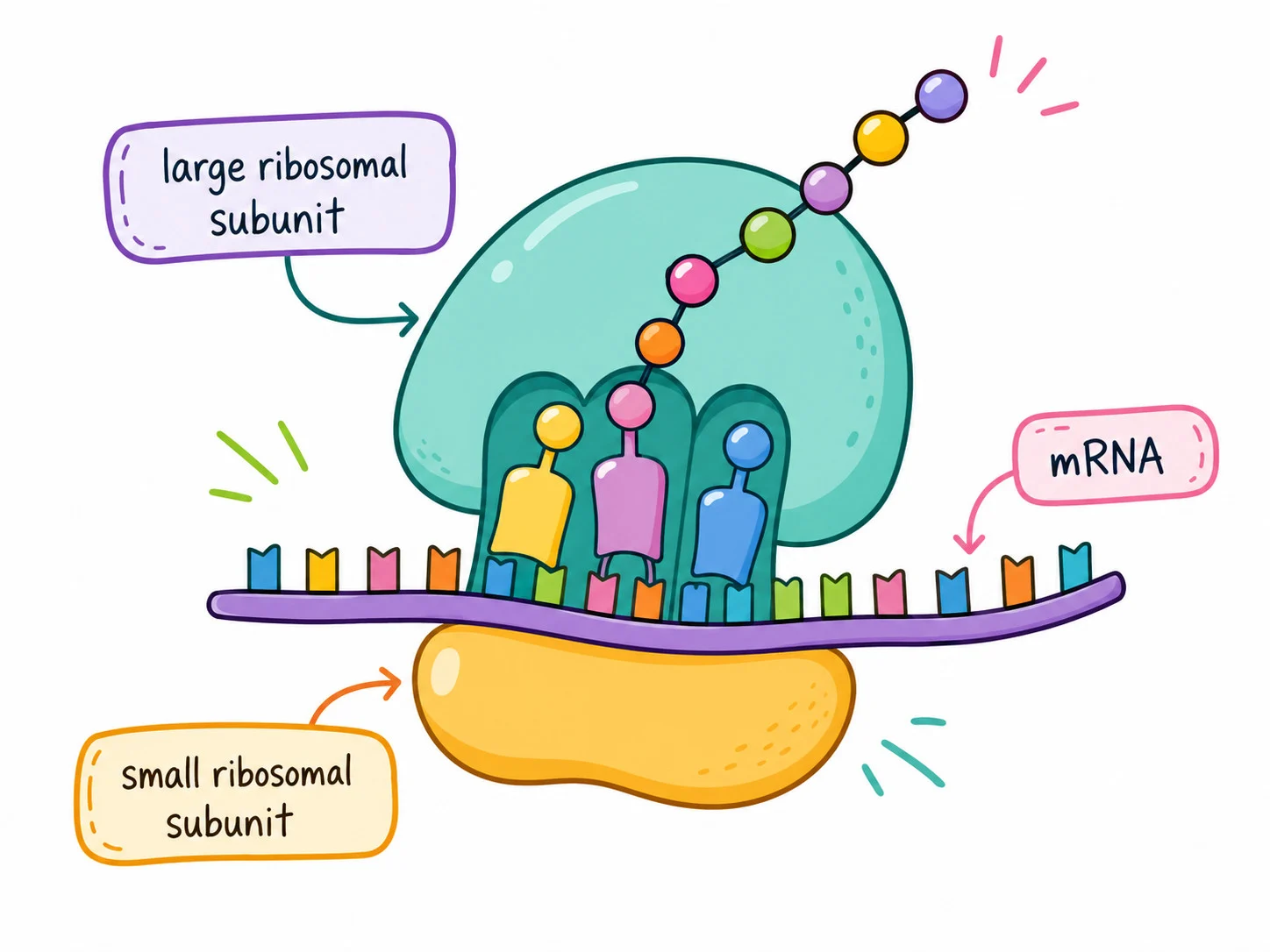
- RNA it is the main component of ribosomes and is also found in the nucleus and cytoplasm.
RNA viruses
- RNA viruses some viruses such as SARS-CoV-2 have RNA as their genetic material and cause COVID-19, Ebola, mumps and influenza.
- Viruses are not classed as living because they cannot replicate alone and have no cellular structure.
Part 2 of 9Nucleotide structure
Nucleotide structure
Polymer
- DNA and RNA are polymers of repeating units called nucleotides.
Components of each nucleotide
Nucleotide structure
A pentose sugar with 5 carbon atoms.
A nitrogen-containing organic base with one or two rings.
A phosphate group, which is acidic and negatively charged.
Nucleotide structure
Bonding
- Bonding the base and the phosphate are both covalently bonded to the sugar.
Bases
- DNA contains adenine (A), guanine (G), cytosine (C) and thymine (T).
- RNA has uracil (U) in place of thymine.
- Adenine and guanine are purines (two rings); cytosine, thymine and uracil are pyrimidines (one ring).
Drawing nucleotides
- Drawing nucleotides pentagons represent sugars, circles with P represent phosphates, rectangles represent bases, solid lines show covalent bonds and dashed lines show hydrogen bonds.
Part 3 of 9Linking nucleotides into strands and directionality of strands
1Condensation
- Condensation the phosphate of one nucleotide bonds to the sugar of the next and a water molecule is released at each bond.
2Phosphodiester bond
Phosphodiester bond the covalent bond between the sugar of one nucleotide and the phosphate of the next.
3Sugar-phosphate backbone
- Sugar-phosphate backbone repeating sugar and phosphate units, with one base attached to each sugar.
4Strand
Strand the polymer of nucleotides, also called a polynucleotide.
5Sequence variety
- Sequence variety any order of the 4 bases is possible because the sugar and phosphate are the same in every nucleotide.
Carbon numbering
- Carbon numbering carbons of the sugar are numbered clockwise from the right, which shows the orientation of the strand.
Linkage
- Linkage the phosphate links carbon-3 of one sugar to carbon-5 of the next.
Ends
- Ends the 5' end carries the phosphate on carbon-5 and the 3' end carries the OH group on carbon-3.
Antiparallel
- Antiparallel in DNA one strand runs 5' to 3' and the other runs 3' to 5'.
Importance
- Importance directionality matters in replication, transcription and translation.
- In transcription the template strand is read 3' to 5' and mRNA is made 5' to 3'.
- Ribosomes translate mRNA in the 5' to 3' direction.
Part 4 of 9DNA structure
DNA structure
Double helix
- DNA is two antiparallel polynucleotide strands wound into a three-dimensional helix.
Backbone
- Backbone each strand has alternating deoxyribose sugars and phosphate groups joined by covalent bonds.
Bases
- Bases they project inwards from the backbone towards the centre of the molecule.
Complementary base pairing
- Complementary base pairing the strands are held together by hydrogen bonds between bases.
- Adenine pairs with thymine by two hydrogen bonds.
- Guanine pairs with cytosine by three hydrogen bonds.
- The number of A equals T and the number of C equals G.
Template
- Template the sequence of one strand determines the other, so DNA can be copied precisely in replication.
Watson and Crick
- Watson and Crick they built the double helix model in 1953 by trial and error.
- Each pair is a purine with a pyrimidine, so every base pair has the same length and the helix has the same shape for any sequence.
- Hydrogen bonds between pairs add to the stability of the helix.
Part 5 of 9DNA compared with RNA and types of RNA
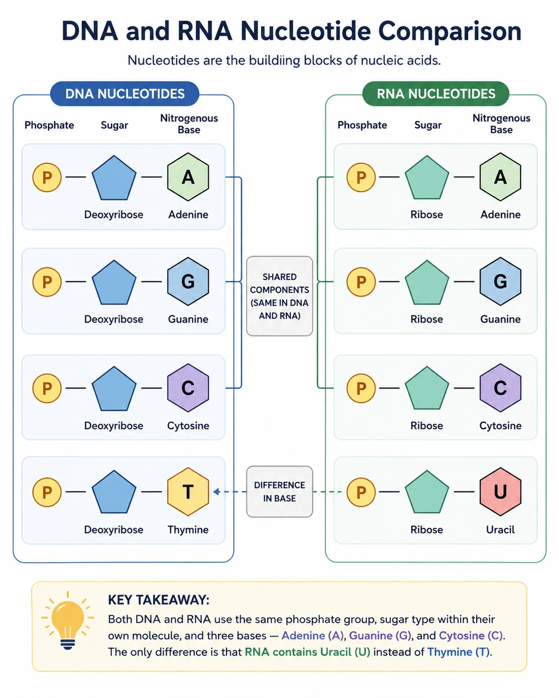
| Feature | DNA | RNA |
|---|---|---|
| Pentose sugar | Deoxyribose | Ribose |
| Bases | A, C, G, T | A, C, G, U |
| Strands | Two (double helix) | Usually one |
| Length | Very long | Short, from about a hundred to a few thousand nucleotides |
DNA compared with RNA and types of RNA
mRNA
- mRNA it is formed in the nucleus and carries the code to ribosomes in the cytoplasm.
tRNA
- tRNA it carries amino acids to ribosomes during protein synthesis.
rRNA
- rRNA it forms part of ribosomes.
Part 6 of 9The genetic code
1Coding strand
- Coding strand one strand of DNA carries the base sequence that enzymes read.
2Gene
- Gene the base sequence of a gene determines the order of amino acids in a protein.
3Codon
Codon a triplet of bases that codes for one amino acid, from the 20 possible amino acids.
4Protein shape
- Protein shape the amino acid sequence determines protein shape and function.
5Universal code
- Universal code almost all organisms use the same codons for the same amino acids, with a few rare exceptions.
- This allows genetic information to be transferred between species and makes genetic engineering possible.
- It is evidence for a universal common ancestor.
6Conserved sequences
Conserved sequences coding and non-coding sequences that have stayed unchanged in all organisms.
- Highly conserved genes code for proteins of transcription and translation and for histones.
- Their similarity shows shared ancestry.
Part 7 of 9DNA as an information store
Capacity
- Capacity four bases can form an almost limitless variety of sequences of different lengths.
Gene numbers
- Gene numbers human about 20 000, dog about 19 000, water flea about 31 000, bacterium (E. coli) about 4 300 and rice about 41 500.
Genome size
- Genome size a human nucleus holds about 3.2 gigabases, about 109 base pairs.
Packaging
- Packaging about 2 metres of DNA fits into a microscopic nucleus, so it stores a vast amount of data very economically.
Part 8 of 9Nucleosomes and molecular visualisation
Nucleosomes and molecular visualisation
Histones
- Histones eukaryotic nuclear DNA is associated with histone proteins to form chromatin, unlike the 'naked' DNA of most prokaryotes.
Nucleosome
- DNA coils twice around a core of eight histone proteins (an octamer), held by one more histone attached to linker DNA.
- Nucleosomes look like a string of beads and supercoil DNA into a compact structure.
- They protect DNA and aid chromosome movement in cell division.
- Histone tails can be tagged chemically to promote or suppress transcription.
Molecular visualisation software
- Molecular visualisation software it displays proteins, DNA, RNA and carbohydrates as 3-D structures.
- It lets researchers relate sequence to structure and function and study interactions.
- Ball-and-stick and ribbon models are used, and structures come from the Protein Data Bank (for example human nucleosome 6T79).
- The viewer shows DNA making two loops around the histone core with histone tails projecting out.
Part 9 of 9Chargaff's data
1Findings
- Chargaff analysed DNA from many organisms in the 1930s and 1940s.
- Purines equal pyrimidines in number.
- Adenine equals thymine and guanine equals cytosine.
- The base composition differs between species.
2Meaning
- Meaning a purine pairs with a pyrimidine, which is the basis of complementary base pairing.
3Tetranucleotide hypothesis
- Levene proposed that nucleic acid was a repeating unit of four different nucleotides.
- Chargaff's data falsified it because it showed species-specific composition, so a repeating tetramer could not carry genetic information.
4Problem of induction
- Problem of induction a hypothesis from past observations can never be proved absolutely true, so theories stay tentative.
- Popper argued that knowledge grows by falsifying existing hypotheses.
Key terms
| Term | Meaning |
|---|---|
| Nucleotide | a pentose sugar, a nitrogenous base and a phosphate group joined by covalent bonds. |
| Phosphodiester bond | the covalent bond joining the sugar of one nucleotide to the phosphate of the next. |
| Antiparallel | running in opposite directions, as the two strands of DNA do. |
| Complementary base pairing | A with T (or U in RNA) and G with C, held by hydrogen bonds. |
| Codon | a triplet of bases that codes for one amino acid. |
| Nucleosome | DNA wound around an octamer of eight histone proteins. |
| Purine | a base with two rings, adenine or guanine. |
| Pyrimidine | a base with one ring, cytosine, thymine or uracil. |
| Conserved sequence | a base sequence that has stayed unchanged across all organisms. |
Avoid these mistakes
1
DNA and RNA differ only in having thymine or uracil — they also differ in sugar, strand number and length.
2
A purine can pair with another purine — only a purine with a pyrimidine fits, so A pairs with T and G with C.
3
Hydrogen bonds join nucleotides in a strand — covalent phosphodiester bonds join nucleotides, hydrogen bonds join the two strands.
4
Both DNA strands run the same way — they are antiparallel, 5' to 3' against 3' to 5'.
Marked practiceQuizzes for this lesson
Last updated:
\n