A2.2Theme A — Unity and diversity
Cell structure — IB Biology HL Revision Notes & Topical Questions
10 min read 12 parts Lesson 8 of 14 2 quizzes
Curriculum Equation Key قوانين الرقم الهيدروجيني والاتزان الأيوني - pH & Acid-Base Equilibrium FormulasFast Exam Solution
pH = -log[H⁺] | pOH = -log[OH⁻] | pH + pOH = 14 | [H⁺][OH⁻] = 10⁻¹⁴
Word Equation & Conditions: الرقم الهيدروجيني = -لوغاريتم تركيز أيون الهيدروجين | pH + pOH = 14
Location & Specifications: الاتزان الأيوني، كيمياء 3 ثانوي، التحصيلي، و AP Chemistry.
Lesson summary
A2.2 Cell structure — IB BIOLOGY HL notes: Describe cell theory, microscopy, prokaryotic and eukaryotic structure, the functions of life and the evolution of eukaryotes and multicellularity.Cell theory — Hooke named cells in the 1660s from cork, and Schleiden and Schwann proposed…
- Features of all cells
- Deductive reasoning
- Magnification
- Eyepiece graticule
- Blurry image
- Resolution
Section 1 of 12Cell theory and microscopy skills, using an optical microscope
Cell theory and microscopy skills, using an optical microscope
Cell theory
- Hooke named cells in the 1660s from cork, and Schleiden and Schwann proposed cell theory in 1837.
- All living organisms are made of one or more cells.
- The cell is the basic unit of structure and organisation.
- New cells come from pre-existing cells.
Features of all cells
- Features of all cells a membrane, genetic material and enzyme-catalysed reactions.
Deductive reasoning
- Deductive reasoning a newly discovered organism can be predicted to consist of one or more cells.
Magnification
Magnification image size ÷ actual size, and it has no units.
- All measurements must be converted to the same unit before calculating.
- 1 mm = 1000 µm and 1 µm = 1000 nm.
Eyepiece graticule
Eyepiece graticule a scale in the eyepiece with no fixed units, calibrated for each objective lens against a stage micrometer.
Scale bar
Scale bar a line representing the actual size on a drawing or micrograph.
- Measure the bar in mm, convert to the units on the bar, then use image size ÷ actual size.
- A scale bar on a drawing should represent about 20% of the specimen length.
Quantitative observations are numerical measurements, while qualitative observations are non-numerical such as colour.
1
Make a temporary mount: add liquid to a slide with a pipette, or cut a very thin layer of solid tissue, and add stain if needed.
2
Lower a coverslip gently to remove air bubbles.
3
Place the slide on the stage and clip it in place.
4
Start with the low power objective to find the specimen and to protect the lens and slide.
5
Focus with the coarse knob, then use only the fine knob at higher power.
6
Move to the 10× then 40× objective without lowering the stage.
- Blurry image
- switch to a lower power, check that the specimen is thin enough for light to pass, and check for contamination.
Section 2 of 12Light and electron microscopes
Magnification
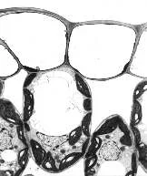
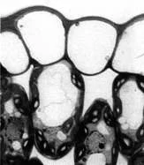
Magnification how many times larger the image is than the object.
Resolution
Resolution the ability to distinguish two close objects as separate.
| Feature | Light microscope | Electron microscope |
|---|---|---|
| Radiation | Light, wavelength 500–650 nm | Electron beam, much shorter wavelength |
| Resolution | About 200 nm (0.2 µm) | About 0.2 nm to 0.5 nm |
| Useful magnification | Up to about ×1500 to ×2000 | Up to about ×500 000 to ×1 500 000 |
| Specimens | Living or dead | Dead, in a vacuum |
| Colour | Colour image | No colour image |
| Preparation | Simple | Complex, can introduce artefacts |
| Size | Small and portable | Large and fixed |
Light microscope
- Light microscope shows eukaryotic cells, nuclei and possibly mitochondria and chloroplasts, but not ribosomes, endoplasmic reticulum or lysosomes.
TEM
- TEM a beam of electrons is transmitted through a very thin section and denser parts absorb more electrons and look darker.
- It gives high resolution of internal structures but cannot show live material and can produce artefacts from preparation and staining.
SEM
- SEM a beam scans the surface and reflected electrons form a 3-D image.
- It can be used on thick or 3-D specimens but has lower resolution than TEM.
Section 3 of 12Developments in microscopy
1
Light microscopy
- Light microscopy condenser lenses direct light through the specimen.
- Fluorescent stains bind to specific structures and show more detail under UV.
- Immunofluorescence uses antibodies carrying fluorescent dyes to find specific molecules such as virus proteins.
2
Cryogenic electron microscopy
- Cryogenic electron microscopy solutions of molecules are flash-frozen and imaged, and software reconstructs 3-D models.
- It improved understanding of virus structure, membranes and protein synthesis.
3
Freeze fracture
- Freeze fracture a sample is frozen in liquid nitrogen and fractured in a vacuum to give a planar view of membrane organisation.
Section 4 of 12General cell structure and prokaryotic cells
General cell structure and prokaryotic cells
Features common to all cells
- DNA is the genetic material, allowing new cells to form and controlling enzyme and protein production.
- Cytoplasm is mainly water with ions, called cytosol, and is where many reactions occur.
- The plasma membrane is a lipid bilayer that controls what enters and leaves and has proteins for recognition, communication and transport.
Prokaryotes
- Prokaryotes the first organisms, small (0.1–5.0 µm), with no nucleus and no membrane-bound organelles.
- Domains are Bacteria (for example E. coli) and Archaea, which live in extreme conditions and include methanogens.
Prokaryotic structures
- 70S ribosomes read mRNA in translation, smaller than the 80S ribosomes of eukaryotes.
- Naked circular DNA lies in the nucleoid, with small plasmids that can carry genes such as antibiotic resistance.
- Archaea have a monolayer plasma membrane.
- Most have a cell wall of peptidoglycan that protects, keeps shape and prevents bursting.
Gram stain
- Gram-positive bacteria such as Bacillus and Staphylococcus retain crystal violet, and Gram-negative bacteria do not.
Additional structures
- A capsule protects against drying and immune attack.
- Flagella rotate like propellers for movement.
- Pili are shorter and thinner and aid adhesion, movement, avoiding white blood cells and conjugation.
Section 5 of 12Eukaryotic cell structure
- Compartmentalisation
- membrane-bound organelles let enzymes and substrates be concentrated, keep damaging substances apart, maintain optimal conditions such as pH and allow organelle numbers to change.
- (digestive enzymes in lysosomes)
- membrane-bound organelles let enzymes and substrates be concentrated, keep damaging substances apart, maintain optimal conditions such as pH and allow organelle numbers to change.
| Organelle | Structure | Function |
|---|---|---|
| Plasma membrane | Phospholipid bilayer about 10 nm wide, partially permeable | Controls exchange of materials |
| Nucleus | Double membrane with pores, chromatin, nucleolus | Holds chromosomes of linear DNA wound on histones; nucleolus makes ribosomes |
| Rough ER | Cisternae continuous with the nuclear envelope, covered with 80S ribosomes | Processes proteins, which bud off in vesicles to the Golgi |
| Ribosomes (80S) | rRNA and protein, free or on rough ER | Site of translation |
| Mitochondria | Double membrane, inner folds (cristae), matrix with DNA and ribosomes | Aerobic respiration to make ATP |
| Golgi apparatus | Flattened sacs (cisternae) | Modifies proteins and lipids and packages them into vesicles for export, lysosomes or organelles |
| Vesicles and lysosomes | Membrane-bound sacs; lysosomes hold hydrolytic enzymes | Transport and storage; lysosomes break down waste and are used in immune cells and apoptosis |
| Microtubules | Tubulin dimers in protofilaments, about 25 nm wide | Cytoskeleton for support and movement |
Eukaryotic cell structure
Nuclear pores let mRNA and ribosomes out and enzymes and signalling molecules in, and red blood cells have no nucleus.
Plant structures
- Chloroplasts are larger than mitochondria with a double membrane;
- thylakoids stack into grana joined by lamellae;
- the light-dependent stage is in the thylakoids and the Calvin cycle in the stroma;
- they contain DNA and ribosomes.
- A large permanent vacuole is surrounded by the tonoplast, while animal vacuoles are small and temporary.
- The cell wall is outside the membrane, made of cellulose in plants and peptidoglycan in most bacteria, and is freely permeable.
- Plasmodesmata are cytoplasm threads that connect neighbouring plant cells.
Section 6 of 12Functions of life and unicellular organisms
Functions of life and unicellular organisms
Metabolism
- Metabolism all enzyme-catalysed reactions including respiration.
Reproduction
- Reproduction production of offspring, sexual or asexual.
Homeostasis
- Homeostasis keeping internal conditions within tolerable limits.
Growth
- Growth a permanent increase in size.
Response
- Response reacting to internal or external stimuli to improve survival.
Excretion
- Excretion removal of metabolic waste such as carbon dioxide.
Nutrition
- Nutrition obtaining energy and nutrients by absorbing organic matter or synthesising it, for example by photosynthesis.
Unicellular organisms
- Unicellular organisms they use mitochondria for energy, the membrane to control exchange, ribosomes for proteins, vacuoles for digestion and waste storage,
- and cilia or flagella for movement.
Section 7 of 12Comparing eukaryotic kingdoms and atypical cells
| Feature | Animal | Plant | Fungi |
|---|---|---|---|
| Cell wall | None | Cellulose | Glucans, chitin and glycoproteins |
| Vacuoles | Small, temporary | Large, permanent | Small, non-permanent |
| Chloroplasts | None | Many | None |
| Centrioles | Present | Absent | Absent |
| Cilia and flagella | Can be present | Absent | Absent in true fungi |
| Carbohydrate store | Glycogen | Starch | Glycogen |
| Shape | Flexible | Fixed | Can vary |
- Atypical cells challenge cell theory.
- Striated muscle fibres are fused cells up to 300 mm long with many nuclei in one membrane (sarcolemma), against 100–150 µm for a cardiac cell.
- Aseptate fungal hyphae have no septa, so they are multinucleate with continuous cytoplasm.
- Red blood cells lack a nucleus so they can carry more haemoglobin, and their biconcave shape gives a large surface area.
- Phloem sieve tubes lack nuclei, mitochondria and ribosomes and survive because companion cells maintain their cytoplasm.
Section 8 of 12Interpreting micrographs and biological drawings
- Identifying a cell
Interpreting micrographs and biological drawings
Decide whether it is prokaryotic or eukaryotic by looking for a nucleus.
Decide plant or animal by looking for a cell wall or large vacuole.
Identify the organelles and deduce their functions.
Features of organelles
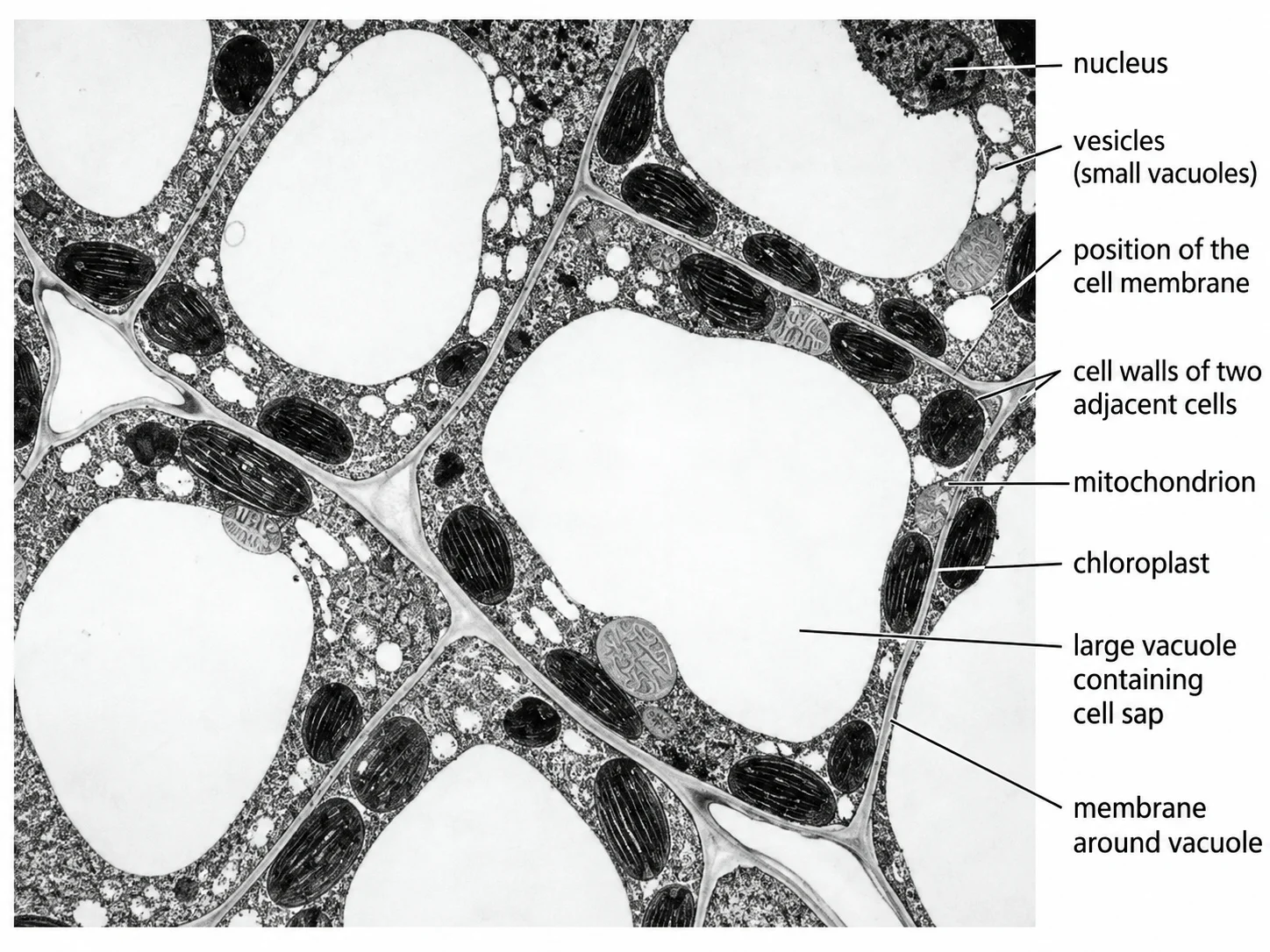
- Chloroplast — oval, double membrane, stacks of thylakoids, larger than mitochondria.
- Nucleus — roughly spherical with a nuclear envelope and a dark nucleolus.
- Vacuole — a large, pale space.
- Mitochondrion — oval with a double membrane and sometimes visible cristae.
- Cell wall — at the perimeter of the cell.
Deduction
- Deduction many mitochondria mean a high energy demand and many lysosomes mean much digestion,
- as in a macrophage; microvilli on a brush border mean a large absorbing surface.
Drawing conventions
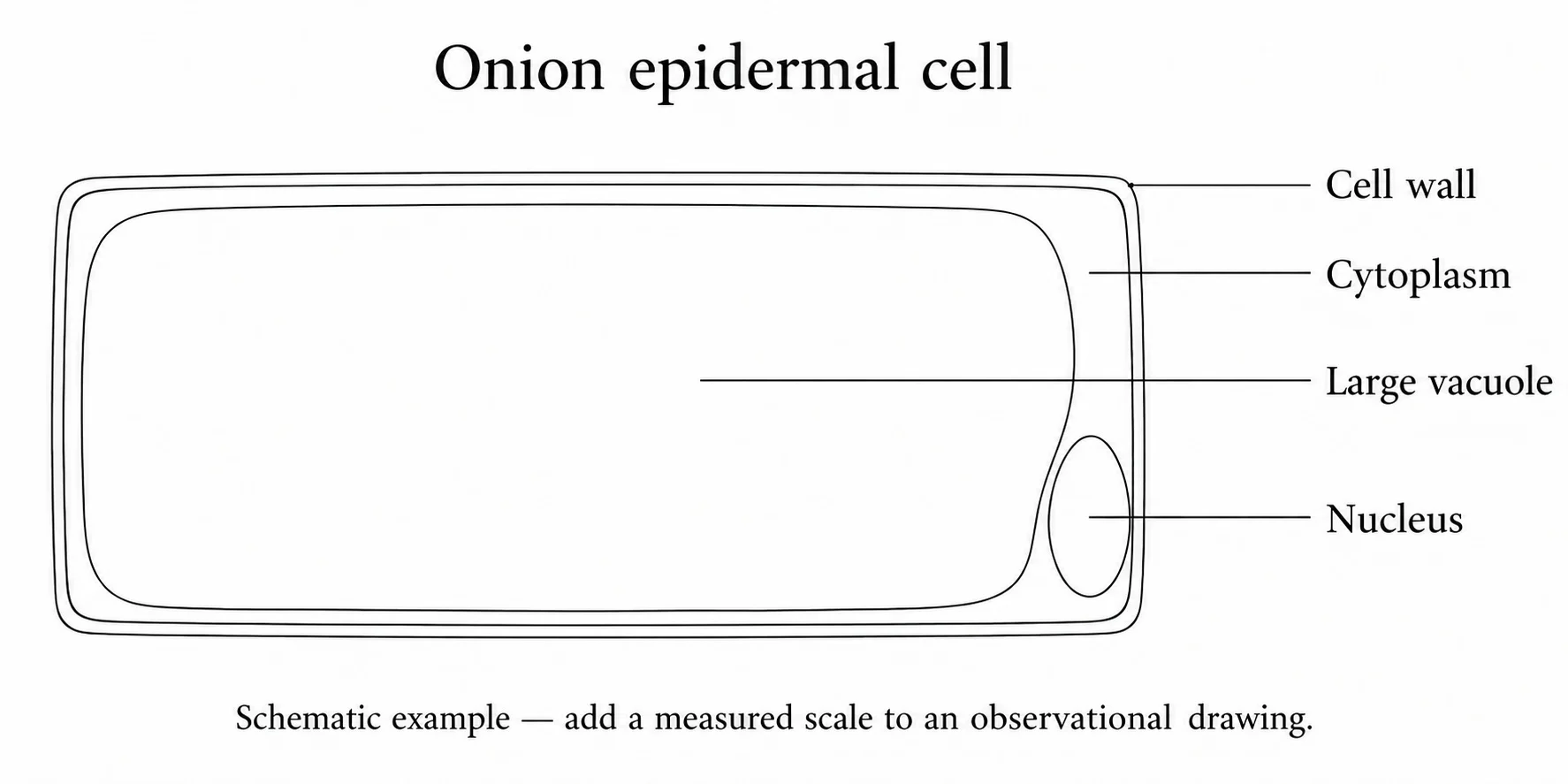
- Give a title and record the magnification, and add a scale bar where possible.
- Use a sharp HB pencil on plain white paper with clear single lines, no shading and correct proportions.
- Draw large, include only well-defined structures and draw only what is seen.
- Use ruled label lines that do not cross, have no arrowheads and touch the part labelled.
- Plan diagrams of tissues show no individual cells.
Section 9 of 12Endosymbiotic theory
Endosymbiosis
Endosymbiosis one organism living inside another, where the engulfed organism is not digested because the relationship benefits both.
Eukaryotic origin
- Eukaryotic origin eukaryotes evolved from a unicellular ancestor with a nucleus.
- Membrane infoldings overcame a small surface area to volume ratio and formed the nucleus and rough ER.
- A larger anaerobic cell engulfed a smaller aerobic prokaryote, which gave a ready supply of ATP and became the mitochondrion.
- A heterotroph then engulfed a photosynthetic prokaryote, which supplied carbohydrates and became the chloroplast.
Evidence from mitochondria and chloroplasts
- They divide by binary fission like bacteria.
- They contain their own naked circular DNA, much smaller than a bacterial genome.
- They transcribe mRNA and have 70S ribosomes to make their own proteins.
- They have double membranes and their own membranes like prokaryotes.
Nature of science
- Nature of science a theory is stronger the more observations it explains and predicts, and it must be adjusted if new observations do not fit.
Section 10 of 12Cell differentiation and multicellularity
Compartmentalisation
- Compartmentalisation it let single cells develop specialised regions such as the nucleus and energy-producing mitochondria,
- but unicellular organisms remain limited.
Differentiation
- Differentiation cells of a multicellular organism become specialised by expressing different genes.
- All cells have an identical genome, and the genes switched on depend on changes in the environment.
- Once specialised the cell is usually fixed in its function.
- Structure fits function through cell shape and organelle content, for example many ribosomes in protein-making cells and a biconcave red blood cell.
Levels of organisation
- Levels of organisation specialised cell, then tissue, then organ, then organ system, then organism.
| Specialised cell | Tissue | Organ | Organ system |
|---|---|---|---|
| Epithelial cell | Epithelial tissue | Stomach | Digestive system |
| Muscle cell | Muscle tissue | Bladder | Urinary system |
| Neurone | Nerve tissue | Brain | Central nervous system |
| Rod and cone cells | Retina | Eye | Visual system |
1Advantages of multicellularity
- Advantages of multicellularity larger size, specialised cell groups performing specific functions and survival in most environments.
2Evolution of multicellularity
- Evolution of multicellularity single cells clumped, aggregates formed specialised cells such as reproductive cells,
- and groups folded into tissues and then organs.
- It evolved many times, and fungi and algae are multicellular and evolved before animals and plants.
Section 11 of 12Key terms
| Term | Meaning |
|---|---|
| Resolution | the ability to distinguish between two objects that are close together. |
| Graticule | an eyepiece scale calibrated against a stage micrometer to measure specimens. |
| Artefact | a structure seen in a micrograph that results from preparation and not from the living cell. |
| Nucleoid | the region of a prokaryotic cell containing the naked circular DNA. |
| Compartmentalisation | separating cell activities in membrane-bound organelles. |
| Endosymbiosis | a relationship in which one organism lives inside another to mutual benefit. |
| Differentiation | development of specialised cells by expressing different genes. |
| Tissue | a group of similar cells working together for a function. |
| Plasmid | a small loop of DNA separate from the main circular DNA of a prokaryote. |
Section 12 of 12Exam technique
1
Magnification and resolution are the same — magnification is enlargement and resolution is the ability to separate close points.
2
Electron microscopes can show living cells — specimens must be dead because of the vacuum.
3
Prokaryotes have no genetic material — they have naked circular DNA in a nucleoid and plasmids.
4
Specialised cells have different genes — all cells share the genome and differ in which genes are expressed.
Marked practiceQuizzes for this lesson
Last updated:
Cambridge 9700 AS Biology
Core Formula
Magnification Triangle & Unit Conversion
Formula: M = I / A ⇔ A = I / M ⇔ I = M × A
Units: 1 mm = 1,000 μm = 1,000,000 nm.
Rule: Convert Image size (ruler mm) to μm by ×1,000 before dividing by Actual size.