B1.2Theme B — Form and function
Proteins — IB Biology HL Revision Notes & Topical Questions
5 min read 10 parts Lesson 3 of 14 2 quizzes
Curriculum Equation Key قوانين الرقم الهيدروجيني والاتزان الأيوني - pH & Acid-Base Equilibrium FormulasFast Exam Solution
pH = -log[H⁺] | pOH = -log[OH⁻] | pH + pOH = 14 | [H⁺][OH⁻] = 10⁻¹⁴
Word Equation & Conditions: الرقم الهيدروجيني = -لوغاريتم تركيز أيون الهيدروجين | pH + pOH = 14
Location & Specifications: الاتزان الأيوني، كيمياء 3 ثانوي، التحصيلي، و AP Chemistry.
Lesson summary
B1.2 Proteins — IB BIOLOGY HL notes: Explain amino acid structure, peptide bonds, the four levels of protein structure, denaturation and the properties of globular and fibrous proteins.Protein = a polymer of amino acid monomers, whose sequence, type and number decide shape and fu…
- Hydrolysis
- Recognition
- Naturally occurring amino acids
- Non-essential
- Primary structure
- Secondary structure
Section 1 of 10Amino acids and the peptide bond
Amino acids and the peptide bond
Protein
Protein a polymer of amino acid monomers, whose sequence, type and number decide shape and function.
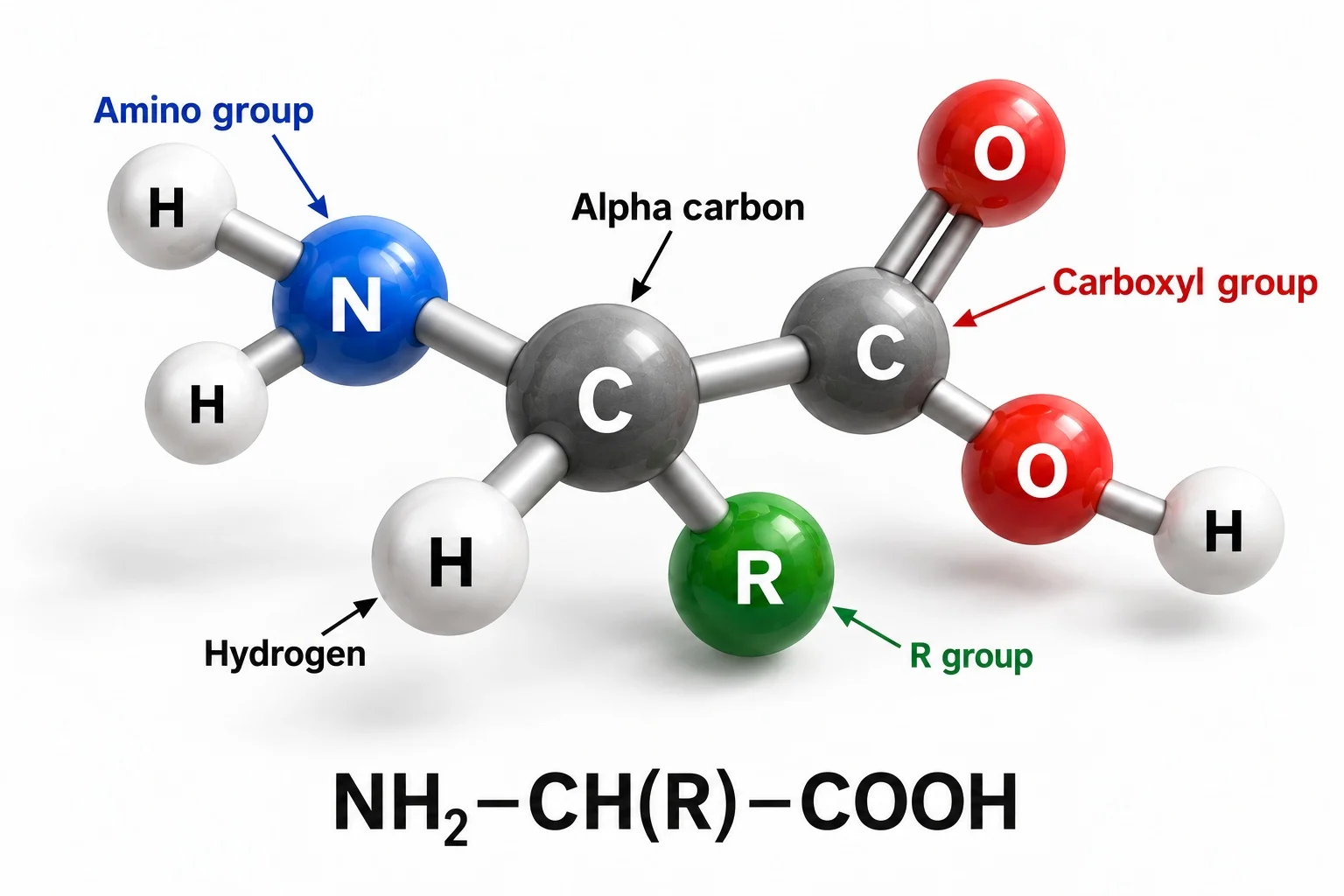
Amino acid structure
- Amino acid structure a central alpha carbon bonded to four groups.
- An amine group, –NH2.
- A carboxyl group, –COOH.
- A hydrogen atom.
- An R group, which differs between the 20 amino acids and decides whether it is acidic, basic, polar or non-polar.
R groups range from hydrogen (glycine) to aromatic rings (phenylalanine).
Peptide bond
- Peptide bond the OH from the carboxyl of one amino acid and an H from the amine of another are lost as water, and the C bonds to the N.
- It is a condensation reaction, and the R group takes no part.
- Two amino acids form a dipeptide and three or more form a polypeptide.
- A protein has one polypeptide chain or several interacting chains.
Hydrolysis
- Hydrolysis adding water breaks peptide bonds and gives amino acids.
Recognition
- Recognition an amino acid has amine and carboxyl groups, and a peptide bond is where N joins a C that has a double bond to O.
Section 2 of 10Essential amino acids
Essential amino acids
Naturally occurring amino acids
- 20 are common in proteins, from around 500 found in nature.
Non-essential
- Non-essential humans make 11 of them from other amino acids.
Essential
- Essential nine must come from the diet.
- A varied balanced diet supplies all nine.
- Meat has all nine, so vegetarians and vegans need a varied diet or supplements.
Section 3 of 10Variety and roles of proteins
Variety and roles of proteins
Variety of polypeptides
- There are 20 amino acids, and lengths range from a few to thousands.
- Each amino acid added at a ribosome is chosen from 20 by the mRNA codon.
- A chain of 50 amino acids has 2050 (about 1.13 × 1065) possible sequences, so variety is effectively infinite for the average length of 300.
Roles of proteins
- Enzymes speed up reactions.
- Blood clotting proteins form a scab over a wound.
- Collagen and keratin strengthen skin, hair, tendons and vessels.
- Haemoglobin transports oxygen.
- The cytoskeleton moves chromosomes in the cell cycle.
- Adhesion proteins hold cells together.
- Hormones are chemical messengers.
- Histones compact DNA.
- Antibodies are the most diverse proteins.
- Channel and carrier proteins control membrane transport.
- Receptors bind hormones, chemicals, light and sound stimuli.
Contractile proteins such as myosin cause movement.
Section 4 of 10Examples of proteins
| Protein | Structure | Function |
|---|---|---|
| Rubisco | Globular, 16 polypeptide chains | Fixes CO2 in photosynthesis; the most abundant enzyme and very slow |
| Insulin | Globular, 2 polypeptide chains | Hormone from pancreatic β cells that binds receptors on liver, fat and muscle cells so glucose is absorbed |
| Immunoglobulins | Globular, Y-shaped with variable binding sites at the tips | Antibodies that bind specific antigens |
| Rhodopsin | Membrane protein in rod cells with a retinal group from vitamin A | A photon changes its shape and triggers an impulse along the optic nerve |
| Collagen | Fibrous, three chains | About 25% of body protein; resists tearing in skin, vessels and connective tissue and reduces brittleness of teeth and bones |
| Spider silk | Rope-like fibrous parts and coiled stretchy parts | Strong as steel but light, does not denature easily, and can be made in goats' milk by genetic engineering |
Section 5 of 10Primary and secondary structure
Primary and secondary structure
Primary structure
Primary structure the sequence of amino acids, set by the DNA and held by peptide bonds.
- The same sequence always folds into the same 3-D shape.
- Describe it as a sequence or order, not just a chain.
Secondary structure
Secondary structure pleating and coiling of the chain, held by weak hydrogen bonds between C=O and N–H groups of the backbone.
- The two forms are the alpha helix and the beta-pleated sheet.
Section 6 of 10Tertiary and quaternary structure
Tertiary and quaternary structure
Tertiary structure
Tertiary structure the 3-D shape into which the secondary structure folds, formed by interactions between R groups and with the surroundings.
- Hydrogen bonds between polar R groups.
- Hydrophobic interactions put non-polar R groups inside the protein.
- Disulfide bridges are covalent bonds between cysteine R groups.
- Ionic bonds form between charged R groups.
Specific shape gives receptor sites on membranes and active sites in enzymes.
Polar amino acids on the outside make proteins soluble, line channels in membranes and keep enzymes soluble.
Non-polar amino acids lie in regions away from water, such as the membrane-embedded regions of integral proteins.
Quaternary structure
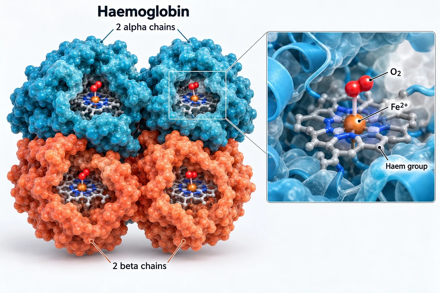
Quaternary structure several polypeptide subunits working as one protein; one-chain proteins have none.
- Conjugated proteins have a non-protein prosthetic group.
- Haemoglobin has four subunits, each with a haem group containing Fe2+.
- Insulin has two subunits joined by disulfide bridges and collagen has three wound in a helix, and neither is conjugated.
| Bond | Level of structure |
|---|---|
| Peptide | Primary |
| Hydrogen (backbone C=O and N–H) | Secondary |
| Hydrogen, ionic, disulfide, hydrophobic interactions (R groups) | Tertiary |
- Nature of science
- cryogenic electron microscopy freezes protein solutions and images them with electrons to rebuild 3-D shapes.
- X-ray crystallography needed crystals, which are slow to make, work only on purified single proteins and some proteins will not form them.
- Cryo-EM shows structure in context, including interactions with other molecules.
Section 7 of 10Globular and fibrous proteins
| Feature | Globular | Fibrous |
|---|---|---|
| Shape | Compact, roughly spherical | Long strands with cross-links |
| Amino acid sequence | Irregular, many types | Limited types, highly repetitive |
| R groups | Non-polar inside, polar outside | Many hydrophobic |
| Solubility | Soluble | Insoluble |
| Function | Metabolic and transport roles | Structural |
| Examples | Enzymes, immunoglobulins, haemoglobin, insulin | Keratin, collagen |
Insulin
- Insulin it was the first protein sequenced.
- Polypeptide A has 21 amino acid residues and B has 30, joined by three disulfide bridges.
Collagen
- It forms tendons, cartilage, ligaments, bones, teeth, skin, vessel walls and the cornea.
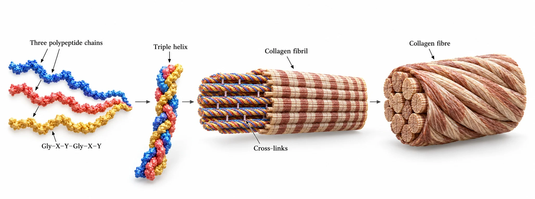
- Three polypeptide chains of about 1000 amino acids, rich in glycine, proline and hydroxyproline, form a triple helix held by hydrogen bonds.
- Covalent cross-links join molecules into fibrils with staggered ends, which gather into fibres aligned with the forces they resist.
Section 8 of 10Denaturation
Environment
- Environment protein shape depends on weak ionic interactions, hydrogen bonds and other forces between R groups.
Denaturation
Denaturation a change in protein shape caused by extremes of temperature or pH, which breaks these bonds but not the peptide bonds.
- It alters function and physical state, and is almost always irreversible.
- Small reversible changes occur, for example haemoglobin at small pH changes.
Egg white
- Egg white heating exposes hydrophobic amino acids of albumin, making it insoluble and forming a solid layer.
Stomach
- Stomach acid at pH 2 denatures dietary proteins, and pepsin has an optimum of pH 2.
Extremophiles
- Thermus aquaticus lives at 80 °C in hot springs with heat-stable proteins.
Experiments
- Experiments denaturation is used to find the optimum pH or temperature of enzymes such as pepsin or lipase.
Drugs
- Drugs protein drugs are injected because stomach acid would denature them.
Section 9 of 10Key terms
| Term | Meaning |
|---|---|
| Peptide bond | the covalent bond between the carboxyl carbon of one amino acid and the amine nitrogen of the next. |
| Primary structure | the sequence of amino acids in a polypeptide. |
| Disulfide bridge | a covalent bond between the R groups of two cysteines. |
| Conjugated protein | a protein with a non-protein prosthetic group. |
| Prosthetic group | a non-protein component, such as haem in haemoglobin. |
| Denaturation | a change in protein shape caused by heat or pH that loses function. |
| Globular protein | a compact, soluble, roughly spherical protein. |
| Fibrous protein | a long, insoluble, repetitive protein with a structural role. |
| Essential amino acid | an amino acid that must come from the diet because the body cannot make it. |
Section 10 of 10Exam technique
1
Denaturation breaks peptide bonds — it breaks the weaker bonds between R groups, and the amino acid sequence stays the same.
2
Hydrogen bonds hold the primary structure — peptide bonds do, and hydrogen bonds act in secondary and tertiary structure.
3
Denatured proteins re-form when cooled — denaturation is almost always irreversible.
4
All proteins have quaternary structure — only those with more than one polypeptide do.
Marked practiceQuizzes for this lesson
Last updated:
\n