B3.2Theme B — Form and function
Transport — IB Biology HL Revision Notes & Topical Questions
10 min read 11 parts Lesson 8 of 14 2 quizzes
Curriculum Equation Key قوانين الرقم الهيدروجيني والاتزان الأيوني - pH & Acid-Base Equilibrium FormulasFast Exam Solution
pH = -log[H⁺] | pOH = -log[OH⁻] | pH + pOH = 14 | [H⁺][OH⁻] = 10⁻¹⁴
Word Equation & Conditions: الرقم الهيدروجيني = -لوغاريتم تركيز أيون الهيدروجين | pH + pOH = 14
Location & Specifications: الاتزان الأيوني، كيمياء 3 ثانوي، التحصيلي، و AP Chemistry.
Lesson summary
B3.2 Transport — IB BIOLOGY HL notes: Explain the structure of blood vessels and the heart, tissue fluid and lymph, coronary heart disease, and transport in xylem and phloem.Blood vessels — arteries, arterioles, capillaries, venules and veins, each with a structure suited to its …
- Identification in micrographs
- Pulse rate
- Consequences
- Correlation coefficient (r)
- Reabsorption
- Single circulation
Part 1 of 9Capillaries
Blood vessels
- Blood vessels arteries, arterioles, capillaries, venules and veins, each with a structure suited to its function.
Capillaries form capillary beds and are the exchange surface between blood and tissues.
- The wall is a single layer of endothelial cells, giving a short diffusion distance.
- Fenestrations (gaps) in some capillaries let plasma leak out to form tissue fluid, but proteins are too large to pass.
- Permeability varies with tissue needs.
- Branching increases surface area and brings the blood close to the cells.
- The narrow lumen makes red blood cells pass in single file, slowing blood and bringing cells close to the wall.
Part 2 of 9Arteries and veins
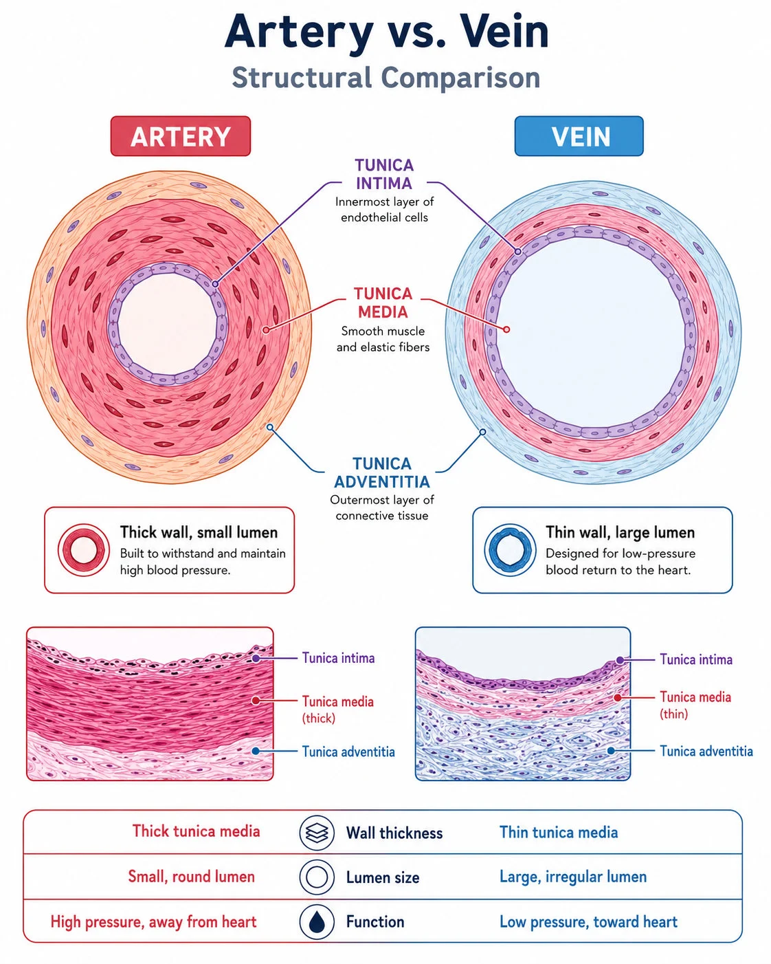
| Feature | Arteries | Veins |
|---|---|---|
| Direction | Blood away from the heart at high pressure | Blood to the heart at low pressure |
| Wall | Thick, with smooth muscle, elastic tissue and collagen | Thin, with a thin middle layer |
| Lumen | Narrow, to maintain pressure | Wide, so a large volume flows with less friction |
| Valves | None | Present, preventing backflow |
| Other | Elastic recoil evens out pressure | Flexible walls are compressed by skeletal muscles |
1
Artery layers
- The inner layer is a smooth endothelium one cell thick that reduces friction.
- The middle layer has smooth muscle, which controls lumen diameter, and elastic tissue, which stretches and recoils.
- The outer layer is collagen and elastic fibres that prevent over-stretching and rupture.
- Further from the heart there is more muscle and less elastic tissue because pressure fluctuates less.
2
Arterial pressure
- Systolic pressure is the peak pressure when blood is forced out and the walls stretch.
- Diastolic pressure is the lowest pressure when the elastic fibres recoil and push blood on.
- Vasoconstriction narrows the lumen and raises pressure, and vasodilation widens it and lowers pressure.
3Veins
- Veins capillaries converge to venules and then veins, and valves catch blood pushed backwards by skeletal muscle movement.
4Identification in micrographs
- Identification in micrographs artery walls are thick with a narrow lumen and veins have thin walls with a wide lumen.
5
Muscle contracts and relaxes, while elastic tissue stretches and recoils.
6Pulse rate
- Pulse rate it is felt where an artery is close to the skin or over a bone.
Arteries and veins
Place two fingers (not the thumb) on the radial artery at the wrist or the carotid artery at the neck.
Count pulses for 60 seconds, or for 30 seconds and double it.
Alternatively use data loggers, smartwatches or fitness bands.
Part 3 of 9Coronary heart disease
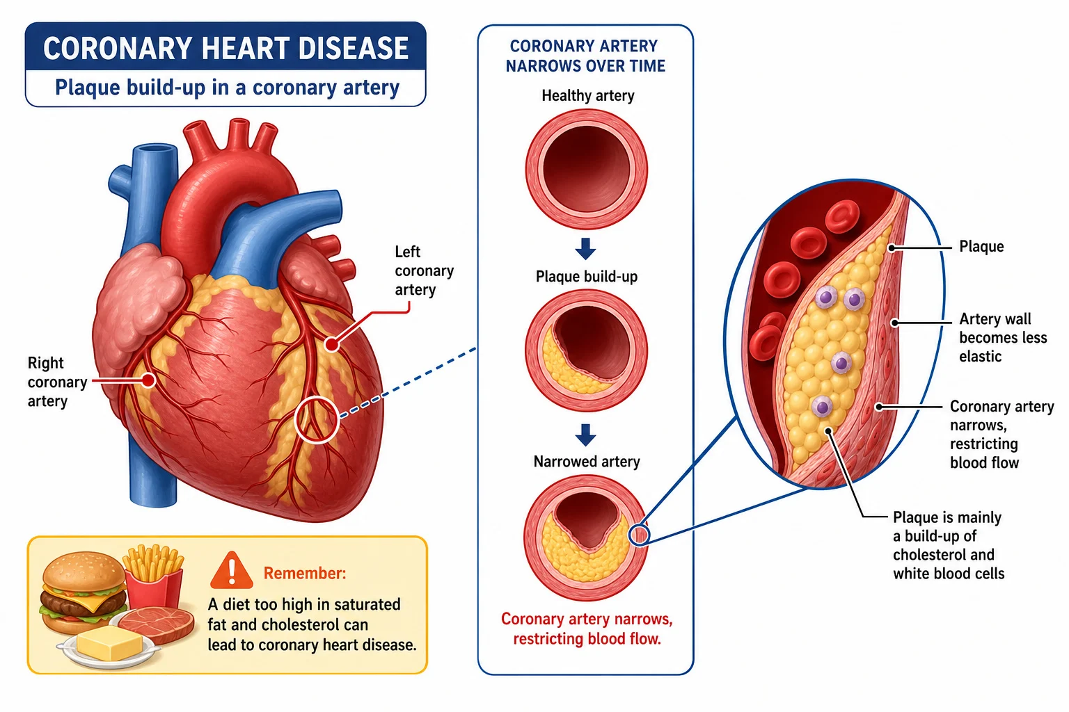
- Atherosclerosis
1
High blood pressure damages the artery wall.
2
Fatty deposits called atheromas build up under the endothelium and narrow the lumen.
3
Fibrous repair tissue is not elastic, so elasticity falls.
4
The lining breaks down into plaques.
5
Rupture causes clotting; a clot in a vessel is a thrombus and a circulating clot is an embolus.
1
Consequences
- Consequences an embolus blocking a small artery starves tissue of oxygen and nutrients,
- and in the coronary arteries heart muscle dies,
- causing a myocardial infarction.
2
Treatment
- Treatment bypass surgery uses leg vessels to route blood around a blockage.
3
Evaluating epidemiological data
- Epidemiological studies show correlation, not causation, and clinical studies of a few patients may be unrepresentative with no controlled experiment.
- Controlled diet experiments on people would be unethical.
- Sample groups must be representative, large and varied in demographics, and must be human.
- Use statistical analysis such as error bars and means to test significance.
- Include a control group, repeat studies and control variables such as prior health, exercise, stress, age and sex.
- Researchers should be unbiased and data collection accurate, because questionnaire answers may be untrue.
4
Correlation coefficient (r)
Correlation coefficient (r) the strength of a relationship, from −1 to +1.
- Positive correlation means both variables rise, negative correlation means one rises as the other falls, and 0 means none.
- Even strong correlation does not prove cause.
Part 4 of 9Tissue fluid and lymph
Plasma
- Plasma about 55% of blood, 95% water, and a solvent carrying dissolved substances.
Tissue fluid forms by pressure filtration when high pressure at the arterial end forces plasma out of capillaries.
- Proteins, red blood cells and platelets stay in the blood, while some flexible white blood cells (phagocytes) leave.
- It bathes cells and exchanges substances, so oxygen, glucose and amino acids are lower and carbon dioxide higher than in plasma.
Reabsorption
- Reabsorption at the venous end pressure is lower because of distance and slow flow.
- Proteins left in the capillary create a solute gradient, so water returns by osmosis.
- About 90% of the fluid returns and about 10% goes to lymph vessels.
Lymph
- Lymph capillaries have closed ends and large pores, and form part of the lymphatic system separate from the circulation.
- Lymph moves through ducts by body movement, with valves preventing backflow, and re-enters the blood through veins near the heart.
- Lymph nodes contain macrophages that engulf bacteria.
- Lymph returns escaped plasma proteins, because leaving them would raise tissue fluid solutes and stop water reabsorption.
- Lymph carries lipids from the intestine after digestion.
- Sedentary travellers can have swollen lower limbs because lymph accumulates.
| Substance | Plasma | Tissue fluid |
|---|---|---|
| Blood cells | Red cells, phagocytes, lymphocytes, platelets | Phagocytes |
| Proteins | Many large | Fewer, smaller |
| Glucose, amino acids, oxygen | Higher | Lower |
| Carbon dioxide | Lower | Higher |
Part 5 of 9Circulatory systems and the heart
Circulatory systems and the heart
Single circulation
- Single circulation bony fish have a two-chambered heart and blood passes through it once per circuit, going to the gills and then the body.
Double circulation
- Double circulation mammals have a four-chambered heart and blood passes through it twice per circuit.
- The right side pumps deoxygenated blood to the lungs (pulmonary circulation).
- The left side pumps oxygenated blood at high pressure around the body (systemic circulation).
- Advantages are separate oxygenated and deoxygenated blood, high pressure to the body and lower pressure in delicate lung vessels.
Heart structure
- Cardiac muscle contracts repeatedly without fatigue.
- Atria have thin walls and receive blood, and ventricles have thick walls, the left thicker than the right.
- The septum prevents mixing of blood.
- The vena cava and pulmonary vein bring blood in, and the pulmonary artery and aorta take blood out.
- Coronary arteries supply the heart muscle with oxygenated blood.
Valves open when pressure behind is greater than in front and close when pressure in front is greater.
- Atrioventricular valves are the tricuspid on the right and the bicuspid on the left.
- Semilunar valves are the pulmonary valve and the aortic valve.
Blood path
- Blood path right atrium, right ventricle, pulmonary artery, lungs, pulmonary vein, left atrium, left ventricle, aorta.
Part 6 of 9Cardiac cycle
1Myogenic control
- Myogenic control the heart beats without nervous stimulation, at about 60 beats per minute.
2
Conduction
1
The sinoatrial node (SAN) in the right atrium starts a wave of depolarisation that makes the atria contract.
2
Non-conducting tissue delays the wave so the atria finish contracting.
3
The atrioventricular node (AVN) passes the wave on.
4
The bundle of His in the septum and Purkyne fibres carry it around the ventricle walls.
5
The ventricles depolarise from the base upwards and contract.
- Cardiac cycle
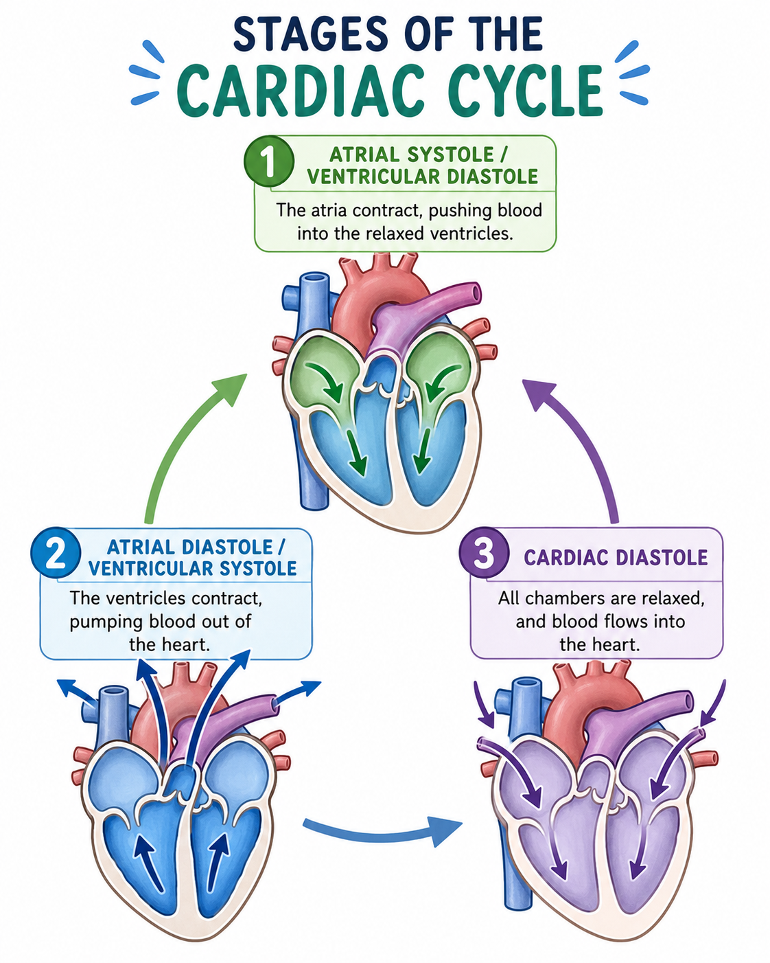
- systole is contraction and diastole is relaxation, in a continuous cycle on both sides at once.
| Stage | AV valves | Semilunar valves |
|---|---|---|
| Atrial systole | Open | Closed |
| Ventricular systole | Closed | Open |
| Diastole | Open | Closed |
1Atrial systole
- Atrial systole atrial pressure rises above ventricular pressure, forcing blood into the relaxed ventricles.
2Ventricular systole
- Ventricular systole ventricular pressure rises above atrial pressure, closing the AV valves,
- and above arterial pressure, opening the semilunar valves.
3Diastole
- Diastole ventricular pressure falls below arterial pressure, closing the semilunar valves,
- and blood flows passively into the ventricles when atrial pressure exceeds ventricular pressure.
4
Pressure graph points
- A: atrial pressure is higher than ventricular and the AV valve is open.
- B: ventricular pressure exceeds atrial pressure and the AV valve shuts.
- C: ventricular pressure exceeds aortic pressure and the aortic valve opens.
- D: ventricular pressure falls below aortic pressure and the aortic valve closes.
- E: atrial pressure exceeds ventricular pressure and the AV valve opens.
5Heart rate
60 ÷ the time of one cycle in seconds.
Part 7 of 9Transpiration stream and xylem
- Cohesion-tension theory
1
Water evaporates from the leaf cell walls into air spaces.
2
More water is drawn from the nearest xylem vessels, helped by adhesion to cell walls and capillary action.
3
This creates tension (negative pressure) in the xylem.
4
Cohesion between water molecules transmits the pull down the stem to the roots.
5
Water is lifted against gravity as a continuous transpiration stream.
Benefits
- Benefits evaporative cooling, mineral ion uptake and turgor that supports leaves and non-woody stems.
Xylem vessels
- They are long, continuous, hollow tubes of dead cells with no end walls or cell contents, so flow is unimpeded.
- Walls thickened with cellulose and lignin resist tension without collapsing.
- Pits let water move sideways between vessels, so flow continues if one is damaged.
Part 8 of 9Root and stem structure and root pressure
Root and stem structure and root pressure
Dicotyledonous stem
- Dicotyledonous stem epidermis protects and prevents water loss, parenchyma forms the cortex and pith and stores starch,
- and vascular bundles form a ring.
- Xylem is on the outer side of each bundle and phloem on the inner side.
Dicotyledonous root
- Dicotyledonous root xylem is central in a cross shape, with phloem between the arms, surrounded by endodermis, cortex and epidermis with root hairs.
Plan diagrams show tissue outlines only, with continuous lines, no shading, good proportions, labels, a scale and function annotations.
Root pressure
1
Minerals are actively transported into root cells, lowering their water potential.
2
Water enters by osmosis from the soil.
3
Minerals pass from cell to cell to the xylem and lower its water potential.
4
Water enters the xylem and creates positive pressure that pushes water up.
- It matters when transpiration is low, such as in high humidity or before deciduous leaves open in spring.
Part 9 of 9Translocation in phloem
Translocation
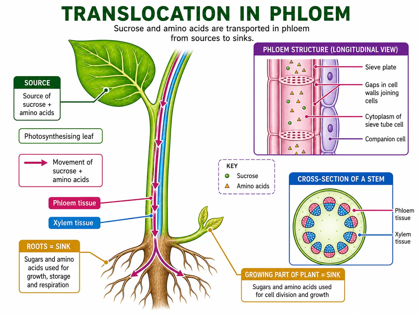
Translocation transport of organic solutes in phloem, as phloem sap of sucrose, water and amino acids (assimilates).
Sources
- Sources mature green leaves and stems, storage organs at the start of growth, and germinating seeds.
Sinks
- Sinks dividing meristems, growing roots, young leaves in bud and storage organs such as seeds and fruits.
- A source can become a sink and the reverse, so direction can be up or down.
| Phloem cell | Structure | Function |
|---|---|---|
| Sieve tube element | Sieve plates | Allow continuous flow of assimilates |
| Sieve tube element | Cellulose wall | Withstands hydrostatic pressure |
| Sieve tube element | No nucleus, vacuole or ribosomes, thin cytoplasm | Maximises space and reduces friction |
| Companion cell | Many mitochondria | ATP for active loading and unloading |
| Companion cell | Transport proteins in the membrane | Move assimilates in and out of the sieve tube |
| Companion cell | Plasmodesmata | Link to sieve tubes |
| Companion cell | Nucleus and organelles | Metabolic support for the sieve tube |
- Mass flow
1
Sucrose is actively loaded into the phloem at the source.
2
The low water potential draws water in by osmosis, including from xylem.
3
Hydrostatic pressure rises, forming a gradient from source to sink.
4
Sap flows towards the sink.
5
Sucrose is unloaded at the sink, water follows by osmosis and the gradient is maintained.
Key terms
| Term | Meaning |
|---|---|
| Pressure filtration | forcing of fluid out of capillaries by high blood pressure. |
| Myogenic | able to contract without nervous or hormonal stimulation. |
| Atherosclerosis | build-up of fatty deposits in artery walls that narrows the lumen. |
| Transpiration pull | the tension in xylem created by evaporation from leaves. |
| Root pressure | positive pressure pushing water up the xylem, from active uptake of minerals. |
| Translocation | movement of organic solutes in phloem. |
| Source | a region where assimilates are loaded into the phloem. |
| Sink | a region where assimilates are unloaded and used or stored. |
| Systole | contraction of the heart, and diastole = its relaxation. |
Avoid these mistakes
1
Arteries carry oxygenated blood only — arteries carry blood away from the heart, and the pulmonary artery carries deoxygenated blood.
2
Elastic tissue contracts — muscle contracts and relaxes, and elastic tissue stretches and recoils.
3
Water moves up xylem by osmosis — it moves by cohesion-tension, and transpiration is diffusion of vapour.
4
Phloem transport needs no energy — loading and unloading at sources and sinks are active.
5
Correlation proves cause — a correlation, even a strong one, does not prove causation.
Marked practiceQuizzes for this lesson
Last updated:
\n