2.5Theme B — Form and function
Cell Specialisation — IB Biology SL Revision Notes & Topical Questions
5 min read 11 parts Lesson 12 of 13 1 quiz
Curriculum Equation Key قوانين الرقم الهيدروجيني والاتزان الأيوني - pH & Acid-Base Equilibrium FormulasFast Exam Solution
pH = -log[H⁺] | pOH = -log[OH⁻] | pH + pOH = 14 | [H⁺][OH⁻] = 10⁻¹⁴
Word Equation & Conditions: الرقم الهيدروجيني = -لوغاريتم تركيز أيون الهيدروجين | pH + pOH = 14
Location & Specifications: الاتزان الأيوني، كيمياء 3 ثانوي، التحصيلي، و AP Chemistry.
Lesson summary
2.5 Cell Specialisation — IB BIOLOGY SL notes: Explain how differentiation arises from gene expression and morphogens, compare stem cells, and calculate SA:V ratios to explain cell size.After fertilisation the zygote divides by mitosis to form a ball of totipotent cells.A blastoc…
- Unipotent cells
- Stem cell niche
- Therapeutic use
- Division of labour
- Red blood cell
- Plasma cell
Section 1 of 11Development of specialised cells
1
After fertilisation the zygote divides by mitosis to form a ball of totipotent cells.
2
A blastocyst forms, and the inner cell mass cells are pluripotent.
3
Morphogen gradients give cells positional information.
4
Cells switch particular genes on and others off.
5
Cells differentiate into specialised types with specific structures and proteins.
6
Some multipotent stem cells stay in niches to replace cells throughout life.
- Gene expression = the process by which a gene's information is used to make a product, usually a polypeptide
- In differentiation, different cell types express different genes.
Section 2 of 11Morphogens
- Morphogen = a signalling chemical that diffuses through the early embryo and forms a concentration gradient
- Examples occur in Drosophila and in the vertebrate limb.
- The concentration a cell receives decides which genes it expresses.
1
Source cells secrete the morphogen.
2
The morphogen diffuses, forming a gradient from high to low concentration.
3
Cells bind the morphogen at different concentrations.
4
Different transcription factors are activated, switching on different genes.
5
Cells differentiate into different types according to their position.
Section 3 of 11Stem cell types and niches
| Type | Potency | Source | Example use |
|---|---|---|---|
| Zygote and morula cells | Totipotent | 1-4 day embryo | Can form the whole organism and placenta |
| Embryonic stem cells | Pluripotent | Inner cell mass of the blastocyst | Research and potential tissue replacement |
| Adult (somatic) stem cells | Multipotent | Bone marrow and skin | Bone marrow transplants for leukaemia |
Stem cell types and niches
Unipotent cells
- Unipotent cells they divide but produce only their own cell type, such as cardiomyocytes making new heart muscle cells
- They have the lowest potency, below multipotent cells, and most cells in an animal body are unipotent.
Stem cell niche
- Stem cell niche bone marrow makes blood cells and hair follicles make hair and skin
- A niche keeps its stem cells inactive until they are needed.
- It also stimulates them to divide and differentiate when replacements are required.
Therapeutic use
- Therapeutic use a bone marrow transplant replaces blood cells after cancer treatment
Ethics
- Ethics embryonic stem cells raise issues because embryos are destroyed
Section 4 of 11Division of labour and cell structure
Division of labour and cell structure
Division of labour
- Division of labour differentiated cells take on particular roles so each works more efficiently
- Specialisation changes cell shape and the organelles present.
- Cells that secrete a lot of protein contain many ribosomes, extensive rough ER and Golgi.
Red blood cell
- Red blood cell no nucleus or mitochondria, biconcave and flexible for capillaries
Neuron
- Neuron a large cell body makes the proteins needed to maintain a very long axon
Sperm
- Sperm a tail with mitochondria
Egg
- Egg cortical granules and large stores
Plasma cell
- Plasma cell an activated white blood cell enlarges to hold more rough ER and Golgi for antibody production
Muscle fibre
- Muscle fibre large and long so it generates strong forces when it contracts
Section 5 of 11Sizes of specialised cells
| Cell | Approximate size | Adaptation |
|---|---|---|
| Red blood cell | 7-8 µm diameter | Biconcave with no nucleus, giving more haemoglobin and a large SA:V |
| Sperm | About 50 µm long | Flagellum, many mitochondria and acrosome |
| Egg (human) | About 110 µm | Large cytoplasm with nutrients |
| Motor neuron | Axon up to about 1 m | Long axon for rapid signalling |
| Type I pneumocyte | About 0.15 µm thick | Short diffusion distance |
- Typical range
- most cells are 10-100 µm, with limits set by SA:V and diffusion distance
Section 6 of 11Calculating SA:V
Cube
- Cube surface area = 6 × L2, volume = L3, so SA:V = 6 / L
Sphere
- Sphere surface area = 4πr2, volume = (4/3)πr3, so SA:V = 3 / r
Growth
- Growth as a cell grows, volume increases faster than surface area, so SA:V decreases
Metabolism and exchange
- Metabolism and exchange the rate of metabolism depends on volume and the rate of exchange depends on surface area
Heat loss
- Heat loss it also depends on SA:V, so small animals lose heat more rapidly
Section 7 of 11Limits on cell size and multicellular solutions
Limits on cell size and multicellular solutions
Small cells
- Small cells high SA:V and a short diffusion distance to every organelle
Large cells
- Large cells low SA:V, so exchange is too slow and wastes and heat build up
Division
- Division growth stops and the cell divides once SA:V becomes too small
Multicellular exchange systems
- Multicellular exchange systems large organisms have a low SA:V and long diffusion distances
- They evolved gas exchange and digestive systems as exchange surfaces.
- They evolved circulatory systems to carry substances to internal tissues.
Adaptations that raise SA:V
- Adaptations that raise SA:V flattening in red blood cells, microvilli in the ileum epithelium, elongation and small size
Section 8 of 11Agar cube diffusion model
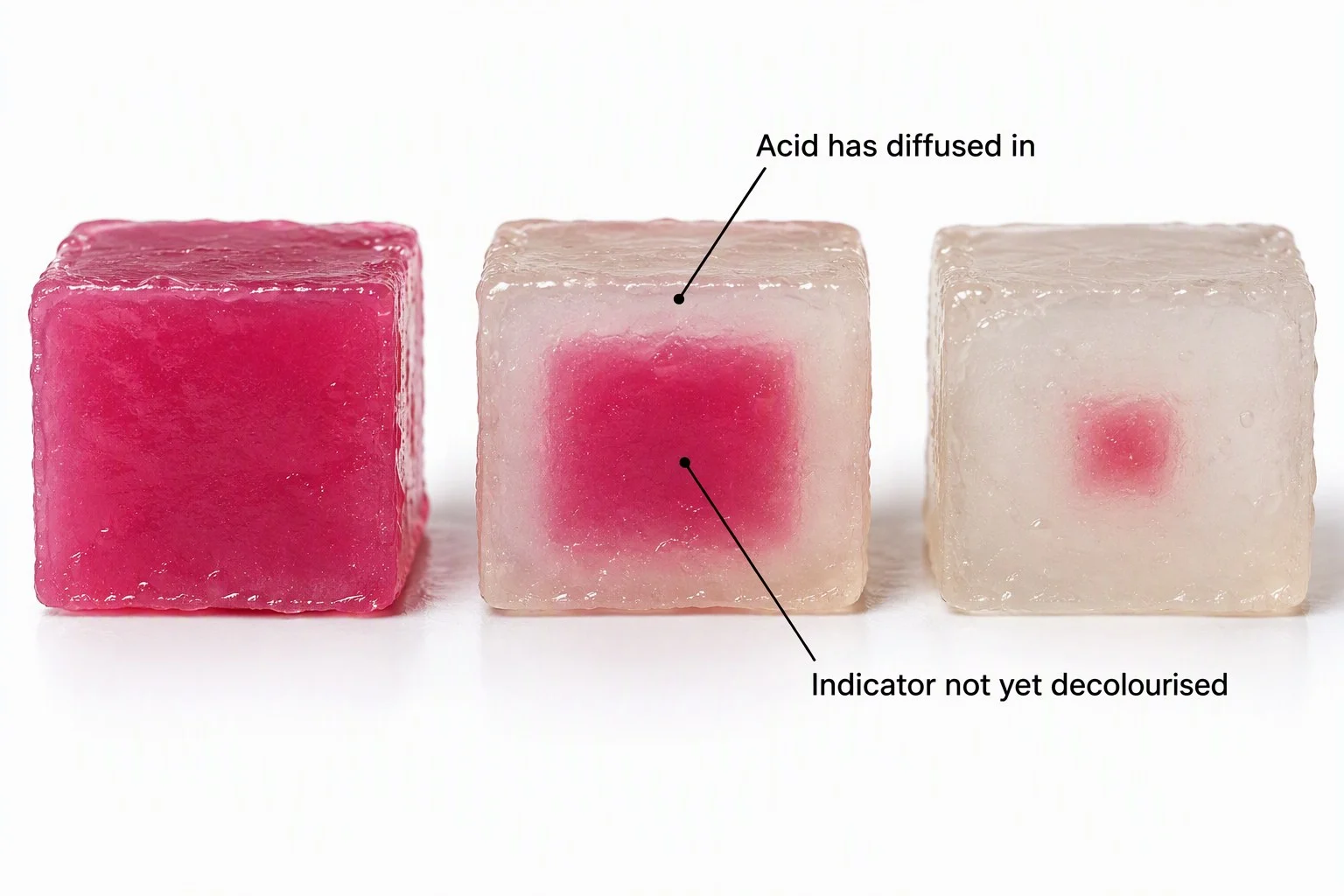
1
Cut agar containing indicator, for example with dilute NaOH, into cubes of different sizes.
2
Calculate the surface area, volume and SA:V of each cube.
3
Place the cubes in equal volumes of dilute acid.
4
Measure the time for complete colour change, or the depth of change in a fixed time.
5
Convert the times to rates and plot rate against SA:V.
Result
- Result smaller cubes, with a higher SA:V, are fully penetrated faster
Limitation
- Limitation the cubes model cells in a simplified form and do not match real shapes exactly
Section 9 of 11More from the specification
1
Differentiation of Eukaryotic Cells
- In multicellular organisms, unspecialised embryonic cells undergo selective gene expression to allow development into specialised eukaryotic cells with distinct structures.
2
Division of Labour and Efficiency
- Cellular specialisation means distinct cell types perform dedicated physiological tasks, enhancing the organism's ability to satisfy metabolic requirements and adapt to its environment.
3
Structural Adaptations to Function
- Specialised eukaryotic cells (such as neurones, erythrocytes,
- and xylem vessels) develop distinct morphological adaptations that optimize their primary biological function.
4
Metabolic Requirements in Multicellular Systems
- As organisms increase in size, simple diffusion is insufficient to meet metabolic requirements, requiring specialized eukaryotic cells organized into organ systems for transport and exchange.
5
Differentiation of Eukaryotic Cells
- In multicellular organisms, unspecialised embryonic cells undergo selective gene expression to allow development into specialised eukaryotic cells with distinct structures.
6
Division of Labour and Efficiency
- Cellular specialisation means distinct cell types perform dedicated physiological tasks, enhancing the organism's ability to satisfy metabolic requirements and adapt to its environment.
7
Structural Adaptations to Function
- Specialised eukaryotic cells (such as neurones, erythrocytes,
- and xylem vessels) develop distinct morphological adaptations that optimize their primary biological function.
8
Metabolic Requirements in Multicellular Systems
- As organisms increase in size, simple diffusion is insufficient to meet metabolic requirements, requiring specialized eukaryotic cells organized into organ systems for transport and exchange.
9
Development
- The changes as an organism becomes more complex and mature.
Section 10 of 11Key terms
| Term | Meaning |
|---|---|
| Zygote | The first diploid cell of a new organism, formed by fusion of gametes, which is totipotent. |
| Differentiation | The process by which unspecialised cells become specialised by expressing some genes and not others. |
| Morphogen | A signalling chemical that forms a concentration gradient and gives cells positional information. |
| Stem cell | An unspecialised cell that can self-renew and differentiate into specialised cell types. |
| Stem cell niche | The specific location where stem cells are found and maintained, such as bone marrow. |
| Totipotent | Able to differentiate into any cell type, including extra-embryonic cells. |
| Multipotent | Able to differentiate into a limited range of related cell types. |
| Surface area-to-volume ratio | The surface area of a cell divided by its volume, which decreases as the cell grows. |
Section 11 of 11Exam technique
1
Using volume instead of surface area to explain exchange rate — exchange depends on surface area and metabolism on volume
2
Saying SA:V increases as a cell grows — it decreases
3
Saying all stem cells are embryonic — adult multipotent stem cells exist in niches such as bone marrow
4
Saying morphogens act by moving the cells — they form a gradient that switches genes on or off
Marked practiceQuizzes for this lesson
Last updated:
Cambridge 9700 AS Biology
Core Formula
Magnification Triangle & Unit Conversion
Formula: M = I / A ⇔ A = I / M ⇔ I = M × A
Units: 1 mm = 1,000 μm = 1,000,000 nm.
Rule: Convert Image size (ruler mm) to μm by ×1,000 before dividing by Actual size.