B3.2Theme B — Form and function
Transport — IB Biology SL Revision Notes & Topical Questions
5 min read 13 parts Lesson 8 of 13 2 quizzes
Curriculum Equation Key قوانين الرقم الهيدروجيني والاتزان الأيوني - pH & Acid-Base Equilibrium FormulasFast Exam Solution
pH = -log[H⁺] | pOH = -log[OH⁻] | pH + pOH = 14 | [H⁺][OH⁻] = 10⁻¹⁴
Word Equation & Conditions: الرقم الهيدروجيني = -لوغاريتم تركيز أيون الهيدروجين | pH + pOH = 14
Location & Specifications: الاتزان الأيوني، كيمياء 3 ثانوي، التحصيلي، و AP Chemistry.
Lesson summary
B3.2 Transport — IB BIOLOGY SL notes: Relate the structure of blood vessels and xylem to transport, and evaluate the evidence on coronary heart disease.Blood vessel types — arteries, arterioles, capillaries, venules and veinsCapillary — the exchange surface, forming capillary bed…
- Fenestrations
- Elastic recoil
- Distance from the heart
- Vasoconstriction
- Vasodilation
- Terminology
Part 1 of 11Capillaries
Capillaries
Blood vessel types
- Blood vessel types arteries, arterioles, capillaries, venules and veins
Capillary
- Capillary the exchange surface, forming capillary beds between cells
- The wall is a single layer of endothelial cells, so the diffusion distance is very short.
- The narrow lumen makes red blood cells pass in single file, which slows the blood and gives time for diffusion.
- Branching beds put every cell near a capillary and give a huge total surface area.
- Blood flow is slow, about 0.5 mm s-1.
Fenestrations
- Fenestrations pores in some capillaries, such as the kidney glomerulus
- Plasma leaks out through them to form tissue fluid.
- Large proteins stay in the blood.
- Tissue fluid carries oxygen, glucose and other small molecules to the cells.
Part 2 of 11Arteries
Artery
- Artery carries blood away from the heart at high pressure
- Inner layersmooth endothelium that reduces friction.
- Middle layerthick smooth muscle and elastic tissue.
- Outer layercollagen and elastic fibres that stop the wall bursting or over-stretching.
- The lumen is narrow, which helps maintain pressure.
Elastic recoil
- Elastic recoil the wall stretches when the ventricles push blood out and recoils during diastole
- This keeps the pressure high through the whole heart cycle.
- Arterial pressure in humans peaks at about 16 kPa.
Distance from the heart
- Distance from the heart arteries further away have more smooth muscle and less elastic tissue
Vasoconstriction
- Vasoconstriction smooth muscle narrows the lumen, which raises pressure and reduces flow to a tissue
Vasodilation
- Vasodilation smooth muscle relaxes and widens the lumen, which lowers pressure and increases flow to active tissues
Terminology
- Terminology muscle contracts and relaxes, whereas elastic tissue stretches and recoils
Part 3 of 11Veins
Vein
- Vein carries blood to the heart at low pressure
- Capillaries converge into venules, which deliver blood to veins.
- The wall is thin because there is no need to withstand high pressure.
- The wall is flexible, so surrounding muscles can compress it.
- The lumen is wide, which lowers friction and keeps the volume flow equal to that in arteries.
Valves
- Valves pocket-shaped flaps that close if blood flows backwards
- Blood pushed backwards is caught and moved forwards by the next muscle movement.
Muscle pump
- Muscle pump skeletal muscles squeeze the veins and push blood towards the heart
Pressure
- Pressure highest in the arteries, lower in the capillaries and lowest in the veins
Part 4 of 11Identifying vessels
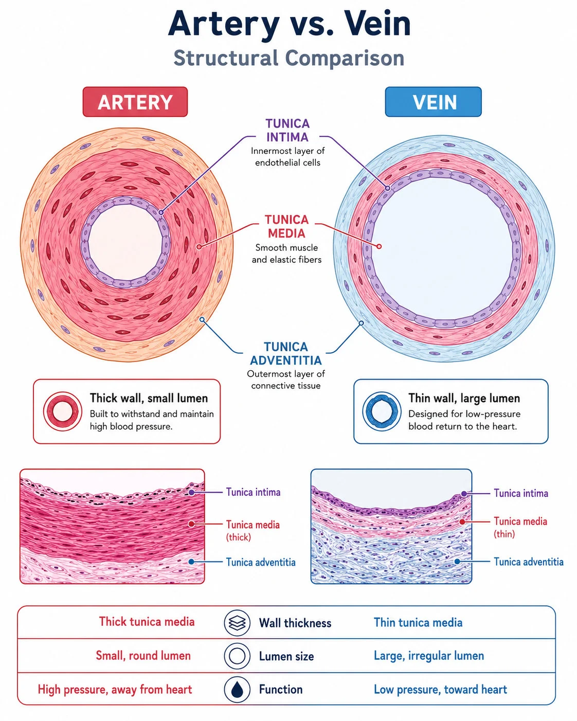
| Feature | Artery | Capillary | Vein |
|---|---|---|---|
| Wall thickness | Thick | One cell | Thin |
| Lumen | Narrow relative to the wall and round | Just wide enough for red cells | Wide and often flattened |
| Muscle and elastic fibres | Plenty | None | Little |
| Valves | None, except at the heart exit | None | Present |
| Pressure | High and pulsing | Low | Very low |
| Function | Carry blood from the heart | Exchange with tissues | Return blood to the heart |
- Micrographs
- arteries have a thick wall and narrow lumen and veins have a thin wall and wide lumen
- The names of the tissue layers need not be memorised.
Part 5 of 11Measuring pulse rate
Pulse
- Pulse the pressure wave from the ventricles contracting, felt where an artery is near the skin or passes over bone
- Pulse rate equals heart rate.
Sites
- Sites the radial artery at the wrist, below the base of the thumb, and the carotid artery at the side of the neck
1
Sit quietly for five minutes.
2
Place two fingers on the radial or carotid artery, never the thumb, which has its own pulse.
3
Start the timer.
4
Count the beats for 60 s, or for 30 s and double it.
5
Repeat three times and take the mean.
- Digital devices
- data loggers, smartwatches and fitness bands sense blood flow in the radial artery and give continuous records during and after exercise
Part 6 of 11Coronary heart disease
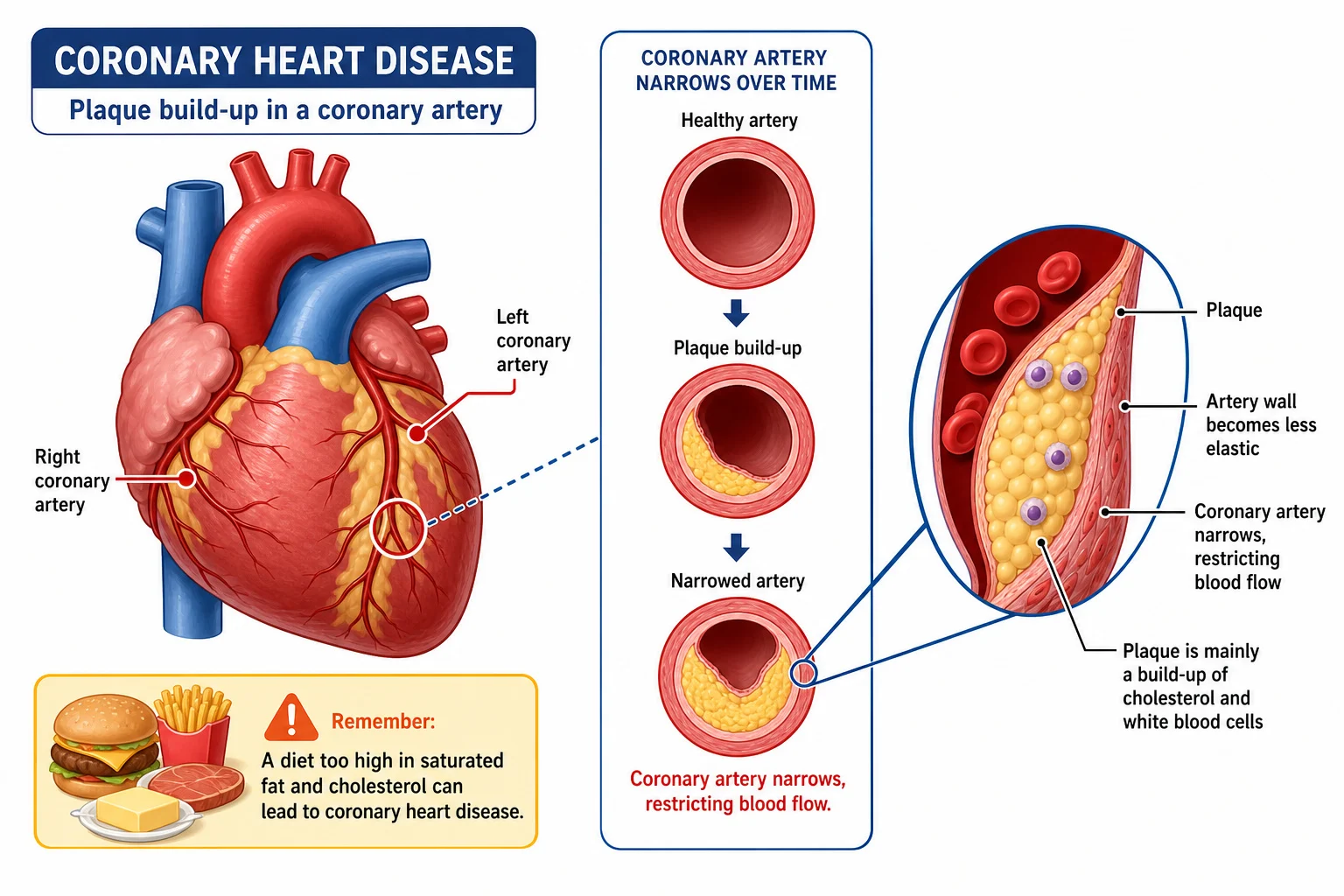
- Occlusion the narrowing of an artery by a blockage
1
High blood pressure damages the artery lining.
2
Fatty deposits called atheromas build up under the endothelium.
3
The lumen narrows and the blood pressure rises, causing more damage.
4
Fibrous repair tissue forms and lowers the elasticity of the wall.
5
The smooth lining breaks down into plaques.
6
A blood clot (thrombus) forms and may break off to become an embolus.
7
A blocked coronary artery starves the heart muscle of oxygen and the cells die, causing a myocardial infarction.
Treatment
- Treatment bypass surgery grafts a leg vein to route blood round the blockage
Risk factors
- Risk factors smoking, high blood pressure, obesity, a diet high in saturated fat and lack of exercise
- Aspirin and statins are used to lower the risk of clots and plaque.
Part 7 of 11Evaluating heart disease data
1Epidemiological studies
- Epidemiological studies they show correlation and not proof of cause
2Controlled diet trials
- Controlled diet trials they would be unethical because of the risk of harm
3
Evaluation checklist
- A sample large enough and representative of the population, and human.
- Statistical analysis, such as error bars or a test of significance.
- A control group.
- Repeats and several studies giving the same result.
- Control of confounding variables such as age, sex, exercise and stress.
- Accurate data, since self-reported diet or exercise may be untrue.
- No bias from the researchers.
4Correlation coefficient r
- Correlation coefficient r near +1 or -1 shows a strong link and near 0 shows no link
- A correlation does not prove cause.
- Positive means B rises as A rises and negative means B falls as A rises.
Part 8 of 11Transpiration stream
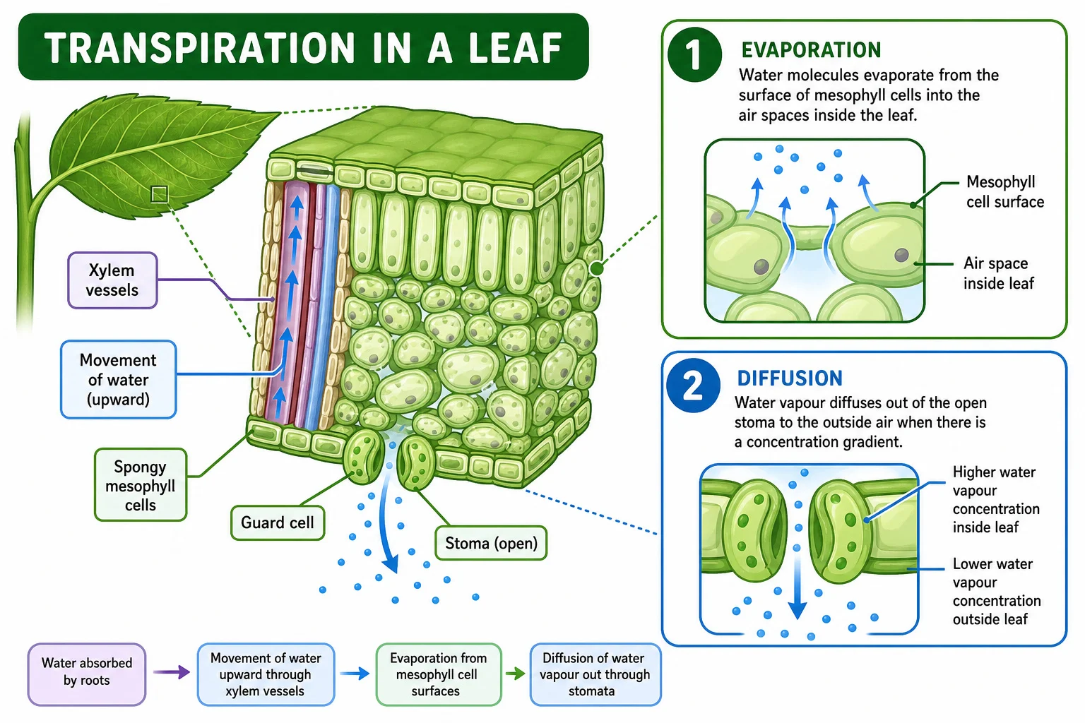
Transpiration pull
- Transpiration pull evaporation from the leaf mesophyll walls draws water out of the xylem
- Adhesion moves water through the cell walls.
- Water loss creates tension in the xylem.
- Cohesion transmits the pull down the whole column to the roots.
Cohesion-tension theory
- Cohesion-tension theory the continuous upward flow of water in the xylem is the transpiration stream
- It is passive, as the plant spends no energy.
1
Water evaporates from the leaf mesophyll walls.
2
Tension pulls water out of the leaf xylem.
3
Cohesion pulls the water column upward.
4
Adhesion to the lignified walls supports the column.
5
Water enters the root xylem to replace the losses.
Importance
- Importance evaporative cooling, uptake of mineral ions and turgor support for leaves and non-woody stems
Compared with blood
- Compared with blood blood is pushed by the heart in a closed loop and xylem water is pulled in one direction from root to leaf
Part 9 of 11Xylem vessel adaptations
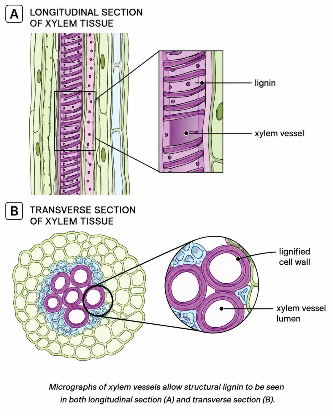
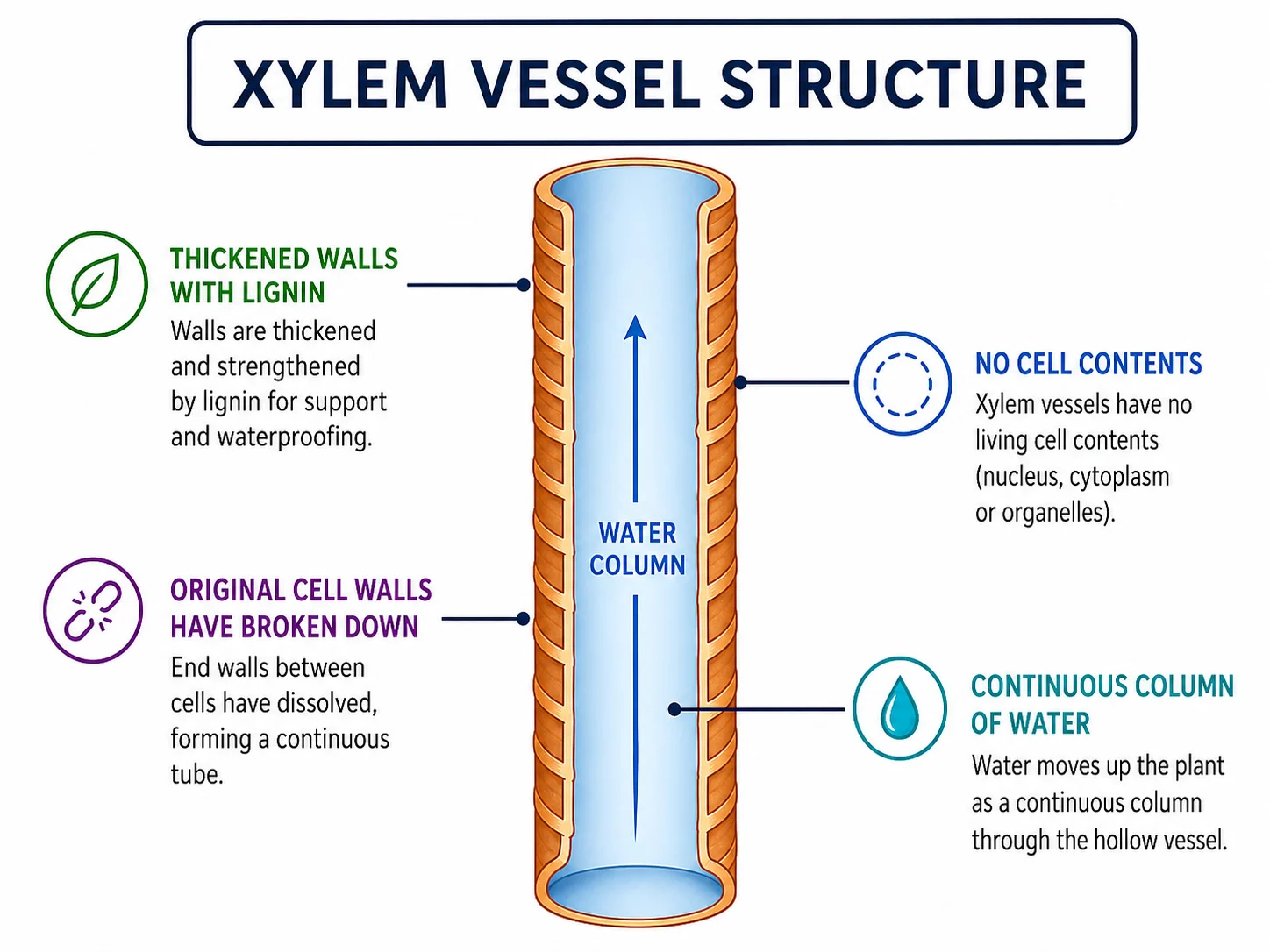
Xylem vessel adaptations
Mature xylem
- Mature xylem long, continuous hollow tubes of dead cells with no cell contents or end walls, so flow is unimpeded
Lignin
- Lignin the walls are thickened with cellulose and lignin
- This makes the vessels tough enough to withstand tension without collapsing.
Pits
- Pits tiny pores in the walls
- Water can move sideways between vessels.
- If a vessel is damaged the water can still reach the leaves.
Function
- Function carries water and mineral ions from the roots
Part 10 of 11Stem and root plan diagrams
| Feature | Dicot stem | Dicot root |
|---|---|---|
| Vascular tissue | Ring of vascular bundles | Central core |
| Xylem | Inner part of each bundle | Central X or star shape |
| Phloem | Outer part of each bundle | Between the arms of the xylem |
| Outer layer | Epidermis that prevents water loss | Epidermis with root hairs that absorb water and minerals |
| Tissue filling | Cortex and pith of parenchyma | Large cortex of parenchyma |
Endodermis
- Endodermis in the root, forms the boundary between the vascular tissue and the cortex
Parenchyma
- Parenchyma stores starch and other substances
Drawing rules
- Outline the tissues and never draw individual cells.
- Use clear continuous lines with no shading.
- Make the drawing large, add a scale and label with functions.
Part 11 of 11More from the specification
- Adaptation
- The process by which populations become better suited to their environment.
Key terms
| Term | Meaning |
|---|---|
| Capillary | A vessel with a wall one cell thick where exchange between blood and tissues occurs. |
| Tissue fluid | Plasma that has leaked out of capillaries and surrounds the cells. |
| Vasoconstriction | Narrowing of a vessel lumen by smooth muscle, raising pressure. |
| Atheroma | A fatty deposit under the endothelium of an artery. |
| Embolus | A clot that has broken free and travels in the blood. |
| Transpiration pull | The pull on water in the xylem caused by evaporation from the leaves. |
| Lignin | A strengthening polymer in xylem walls that prevents collapse under tension. |
| Pit | A pore in a xylem wall that lets water pass sideways between vessels. |
Avoid these mistakes
1
Saying arteries always carry oxygenated blood — they carry blood away from the heart, and the pulmonary artery carries deoxygenated blood
2
Saying muscle stretches and recoils — muscle contracts and relaxes and elastic tissue stretches and recoils
3
Saying a correlation proves cause — other variables may explain the link
4
Saying xylem vessels are living cells — mature vessels are dead and empty
Marked practiceQuizzes for this lesson
Last updated:
\n