D2.1Theme D — Continuity and change
Cell and nuclear division — IB Biology HL Revision Notes & Topical Questions
10 min read 12 parts Lesson 5 of 18 2 quizzes
Curriculum Equation Key قوانين الرقم الهيدروجيني والاتزان الأيوني - pH & Acid-Base Equilibrium FormulasFast Exam Solution
pH = -log[H⁺] | pOH = -log[OH⁻] | pH + pOH = 14 | [H⁺][OH⁻] = 10⁻¹⁴
Word Equation & Conditions: الرقم الهيدروجيني = -لوغاريتم تركيز أيون الهيدروجين | pH + pOH = 14
Location & Specifications: الاتزان الأيوني، كيمياء 3 ثانوي، التحصيلي، و AP Chemistry.
Lesson summary
D2.1 Cell and nuclear division — IB BIOLOGY HL notes: Explain cytokinesis, mitosis, meiosis, the cell cycle and its control, and tumour formation.Cell division — new cells come from pre-existing cells, so a multicellular organism arises from one cell dividing many times, first fo…
- Usually equal
- DNA replication
- Condensation
- Interphase
- Prophase versus telophase
- Meiosis I (reduction division)
Section 1 of 12Cell division and cytokinesis
Cell division
- Cell division new cells come from pre-existing cells,
- so a multicellular organism arises from one cell dividing many times, first forming embryonic stem cells that specialise.
- A dividing cell is a parent (mother) cell and produces two daughter cells.
- One type of division gives daughter cells genetically identical to each other and the parent,
- and the other gives genetically different cells, an important source of variation.
Cytokinesis
Cytokinesis division of the cytoplasm after the nucleus divides, so it occurs only after nuclear division.
| Cell type | Process |
|---|---|
| Animal | A contractile ring of actin and myosin forms at the equator under the plasma membrane and pulls it inwards, forming a cleavage furrow that separates the daughter cells |
| Plant | A cell plate forms at the equator from vesicles of carbohydrates, lipids and proteins that fuse into two membranes; other vesicles deposit pectin and cellulose by exocytosis to form new cell walls |
Section 2 of 12Equal and unequal cytokinesis, mitosis and meiosis, and chromosomes
Usually equal
- Usually equal it gives daughter cells of similar size.
- Each daughter cell must receive at least one mitochondrion, and plant cells at least one chloroplast,
- because these organelles arise only by dividing existing ones.
Unequal division
- Oogenesis in humans: a primary oocyte divides in puberty into a secondary oocyte and a small polar body,
- and the secondary oocyte divides again into an ovum and another polar body.
- The polar bodies degenerate, and the ovum keeps most of the cytoplasm.
- Budding in yeast gives a small bud.
- Nuclear division must precede cell division to avoid anucleate cells (cells with no nucleus).
| Feature | Mitosis | Meiosis |
|---|---|---|
| Daughter cells | Genetically identical, usually diploid (2n) | Genetically different, haploid (n) |
| Purpose | Growth, repair, cell replacement, asexual reproduction | Making gametes and genetic diversity |
- DNA replication
- in interphase DNA copies to make two identical sister chromatids joined at the centromere.
- At the start of anaphase the centromere splits and each chromatid is again called a chromosome.
- Chromatid, sister chromatids and chromosome are different terms.
Section 3 of 12Condensation and movement of chromosomes, phases of mitosis
1
Condensation
- Condensation human DNA can exceed 50 000 µm but nuclei average less than 5 µm.
- DNA is loosely coiled round histones as chromatin, in nucleosomes of DNA round eight histone proteins.
- In prophase, supercoiling condenses chromatin into chromosomes, aided by histones and enzymes.
2
Movement
- Movement microtubules and motor proteins move chromosomes.
- Microtubules are tubulin fibres of the cytoskeleton that lengthen and shorten as α- and β-tubulin dimers are added or removed.
- Motor proteins carry chromosomes along the microtubules to the equator.
- Mitosis nuclear division giving two genetically identical nuclei with the same chromosome number as the parent, needed in embryonic development, growth, repair and asexual reproduction.
| Phase | Events |
|---|---|
| Prophase | Chromosomes condense and are visible, each of two sister chromatids; centrosomes (replicated in G2) move to opposite poles; spindle fibres emerge; the nuclear envelope breaks into vesicles and the nucleolus disappears |
| Metaphase | Chromosomes line up at the equator (metaphase plate); spindle fibres attach to centromeres at kinetochores, each sister chromatid to a different pole |
| Anaphase | Centromeres divide, spindle fibres shorten and pull the separated chromatids (now chromosomes) to opposite poles |
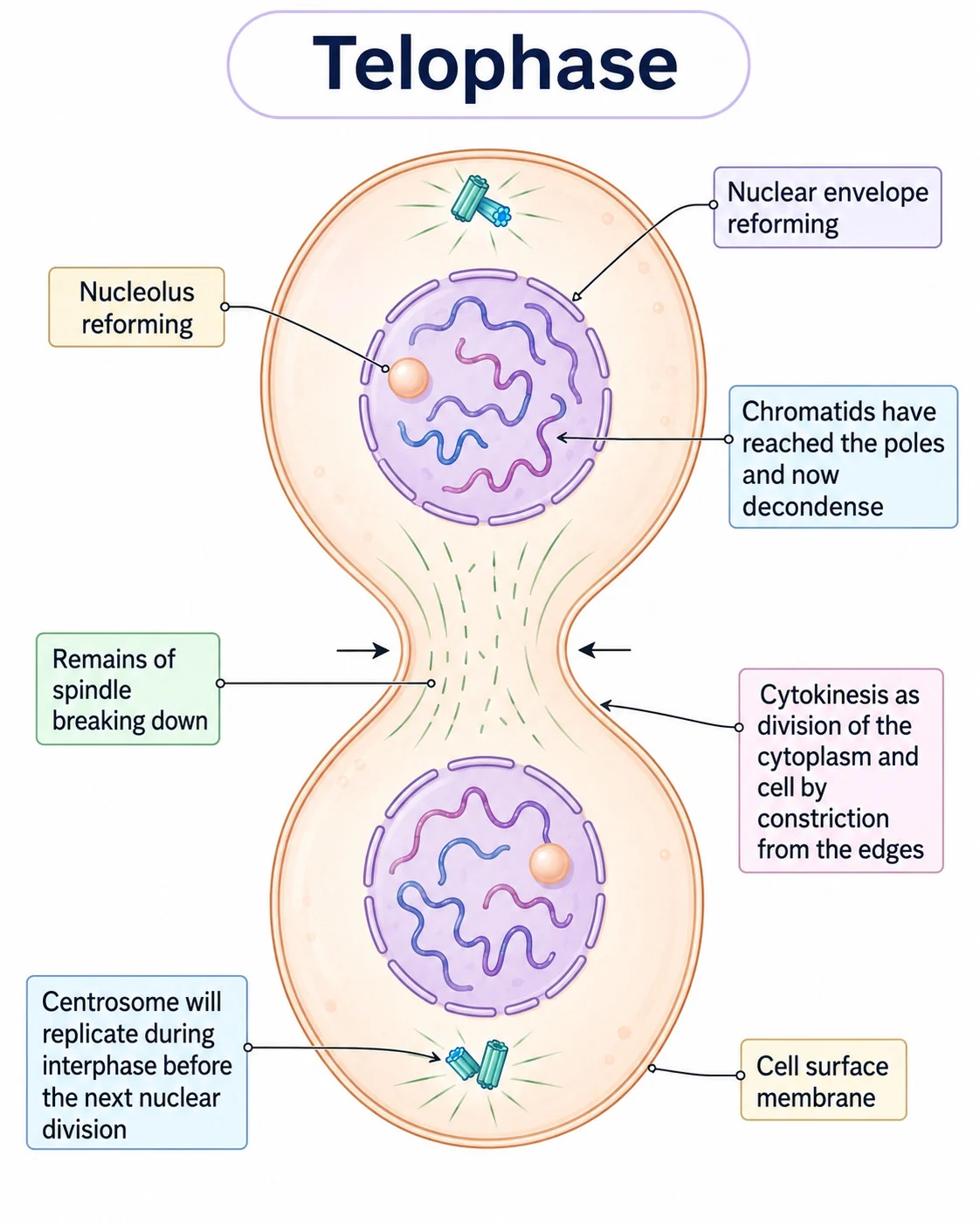
| Telophase | Chromosomes reach the poles and decondense; nuclear envelopes and nucleoli reform; spindle fibres break down |
- Remember the order PMAT.
Section 4 of 12Identifying phases of mitosis
1
Interphase
- Interphase most cells are in it, with visible chromatin but no visible chromosomes and a dark nucleus.
2
Prophase
- Prophase chromosomes visible and the nuclear envelope breaking down.
3
Metaphase
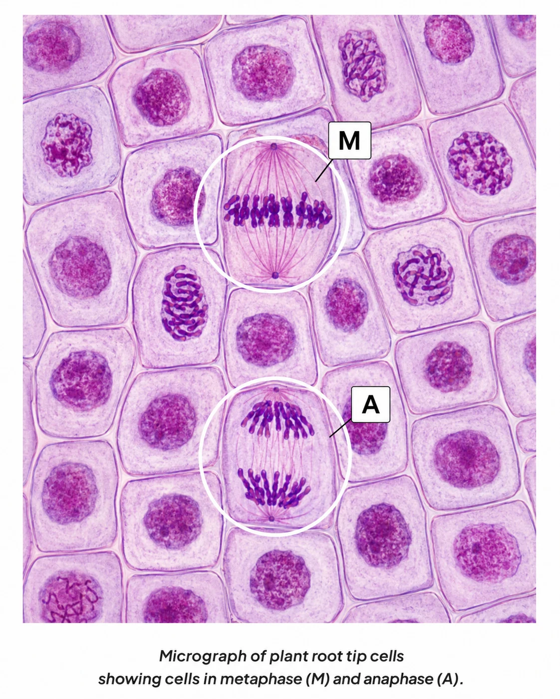
- Metaphase chromosomes in a line across the middle.
4
Anaphase
- Anaphase chromosomes pulled towards the poles, often V-shaped.
5
Telophase
- Telophase chromosomes at the poles, uncoiling, with the nuclear envelope reforming.
6
Prophase versus telophase
- Prophase versus telophase prophase has one group of chromosomes, while telophase has two groups, one at each pole.
7
Cytokinesis
- Cytokinesis a cleavage furrow in animal cells, or a cell plate forming at the metaphase plate in plant cells.
Section 5 of 12Meiosis
1Meiosis I (reduction division)
- Meiosis I (reduction division) the diploid nucleus has replicated its chromosomes.
- Homologous chromosomes pair as a bivalent in prophase I.
- Non-sister chromatids exchange alleles at chiasmata (crossing over), giving recombinant chromosomes.
- Spindle fibres pull homologous chromosomes to opposite poles, giving two haploid nuclei in which each chromosome has two chromatids.
- The chromosome number halves in meiosis I, not meiosis II.
2
No replication occurs between meiosis I and II.
3Meiosis II
- Meiosis II chromatids separate, giving four haploid nuclei with single-chromatid chromosomes.
4
Need in a sexual life cycle
- Asexual offspring are genetically identical to the parent, and sexual offspring are genetically distinct from each other and the parents.
- Fertilisation fuses gamete nuclei into a zygote and doubles the chromosome number.
- Meiosis halves it in animals when gametes form, otherwise the number would double every generation.
Section 6 of 12Non-disjunction and Down syndrome, meiosis as a source of variation
Non-disjunction and Down syndrome, meiosis as a source of variation
Non-disjunction
Non-disjunction failure of chromosomes to separate correctly in anaphase I or anaphase II,
- giving gametes with an extra or a missing copy of a chromosome.
- Fertilisation of such a gamete gives a zygote with the wrong chromosome number.
Down syndrome (trisomy 21)
- Down syndrome (trisomy 21) the 21st pair fails to separate, giving 47 chromosomes with three copies of chromosome 21.
- Features include growth delay, reduced intellectual ability and sight or hearing problems.
Other trisomies
- Patau syndrome (trisomy 13) and Edwards syndrome (trisomy 18) are very serious.
Risk increases with age, especially maternal age, since non-disjunction is more likely in older ova.
Karyotyping
- Karyotyping foetal cells from amniocentesis or chorionic villus sampling show chromosomal abnormalities.
Non-disjunction and Down syndrome, meiosis as a source of variation
Variation helps natural selection and survival of the species.
Crossing over
Non-disjunction and Down syndrome, meiosis as a source of variation
Homologous chromosomes pair by synapsis to form a bivalent of four DNA molecules, one maternal and one paternal chromosome, each with two sister chromatids.
Two non-sister chromatids form a junction, break and rejoin at the same position.
Alleles are exchanged, producing new combinations not in the parent cell.
- Random orientation
- bivalents line up at the equator in metaphase I with random orientation.
- Each homologous chromosome attaches to a different pole, and bivalents assort independently.
- The number of combinations is 2n, with n the number of homologous pairs, so humans (n = 23) give 223 = 8 388 608.
Section 7 of 12Cell proliferation
Proliferation
Proliferation rapid increase in cell number, in early embryos and in plant meristems.
Plant meristems
Plant meristems regions of undifferentiated cells dividing by mitosis.
- Shoot apical meristems and root apical meristems are at the tips, and axillary buds form side branches.
- Lateral meristems (cambium) between xylem and phloem let stems grow in diameter.
Early embryo
- Early embryo a fertilised ovum divides by mitosis into a two-cell embryo, a four-cell embryo and eventually a hollow ball called a blastocyst whose undifferentiated blastomeres form the foetus.
Replacement
- Replacement skin and blood cells are replaced by proliferation.
- Stem cells in the basal layer of the epidermis form new layers to replace those lost.
Wound healing
1
A blood clot seals the wound.
2
Blood vessels dilate to bring fibroblasts and macrophages.
3
Macrophages engulf pathogens.
4
Fibroblasts proliferate, break down fibrin and make a collagen matrix that closes the wound.
Section 8 of 12The cell cycle and control of the cell cycle
1
Cell cycle
Cell cycle the regulated sequence between divisions: interphase (G1, S, G2), nuclear division (mitosis) and cytokinesis.
- Length varies with conditions, cell type and organism,
- such as about 20 hours for onion root tip cells and about 10 hours for human intestinal epithelium.
- Cyclins trigger the movement from one phase to the next.
2
Interphase
- Interphase the longest and most active phase.
- The cell grows in mass and size, makes proteins, replicates DNA and increases mitochondria (and chloroplasts in plants and algae).
- G1: growth, making RNA, enzymes and proteins;
- a signal to divide arrives, and some cells enter G0 and never divide.
- S: DNA replication, leaving each chromosome with two sister chromatids.
- G2: continued growth, DNA checked and errors repaired, and preparations such as tubulin for the spindle.
1
Checkpoints at G1, G2 and M must be passed before the next stage.
2Cyclins and kinases
- Cyclins and kinases four cyclins (D, E, A and B) rise and fall in concentration.
1
A cyclin reaches a threshold concentration.
2
It binds a cyclin-dependent kinase to form an active complex.
3
The complex phosphorylates a target protein, activating a function such as DNA replication.
4
The phosphate is released, the cyclin breaks down and the kinase becomes inactive.
- Result
- key processes such as DNA replication, organelle multiplication and protein synthesis occur at the right time.
Section 9 of 12Mutations, cancer and tumours
Cell cycle genes
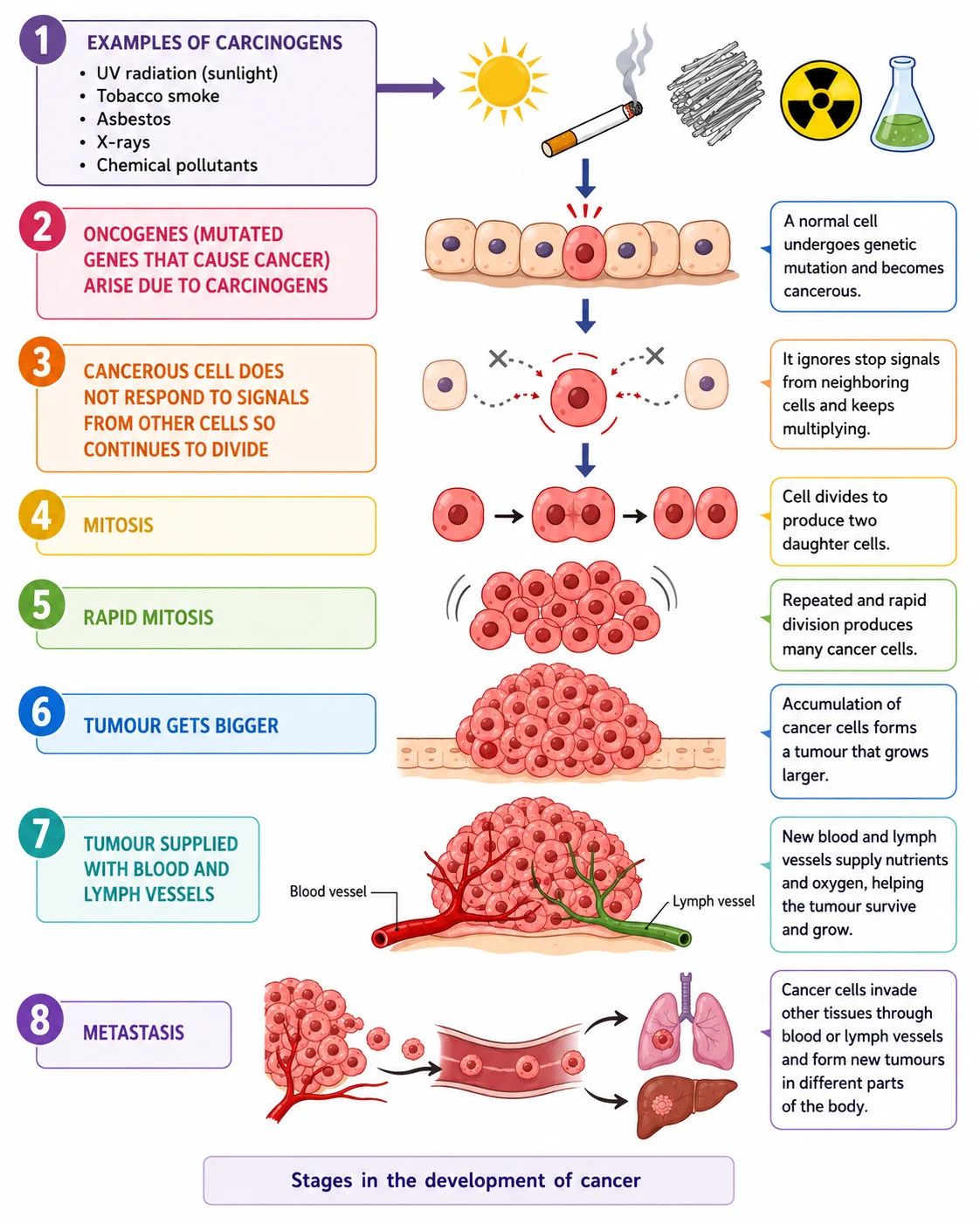
- Proto-oncogenes code for proteins that stimulate normal division, and mutation makes oncogenes that make more product or permanently active proteins.
- Tumour-suppressor genes code for proteins that inhibit division or cause apoptosis if DNA is damaged,
- and mutation gives no, less or permanently inactive protein.
- Either change leads to uncontrolled division and possibly cancer.
Tumour
Tumour an irregular mass of cells from uncontrolled mitosis, with about a thousand million cells when detected.
- All tumours can damage the organ they are in, cause blockages or press on other organs.
| Feature | Malignant | Benign |
|---|---|---|
| Cancerous | Yes | No |
| Growth | Rapid, invading and destroying surrounding tissue | Slow, without invasion |
| Spread | Metastasis to other parts through blood or lymph, forming secondary tumours | No metastasis |
| Blood supply | Cells secrete chemicals that stimulate new blood vessels | Not described |
| After removal | Can grow back | Do not usually grow back |
| Causes | Carcinogens such as UV, X-rays, tobacco, asbestos, processed meat | Inflammation, infection, injury, diet, genetics, toxins, radiation |
| Examples | Primary and secondary tumours | Polyps, non-cancerous brain tumours, warts |
Section 10 of 12Mitotic index
Root tip meristem
- Root tip meristem it lies behind the root cap, with a zone of dividing cells.
Preparing a squash
Mitotic index
Stain the root tip.
Gently squash it to spread the cells in a thin layer.
Observe, draw and annotate cells in the stages of mitosis.
- Mitotic index = number of cells in mitosis ÷ total number of cells, which can be multiplied by 100 for a percentage.
- The formula is not given, so it must be remembered.
Section 11 of 12Key terms
| Term | Meaning |
|---|---|
| Cytokinesis | division of the cytoplasm after nuclear division. |
| Bivalent | a pair of homologous chromosomes paired in prophase I. |
| Crossing over | exchange of alleles between non-sister chromatids. |
| Non-disjunction | failure of chromosomes to separate in meiosis. |
| Cyclin | a protein whose concentration triggers progress through the cell cycle. |
| Proto-oncogene | a gene that stimulates normal cell division. |
| Metastasis | spread of cancer cells from a primary tumour to other parts of the body. |
| Mitotic index | the proportion of cells in a sample that are in mitosis. |
| Meristem | a region of undifferentiated plant cells that divide by mitosis. |
Section 12 of 12Exam technique
1
Chromosome number halves in meiosis II — it halves in meiosis I.
2
Cytokinesis and mitosis are the same — mitosis divides the nucleus and cytokinesis divides the cytoplasm.
3
Benign tumours metastasise — only malignant tumours invade and metastasise.
4
Interphase is a resting phase — it is the longest and most active phase.
Marked practiceQuizzes for this lesson
Last updated:
Cambridge 9700 AS Biology
Core Formula
Magnification Triangle & Unit Conversion
Formula: M = I / A ⇔ A = I / M ⇔ I = M × A
Units: 1 mm = 1,000 μm = 1,000,000 nm.
Rule: Convert Image size (ruler mm) to μm by ×1,000 before dividing by Actual size.