D3.3Theme D — Continuity and change
Homeostasis — IB Biology HL Revision Notes & Topical Questions
10 min read 12 parts Lesson 10 of 18 3 quizzes
Curriculum Equation Key قوانين الرقم الهيدروجيني والاتزان الأيوني - pH & Acid-Base Equilibrium FormulasFast Exam Solution
pH = -log[H⁺] | pOH = -log[OH⁻] | pH + pOH = 14 | [H⁺][OH⁻] = 10⁻¹⁴
Word Equation & Conditions: الرقم الهيدروجيني = -لوغاريتم تركيز أيون الهيدروجين | pH + pOH = 14
Location & Specifications: الاتزان الأيوني، كيمياء 3 ثانوي، التحصيلي، و AP Chemistry.
Lesson summary
D3.3 Homeostasis — IB BIOLOGY HL notes: Explain homeostasis and negative feedback, blood glucose control, thermoregulation, kidney function and osmoregulation, and changes in blood supply to organs.Homeostasis = maintaining a constant internal environment within preset limits, so…
- Insulin resistance
- Why it matters
- Islets of Langerhans
- Core temperature
- Uncoupled respiration
- Kidney roles
Part 1 of 10Homeostasis and negative feedback
Homeostasis
Homeostasis maintaining a constant internal environment within preset limits, so conditions are optimal for enzyme action and cell function.
- Sensory cells detect internal and external conditions and the body responds to keep them constant.
- Controlled factors include core body temperature, blood pH, blood glucose concentration and blood osmotic concentration.
Negative feedback
- Negative feedback most control mechanisms reverse any change and return a value to its set point.
- A receptor detects a change in a physiological factor.
- A coordination system (brain and nervous system) passes information between body parts.
- An effector (muscle or gland) brings about the response.
- An increase leads to a response that decreases the factor, and a decrease leads to a response that increases it.
Positive feedback instead amplifies a change.
Part 2 of 10Diabetes
- Diabetes = failure or deterioration of homeostatic control of blood glucose, so the glucose concentration in blood rises.
- Excess glucose appears in the urine and the kidneys make large volumes of urine, causing thirst through dehydration.
- Glucose stays in the blood rather than entering cells, so respiration falls, causing fatigue.
- Very high levels after a meal can damage organs.
| Feature | Type 1 | Type 2 |
|---|---|---|
| Cause | The pancreas cannot make enough insulin because an autoimmune response destroys islet cells | Cells become resistant to insulin because receptors are fewer or do not respond |
| Onset | Normally childhood | Usually older adults and more common |
| Pancreas | Cannot make insulin | Still makes insulin, then fails to compensate |
| Treatment | Monitoring blood glucose, insulin injections and a modified diet with less carbohydrate | Medication, low carbohydrate diet and exercise |
Insulin resistance
- Insulin resistance the pancreas secretes more and more insulin, then cannot compensate, so glucose uptake falls and blood glucose is uncontrolled.
- Rapidly digested sugary food causes dangerous spikes.
Obesity is a major risk factor, because over-production of insulin on a high carbohydrate diet triggers insulin resistance.
Part 3 of 10Regulation of blood glucose
Why it matters
- Why it matters glucose is needed for respiration and levels must not fall too low,
- and because it is soluble it affects osmotic balance between cells and blood.
Islets of Langerhans
Islets of Langerhans the endocrine tissue of the pancreas (the exocrine tissue makes digestive enzymes).
- α cells secrete glucagon and β cells secrete insulin, and both monitor blood glucose.
| Hormone | Trigger | Effects |
|---|---|---|
| Insulin | Blood glucose rises after a carbohydrate meal | Glucose channels open so glucose enters cells by facilitated diffusion; liver and muscle convert glucose to glycogen (glycogenesis); respiration rate rises; glucose is converted to fatty acids for fat storage; blood glucose falls |
| Glucagon | Blood glucose falls after fasting or exercise | Enzymes hydrolyse glycogen in liver and muscle to glucose (glycogenolysis); respiration rate falls; amino acids are converted to glucose (gluconeogenesis); blood glucose rises |
Insulin's main targets are the liver and muscles.
Spelling
- Spelling glucagon is the hormone and glycogen is the animal storage polysaccharide.
Part 4 of 10Thermoregulation: control and mechanisms
Thermoregulation
Thermoregulation control of internal body temperature by negative feedback.
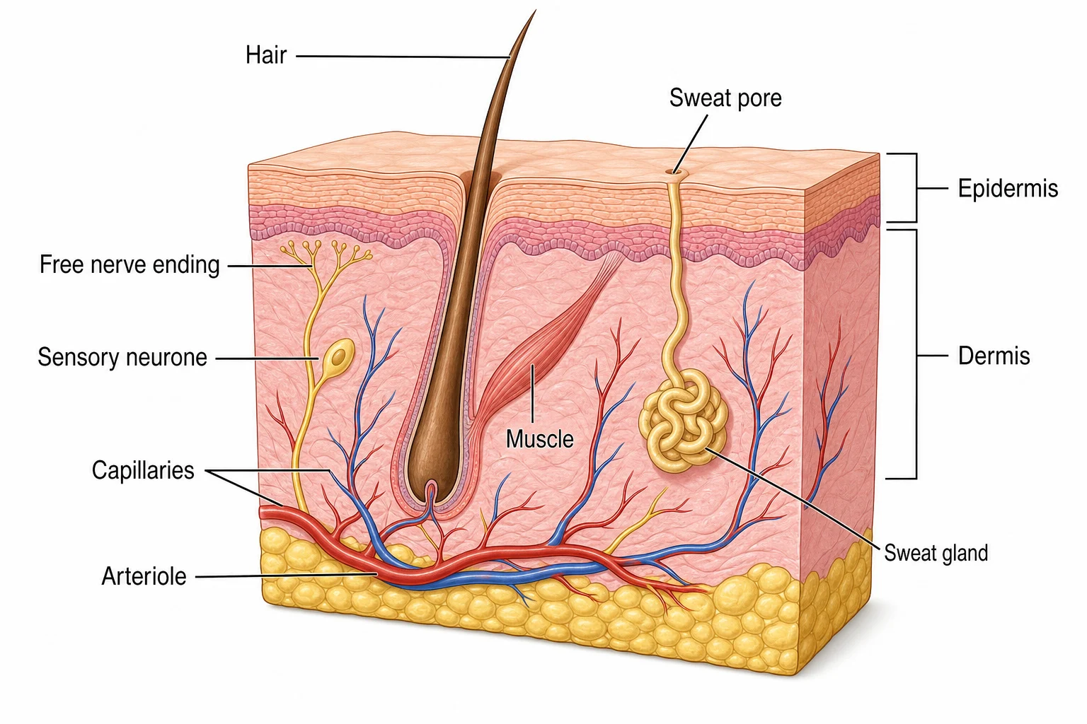
- Peripheral thermoreceptors in the skin monitor external temperature, and receptors in the hypothalamus monitor internal temperature.
- Effectors control heat loss at the skin and heat generation by metabolism.
Core temperature
- Core temperature human enzymes work best at about 37 °C.
- At low temperatures molecules have little kinetic energy, collisions are rare and few enzyme-substrate complexes form.
- At high temperatures enzymes denature and lose their tertiary structure.
Endotherms (mammals and birds) maintain a constant body temperature by physiological means (shivering, altered metabolism) and behavioural means (seeking shade in a burrow, sunbathing).
Effectors
- The hypothalamus releases thyrotropin-releasing hormone,
- which makes the pituitary release thyroid-stimulating hormone,
- which makes the thyroid release thyroxine that raises metabolic rate and heat production.
- Muscle tissue shivers to raise metabolic rate and release heat.
- White adipose tissue under the skin and around organs insulates.
- Brown adipose tissue generates heat before shivering, by non-shivering thermogenesis.
Part 5 of 10Human responses to temperature change
| Response | Rise in temperature | Fall in temperature |
|---|---|---|
| Arterioles | Vasodilation: muscles relax, more blood flows through skin capillaries and more heat is lost by radiation | Vasoconstriction: muscles contract and blood is diverted through deeper shunt vessels, so less heat is lost |
| Sweating | Sweat evaporates and cools the skin by taking heat energy | Not stimulated |
| Hair | Erector muscles relax, hairs lie flat and air circulates | Erector muscles contract and hairs stand up to trap an insulating air layer, though humans have little hair |
| Shivering | Not used | Rapid muscle contraction produces heat that warms the blood |
| Metabolism | Lower | Thyroxine raises basal metabolic rate and heat production |
Uncoupled respiration
- Uncoupled respiration in brown adipose tissue lipids are metabolised and the energy is released entirely as heat instead of making ATP.
- It occurs mainly in newborn infants, who cannot shiver.
Vasodilation and vasoconstriction involve muscles in arterioles, not capillaries, which have no muscles.
Part 6 of 10Excretion and osmoregulation, kidney structure
Excretion and osmoregulation, kidney structure
Kidney roles
- Kidney roles excretion and osmoregulation.
Excretion
Excretion removal of toxic metabolic waste, including nitrogenous waste from excess amino acids and nucleic acids.
- The waste is first ammonia, which is highly toxic and cannot be stored.
- Some organisms convert it into less toxic urea, which can stay in the body at low levels but must be excreted, dissolved in water as urine.
Osmoregulation
Osmoregulation keeping a safe balance of water and solutes in cells (osmotic concentration, in osmoles per litre).
- A cell with lower water potential than its surroundings gains water and may burst, though plant cells are protected by their walls.
- A cell with higher water potential loses water and shrinks.
| Structure | Function |
|---|---|
| Renal artery | Carries blood containing urea and salts to the kidney |
| Renal vein | Carries blood with urea and excess salts removed away |
| Kidney | Filters blood and regulates its water content |
| Ureter | Carries urine to the bladder |
| Bladder | Stores urine temporarily |
| Urethra | Releases urine outside the body |
Excretion and osmoregulation, kidney structure
Regions
- Regions the kidney has a fibrous capsule and then the cortex, the medulla and the renal pelvis.
- Nephrons are the functional units that form urine.
- The cortex holds the glomerulus, Bowman's capsule, proximal convoluted tubule and distal convoluted tubule.
- The medulla holds the loop of Henle and collecting duct.
- The renal pelvis receives all nephrons and joins the ureter.
Blood supply
- Blood supply an afferent arteriole from the renal artery splits into the glomerulus capillaries,
- which rejoin as the efferent arteriole, and blood then flows through capillaries alongside the nephron to the renal vein.
Part 7 of 10Ultrafiltration
Ultrafiltration
- Ultrafiltration high pressure in the glomerulus forces small molecules into the Bowman's capsule.
- The afferent arteriole is wider than the efferent arteriole, raising pressure, which is much higher than in other capillaries.
- Small molecules passchloride ions, sodium ions, glucose, urea and amino acids, forming the glomerular filtrate.
- Large molecules such as proteins stay in the blood, and red and white cells and platelets are too large.
Filtration layers
Ultrafiltration
The capillary endothelium has fenestrations that let fluid but not blood cells through.
The basement membrane of collagen and glycoproteins acts as a sieve that holds back large proteins.
The epithelium of the Bowman's capsule has podocytes with foot-like projections, and the gaps between them let small molecules pass.
| Substance | Blood plasma | Glomerular filtrate |
|---|---|---|
| Urea | 5 | 5 |
| Na+ | 150 | 145 |
| Cl− | 110 | 115 |
| Glucose | 5 | 5 |
| Protein | 740 (mg dm−3) | 5 (mg dm−3) |
Part 8 of 10Selective reabsorption
Selective reabsorption
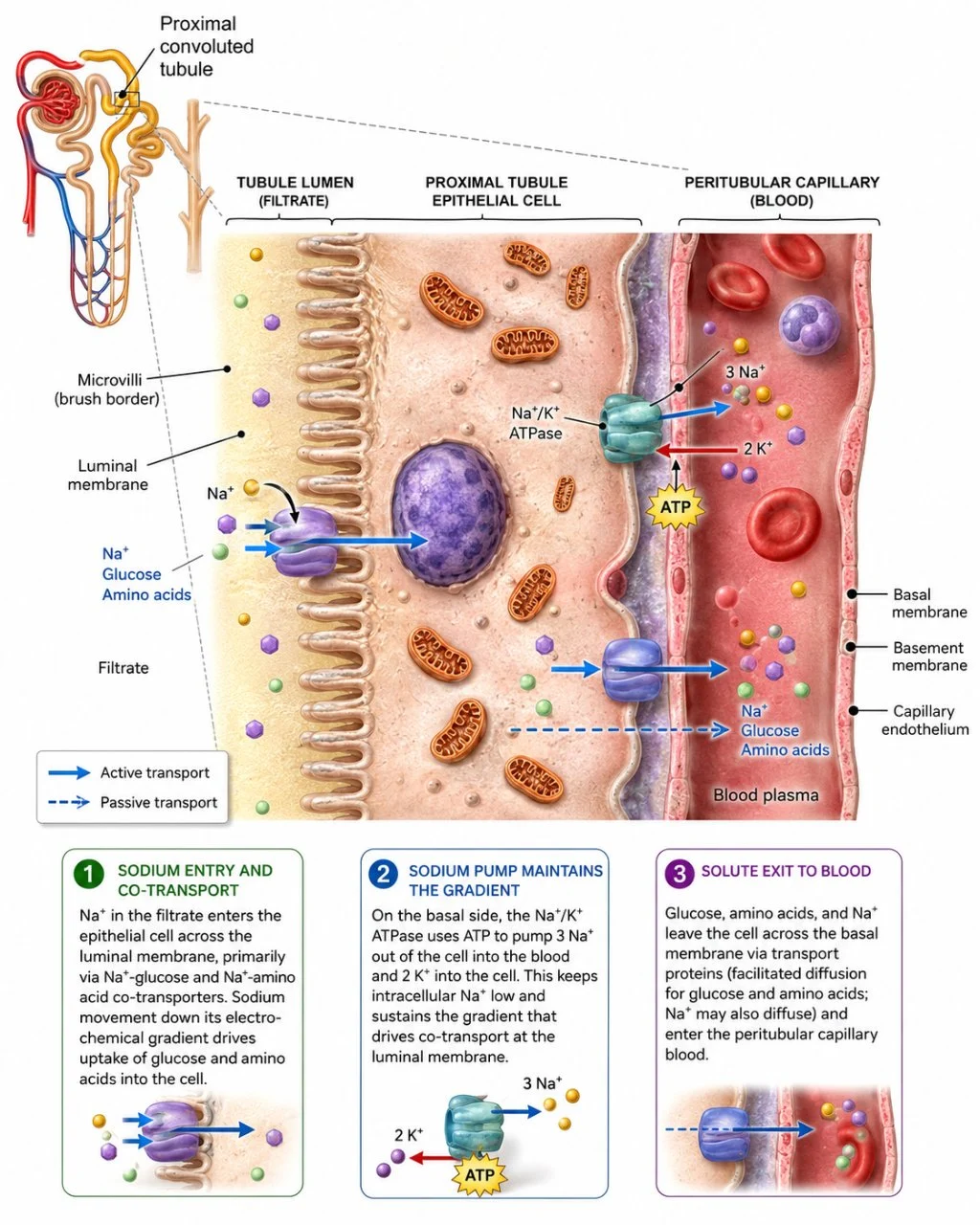
- Selective reabsorption useful substances are reabsorbed into the blood, mostly in the proximal convoluted tubule, with the loop of Henle and collecting duct also reabsorbing water and salts.
- (water, salts, glucose, amino acids)
Adaptations of the proximal convoluted tubule epithelium
| Adaptation | Effect |
|---|---|
| Microvilli on the luminal membrane | Large surface area for reabsorption |
| Co-transporter proteins | Each moves a specific solute such as glucose or an amino acid across the membrane |
| Many mitochondria | Provide energy for Na+–K+ pumps in the basal membranes |
| Tightly packed cells | No fluid passes between cells, so all reabsorbed substances go through the cells |
- Process
1
Sodium ions are actively transported out of the tubule into surrounding tissue.
2
Chloride ions follow by diffusion because of the electrical gradient.
3
Sugars and amino acids move by co-transporter proteins that also move sodium, an active process.
4
Water potential of the tissue falls, so water leaves by osmosis.
5
Urea moves out by diffusion.
6
All these substances pass into nearby capillaries down their gradients.
- Unwanted solutes and toxins that remain are excreted in urine.
Part 9 of 10Loop of Henle
Role
- Role it makes urine more concentrated than blood and so conserves water, and dilute urine can also be made when water intake is high.
Process
1
Na+ and Cl− are pumped out of the ascending limb into the medulla, lowering its water potential.
2
The ascending limb is impermeable to water, so water cannot leave it.
3
The descending limb is permeable to water, so water leaves by osmosis into the low water potential medulla.
4
The descending limb has few transport proteins, so it is not permeable to ions, and the filtrate's water potential falls as it descends.
5
The low water potential of the medulla also allows water reabsorption from the collecting duct.
6
Water and ions pass into capillaries, notably the vasa recta, which also supplies oxygen and removes carbon dioxide.
- Result
- the loop generates a steep water potential gradient across the medulla, which maximises water reabsorption.
Part 10 of 10ADH and osmoregulation, changes in blood supply to organs
- Osmoregulation by the kidney
- the amount of water reabsorbed is regulated by changing the permeability of the distal convoluted tubule and collecting duct walls to water, controlled by antidiuretic hormone (ADH).
- ADH is released from the posterior pituitary, regulated by the hypothalamus, whose osmoreceptor cells monitor blood water content.
| Condition | Response |
|---|---|
| Low blood water content (less water intake, sweating, diarrhoea; high blood solutes) | The hypothalamus makes the pituitary secrete ADH, vesicles with aquaporins fuse with the membrane of collecting duct cells, permeability rises, more water is reabsorbed, and a small volume of concentrated urine is produced |
| High blood water content (more water intake, salt loss in sweat; low blood solutes) | ADH release stops and levels fall, aquaporins are removed by pinching the membrane back into vesicles, less water is reabsorbed, and a large volume of dilute urine is produced |
Gradient
- Gradient the loop of Henle gives the medulla an increasing osmolarity, so water is reabsorbed all along the collecting duct.
Urine
- Urine the remaining filtrate passes to the renal pelvis, then along the ureter to the bladder.
Dehydration follows excessively low water content, and overhydration follows excessively high water content.
- Principle
- the circulatory system supplies oxygen and nutrients and removes waste,
- and blood flow changes with activity by vasodilation or vasoconstriction of arterioles supplying capillary beds.
- the circulatory system supplies oxygen and nutrients and removes waste,
| Organ | Change |
|---|---|
| Skeletal muscles | Low during sleep, higher when awake and upright, and much larger in exercise |
| Gut | Rises soon after a meal and falls during exercise so blood goes to muscles |
| Brain | Relatively constant whatever the activity, rising slightly in REM sleep |
| Kidneys | Little change, a slight rise in sleep and rest and a slight fall in prolonged exercise |
Key terms
| Term | Meaning |
|---|---|
| Homeostasis | maintenance of a constant internal environment within set limits. |
| Glycogenolysis | breakdown of glycogen to glucose. |
| Thermoregulation | control of internal body temperature. |
| Ultrafiltration | filtering of small molecules from blood in the glomerulus under high pressure. |
| Selective reabsorption | return of useful substances from the filtrate to the blood. |
| ADH | antidiuretic hormone that increases water reabsorption in the collecting duct. |
| Nephron | the functional unit of the kidney that forms urine. |
| Aquaporin | a channel protein that lets water cross a membrane. |
| Non-shivering thermogenesis | heat production in brown adipose tissue. |
Avoid these mistakes
1
Negative feedback amplifies change — it reverses change and returns values to the set point.
2
Glucagon and glycogen are the same — glucagon is a hormone and glycogen a storage polysaccharide.
3
Capillaries dilate and constrict — arterioles do, because they contain muscle.
4
Proteins are filtered into the filtrate — large proteins stay in the blood.
Marked practiceQuizzes for this lesson
Last updated:
\n