A2.2Theme A — Unity and diversity
Cell structure — IB Biology SL Revision Notes & Topical Questions
10 min read 13 parts Lesson 8 of 11 2 quizzes
Lesson summary
A2.2 Cell structure — IB BIOLOGY SL notes: Describe cell theory, microscopy and the structure of prokaryotic and eukaryotic cells, including atypical cells.Cell theory — all living organisms are made of one or more cellsCells are the basic functional unit of living organisms.New …
- Shared features
- Deductive reasoning
- Inductive reasoning
- Data types
- Liquid mount
- Solid mount
Section 1 of 13Cell theory
1Cell theory
- Cell theory all living organisms are made of one or more cells
- Cells are the basic functional unit of living organisms.
- New cells are produced from pre-existing cells.
2History
- Hooke named cells in the 1660s from cork, and Schleiden and Schwann proposed cell theory in 1837
3Shared features
- Shared features every cell has a membrane, genetic material and enzyme-catalysed reactions
4Deductive reasoning
- Deductive reasoning moves from a general theory to a specific prediction that is tested
- Cell theory predicts that a newly discovered organism will contain one or more cells.
5Inductive reasoning
- Inductive reasoning many specific observations lead to a general theory
6Data types
- Data types measurements with instruments are quantitative observations and colour or the presence of structures are qualitative
Section 2 of 13Using a light microscope
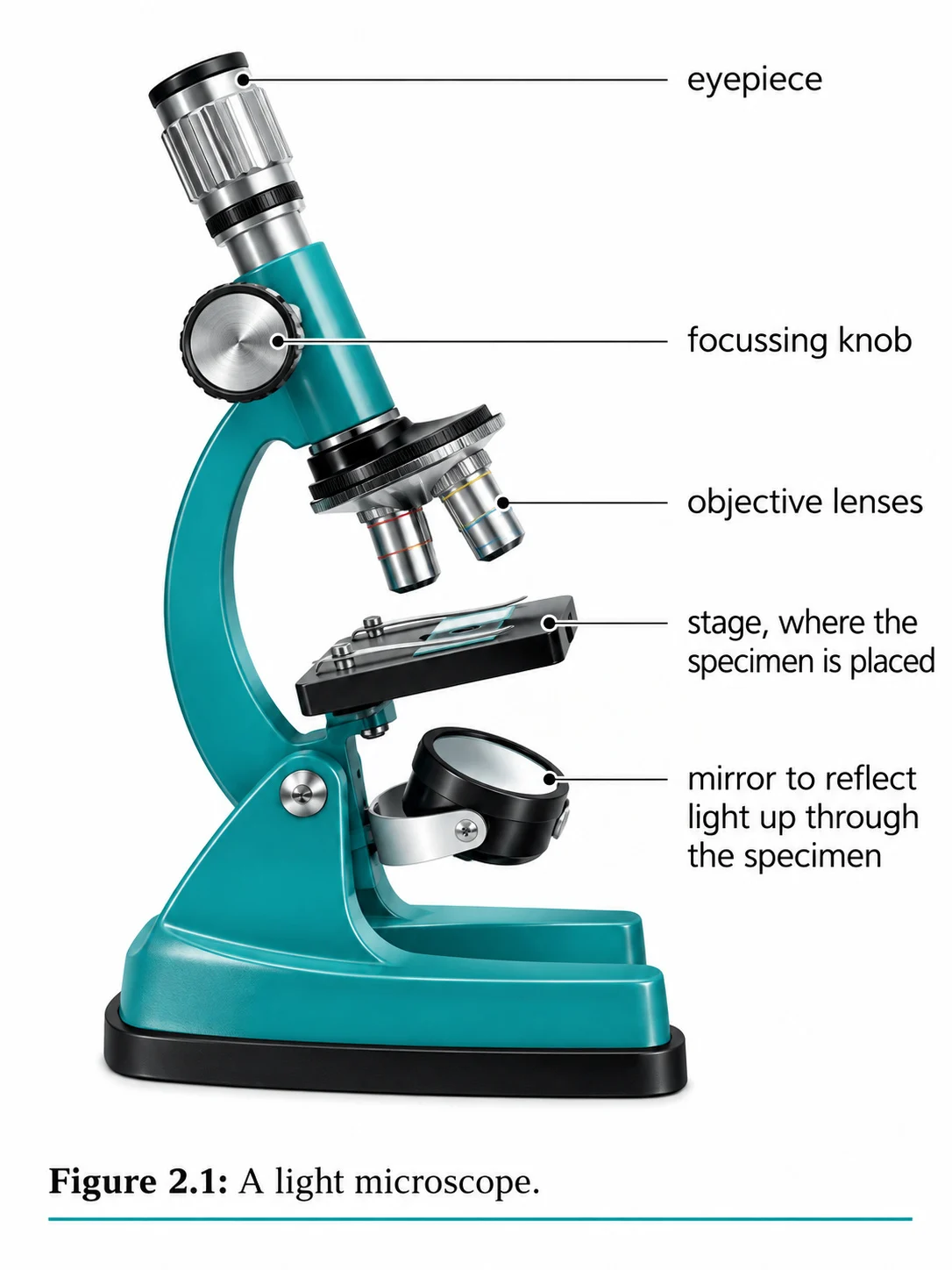
Using a light microscope
Parts
- Parts eyepiece lens, objective lenses, stage with clips, light source, condenser, and coarse and fine focus
Liquid mount
- Liquid mount add drops to the slide with a pipette and lower the coverslip to avoid bubbles
Solid mount
- Solid mount cut a thin layer with a scalpel or forceps, stain if needed and lower the coverslip
- Wear gloves and take care with sharp tools.
1
Place the slide on the stage and fix it with the clips.
2
Start on the low-power objective and use the coarse focus until the specimen is in focus.
3
Move to a higher objective only after focusing, and never move the stage down while changing objective.
4
At high power use only the fine focus.
- Blurry image
- return to low power, check the specimen is thin enough and check for contamination
Section 3 of 13Magnification and measurement
Magnification
Magnification image size / actual size
- Convert all measurements to the same units, and magnification has no units.
- 1 mm = 1000 µm and 1 µm = 1000 nm.
Scale bar
- Scale bar measure the bar with a ruler, convert to the units written on it and divide by the real length
Adding a scale bar
- Make the bar about 20% of the specimen length.
- Write the real length it represents underneath.
Eyepiece graticule
- Eyepiece graticule a scale in the eyepiece with no fixed units
- Calibrate it for each objective lens with a stage micrometer.
1
Place a stage micrometer on the stage and line up the two scales.
2
Calculate the value of one graticule division.
3
Replace the micrometer with the specimen.
4
Count the divisions across the cell.
5
Multiply the divisions by the value of one division to find the actual size.
Section 4 of 13Light and electron microscopes
| Feature | Light | TEM | SEM |
|---|---|---|---|
| Radiation | Light | Electrons | Electrons |
| Resolution | About 200 nm | About 0.2 nm | About 5 nm |
| Image | Colour, 2D | Black and white, 2D section | Black and white, 3D surface |
| Living specimens | Yes | No | No |
| Useful magnification | Up to about ×1500 | Over ×500 000 | High, but below TEM |
| Preparation | Simple and quick | Thin sections and vacuum, artefacts possible | Coated whole specimen and vacuum |
| Portability | Small and portable | Large and fixed | Large and fixed |
Magnification
Magnification how many times larger the image is than the object
Resolution
Resolution the ability to see two close objects as separate
- Light cannot resolve points closer than half its wavelength, which is about 200 nm.
- The shorter wavelength of electrons gives far higher resolution.
Light microscope
- Light microscope shows eukaryotic cells, nuclei and possibly mitochondria and chloroplasts
- It cannot show ribosomes, the endoplasmic reticulum or lysosomes.
TEM
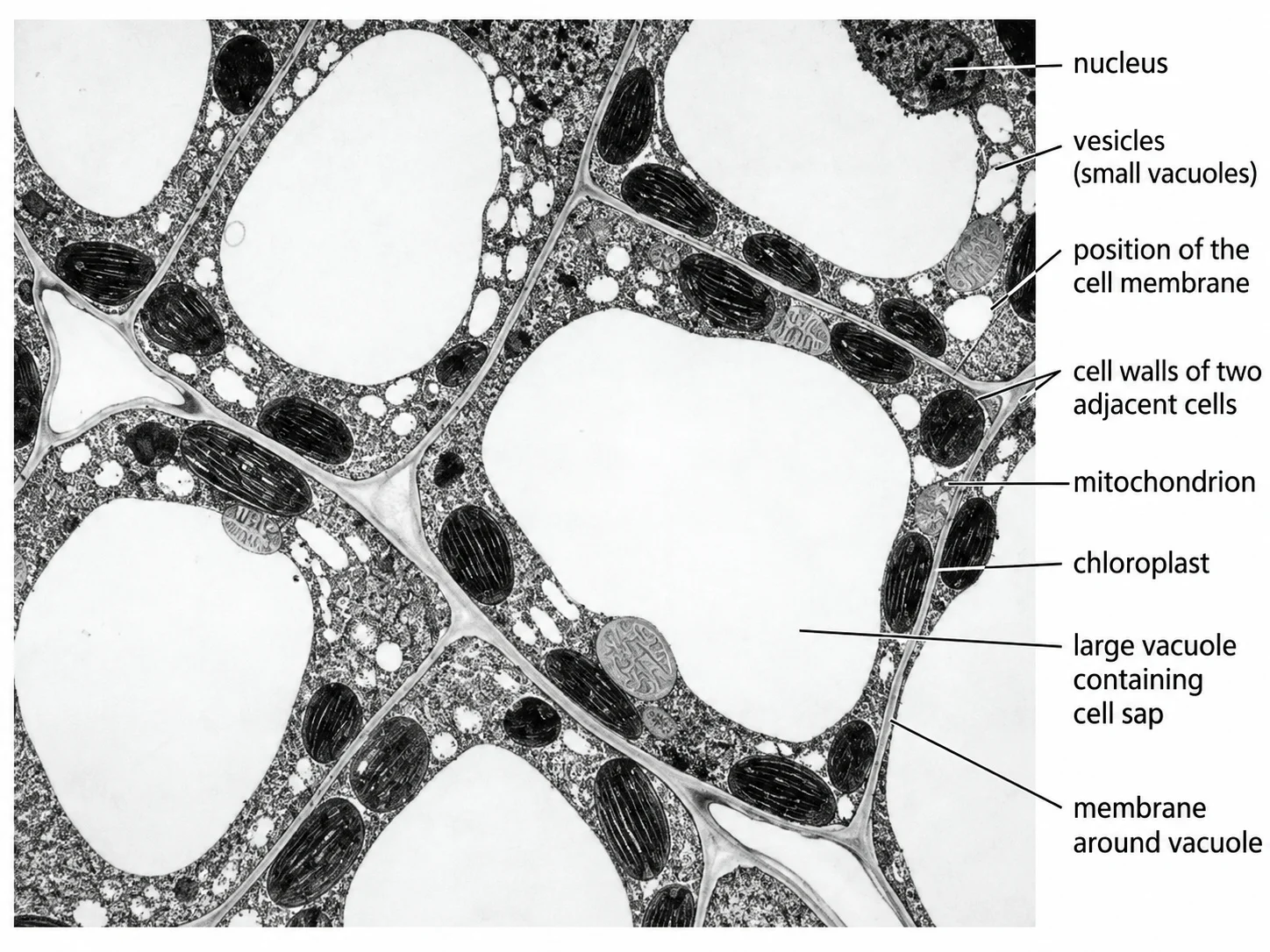
- TEM electrons pass through a thin section and denser parts absorb more, so they appear darker
SEM
- SEM electrons bounce off the surface, giving a 3D image of the surface
Artefacts
- Artefacts features from specimen preparation that look like real structures
Section 5 of 13Developments in microscopy
1Fluorescent stains
- Fluorescent stains dyes bind to specific structures and glow under UV
2Immunofluorescence
- Immunofluorescence antibodies carrying fluorescent dyes bind to chosen proteins, such as virus proteins
3Freeze fracture
- Freeze fracture frozen cells are split along membranes in a vacuum to give a planar view of membrane interiors
- It revealed proteins spanning the bilayer.
4Cryo-electron microscopy
- Cryo-electron microscopy solutions of proteins are flash-frozen and imaged
- Software builds a 3D model from images of individual molecules.
- It has improved knowledge of virus structure, membranes and protein synthesis.
Section 6 of 13Features of all cells and prokaryotes
Features of all cells and prokaryotes
Plasma membrane
- Plasma membrane a phospholipid bilayer that controls what enters and leaves
- Its proteins act in cell recognition, communication and transport.
Cytoplasm
- Cytoplasm watery cytosol with ions, small molecules and macromolecules, where many reactions occur
DNA
- DNA genetic material, which lets new cells form from old ones and controls enzyme production
Prokaryote
Prokaryote a cell without a nucleus or membrane-bound organelles, 0.1-5 µm across
- The two domains are Bacteria, such as E. coli, and Archaea, often found in extreme environments.
- DNA is a naked circular molecule in the nucleoid, with small plasmids that may carry antibiotic resistance.
- Ribosomes are 70S.
- Most have a peptidoglycan wall that gives shape and stops bursting.
Gram positive
- Gram positive a thick wall holds crystal violet and stains purple, as in Staphylococcus
Extras
- Extras a slime capsule resists drying and immune attack, flagella move the cell and pili attach to surfaces and transfer DNA
Section 7 of 13Eukaryotic organelles
| Feature | Prokaryote | Eukaryote |
|---|---|---|
| Nucleus | Absent, DNA in the nucleoid | Present with a double membrane |
| DNA | Circular and naked, with plasmids | Linear, wrapped around histones |
| Ribosomes | 70S | 80S in the cytoplasm |
| Organelles | None membrane-bound | Mitochondria, ER, Golgi and more |
| Cell wall | Peptidoglycan | Cellulose (plants), chitin (fungi), none (animals) |
| Typical size | 1-5 µm | 10-100 µm |
Eukaryotic organelles
Compartmentalisation
- Compartmentalisation gives higher enzyme and substrate concentrations, isolates harmful enzymes and allows separate optimum pH
Nucleus
- Nucleus a double membrane with pores for mRNA and ribosome subunits out and enzymes in
- Chromatin is linear DNA wrapped around histone proteins.
- The nucleolus makes ribosomes.
Rough ER
- Rough ER flattened sacs covered with 80S ribosomes that make proteins for secretion
Golgi apparatus
- Golgi apparatus modifies proteins and lipids and packages them into vesicles
- Cargo includes secreted hormones such as insulin, lysosome enzymes and membrane proteins.
Mitochondrion
- Mitochondrion double membrane with folded cristae, containing the enzymes of aerobic respiration, circular DNA and ribosomes
Lysosome
- Lysosome a vesicle with hydrolytic enzymes that digests worn-out organelles, and is used in immunity and apoptosis
Microtubules
- Microtubules tubulin cylinders about 25 nm wide forming the cytoskeleton for support and movement
Section 8 of 13Plant, animal and fungal cells
| Feature | Plant | Animal | Fungus |
|---|---|---|---|
| Cell wall | Cellulose | None | Chitin, glucans, glycoproteins |
| Vacuole | Large and permanent | Small and temporary | Small and non-permanent |
| Chloroplasts | In photosynthetic cells | None | None |
| Centrioles | Absent | Present | Absent |
| Cilia or flagella | None | Often present | None in true fungi |
| Carbohydrate store | Starch | Glycogen | Glycogen |
Chloroplast
- Chloroplast larger than a mitochondrion with a double membrane
- Thylakoids stack into grana joined by lamellae.
- The light reactions occur in the thylakoids and the Calvin cycle in the stroma.
Vacuole
- Vacuole bounded by the tonoplast and stores sap
Cell wall
- Cell wall extracellular and freely permeable, giving plant cells a fixed shape
- Plasmodesmata link neighbouring plant cells through the walls.
Animal cilia
- Animal cilia move an egg along the oviduct or sweep fluids in the respiratory tract
Fungal shape
- Fungal shape fungal cells have walls but may still change shape
Section 9 of 13Functions of life and atypical cells
Functions of life and atypical cells
Seven functions
- Seven functions metabolism, reproduction, homeostasis, growth, response, excretion and nutrition
Unicellular organisms
- Unicellular organisms carry out every function in one cell
- Paramecium uses cilia to move and a contractile vacuole to remove water.
- Chlamydomonas has a chloroplast, an eyespot and flagella.
Red blood cells
- Red blood cells have no nucleus, giving more room for haemoglobin, and a biconcave shape for a large surface area
Aseptate fungal hyphae
- Aseptate fungal hyphae no cross-walls, so many nuclei share one cytoplasm
Skeletal muscle
- Skeletal muscle long multinucleate fibres formed by the fusion of cells, up to 300 mm long
Phloem sieve tubes
- Phloem sieve tubes no nucleus, mitochondria or ribosomes, so they depend on companion cells
Challenge to cell theory
- Challenge to cell theory these atypical cells question the idea that cells work independently
Section 10 of 13Interpreting micrographs and drawing cells
Interpreting micrographs and drawing cells
Look for a nucleus to decide prokaryote or eukaryote.
Look for a cell wall or large vacuole to decide plant or animal.
Identify the abundant organelles and relate them to function.
Recognising organelles
- Recognising organelles mitochondria are oval with cristae, and chloroplasts are larger with grana
- Nuclei have a membrane and dark nucleolus, and vacuoles look pale.
Examples
- Examples many lysosomes and mitochondria suggest a phagocytic cell such as a macrophage
- Microvilli and many mitochondria suggest an absorptive cell lining the small intestine.
Drawing conventions
- Title, magnification recorded and a scale bar if used.
- A sharp HB pencil with clear single lines and no shading.
- A drawing that fills the space with correct proportions.
- Label lines drawn with a ruler, kept to one side, not crossing and without arrowheads.
Plan diagrams
- Plan diagrams draw tissue outlines only and never individual cells
Section 11 of 13More from the specification
- Electron micrograph
- A photograph taken through an electron microscope.
Section 12 of 13Key terms
| Term | Meaning |
|---|---|
| Cell theory | All organisms are made of cells, cells are the basic unit of life and cells arise from existing cells. |
| Resolution | The smallest distance at which two points can still be seen as separate. |
| Eyepiece graticule | A scale in the eyepiece that is calibrated with a stage micrometer to measure specimens. |
| Prokaryote | A cell without a nucleus or membrane-bound organelles. |
| Eukaryote | A cell with a nucleus and membrane-bound organelles. |
| Nucleoid | The region of a prokaryotic cell holding its naked circular DNA. |
| Compartmentalisation | The division of a cell into membrane-bound regions so that processes stay apart. |
| Atypical cell | A cell, such as a multinucleate one or one without a nucleus, that does not fit the standard pattern. |
Section 13 of 13Exam technique
1
Confusing magnification with resolution — magnification is the enlargement and resolution is the detail that can be separated
2
Mixing units in magnification calculations — convert image and actual size to the same unit first
3
Saying electron microscopes can view living cells — the vacuum means specimens must be dead
4
Saying all eukaryotic cells have a nucleus — mature red blood cells and sieve tube elements do not
Marked practiceQuizzes for this lesson
Last updated:
Cambridge 9700 AS Biology
Core Formula
Magnification Triangle & Unit Conversion
Formula: M = I / A ⇔ A = I / M ⇔ I = M × A
Units: 1 mm = 1,000 μm = 1,000,000 nm.
Rule: Convert Image size (ruler mm) to μm by ×1,000 before dividing by Actual size.