Curriculum
Biology for AP Courses
Unit 1: The Study of Life
0/6
Unit 2: The Chemical Foundation of Life
0/8
Unit 3: Biological Macromolecules
0/12
Unit 4: Cell Structure
0/14
-
Unit 4 overview: Cell Structure
 Preview
-
4.1 Studying Cells
Preview
-
4.1 Studying Cells - Check yourself
 Preview
-
4.2 Prokaryotic Cells
Preview
-
4.2 Prokaryotic Cells - Check yourself
Preview
-
4.3 Eukaryotic Cells
Preview
-
4.3 Eukaryotic Cells - Check yourself
Preview
-
4.4 The Endomembrane System and Proteins
Preview
-
4.4 The Endomembrane System and Proteins - Check yourself
Preview
-
4.5 Cytoskeleton
Preview
-
4.5 Cytoskeleton - Check yourself
Preview
-
4.6 Connections between Cells and Cellular Activities
Preview
-
4.6 Connections between Cells and Cellular Activities - Check yourself
Preview
-
Unit 4 review quiz: Cell Structure
Preview
Unit 5: Structure and Function of Plasma Membranes
0/10
Unit 6: Metabolism
0/12
Unit 7: Cellular Respiration
0/16
Unit 8: Photosynthesis
0/8
Unit 9: Cell Communication
0/10
Unit 10: Cell Reproduction
0/12
Unit 11: Meiosis and Sexual Reproduction
0/6
Unit 12: Mendel's Experiments and Heredity
0/8
Unit 13: Modern Understandings of Inheritance
0/6
Unit 14: DNA Structure and Function
0/14
Unit 15: Genes and Proteins
0/13
Unit 16: Gene Regulation
0/16
Unit 17: Biotechnology and Genomics
0/12
Unit 18: Evolution and Origin of Species
0/8
Unit 19: The Evolution of Populations
0/8
Unit 20: Phylogenies and the History of Life
0/8
Unit 21: Viruses
0/10
Unit 22: Prokaryotes: Bacteria and Archaea
0/12
Unit 23: Plant Form and Physiology
0/14
Unit 24: The Animal Body: Basic Form and Function
0/8
Unit 25: Animal Nutrition and the Digestive System
0/10
Unit 26: The Nervous System
0/12
Unit 27: Sensory Systems
0/12
Unit 28: The Endocrine System
0/12
Unit 29: The Musculoskeletal System
0/10
Unit 30: The Respiratory System
0/10
Unit 31: The Circulatory System
0/10
Unit 32: Osmotic Regulation and Excretion
0/12
Unit 33: The Immune System
0/10
Unit 34: Animal Reproduction and Development
0/16
Unit 35: Ecology and the Biosphere
0/12
Unit 36: Population and Community Ecology
0/16
Unit 37: Ecosystems
0/8
Unit 38: Conservation Biology and Biodiversity
0/10
Text lesson
4.5 Cytoskeleton
4.5 Cytoskeleton — BIOLOGY FOR AP COURSES notes: You will be able to name the three types of cytoskeleton fibers, describe the structure and job of each, explain how flagella and cilia are built, and compare the components of prokaryotic and eukaryotic cells.
1Science Practice Connection for AP Courses2Science Practice Connection for AP Courses
Learning objectives
Learning objectives
In this section you will explore one central question: how do the various components of the cytoskeleton perform their functions?
Connection for AP Courses
Connection for AP Courses
All cells, from simple bacteria to complex eukaryotes, have a cytoskeleton.
It is built from three kinds of protein elements: microfilaments, intermediate filaments and microtubules.
The cytoskeleton gives the cell rigidity and shape, makes cell movement possible, anchors the nucleus and other organelles in place, moves vesicles through the cell,
and pulls replicated chromosomes to the poles of a dividing cell.
The same protein elements also drive the movement of centrioles, flagella and cilia.
This topic supports Big Idea 1 of the AP Biology Curriculum Framework. The table below shows how each framework element connects to the content.
The learning objectives form the basis for the AP Biology course, its inquiry-based lab work, its classroom activities and its exam questions. Each learning objective combines required content with one or more of the seven science practices.
| Framework element | What it means for this topic |
|---|---|
| Big Idea 1 | Evolution drives the diversity and unity of life. |
| Enduring understanding 1.B | Organisms are linked by lines of descent from a common ancestor. |
| Essential knowledge 1.B.1 | Organisms share many core processes and features that evolved and are widely spread among organisms today. |
| Science practice 7.2 | Connect concepts within and across domains to generalize or extend enduring understandings and big ideas. |
| Learning objective 1.15 | Describe examples of conserved core biological processes and features, and explain how these shared features support common ancestry. |
1Microfilaments2Microfilaments3Microfilaments4Microfilaments5Microfilaments
Microfilaments
1
Imagine removing every organelle from a cell.
2
The plasma membrane and cytoplasm would still be there, and the cytoplasm would still hold ions, organic molecules and a network of protein fibers.
3
These fibers help maintain cell shape, hold some organelles in fixed positions, allow cytoplasm and vesicles to move inside the cell,
and let cells in multicellular organisms move.
4
This network is the cytoskeleton, and it has three fiber types: microfilaments, intermediate filaments and microtubules.
5
The diagram below shows the three fiber types inside a cell. Microfilaments form a supporting layer just inside the membrane, intermediate filaments spread through the whole cell, and microtubules sit in the interior. The lower drawing shows microtubules forming two spindle-like groups in a dividing cell.
1 / 5Next ▶
Two cell drawings with the three cytoskeleton fiber types labeled: microfilaments near the membrane, intermediate filaments throughout the cell, and microtubules in the interior. The lower drawing shows microtubules pulling into two groups in a dividing cell. Image: OpenStax, Biology for AP Courses, CC BY 4.0.
Microfilaments
 ⤢ Enlarge
Microfilaments
Microfilaments are the narrowest cytoskeleton fibers, about 7 nm in diameter. Each one is made of two strands of the globular protein actin twisted around each other
which is why microfilaments are also called actin filaments.
◀ Previous2 / 5Next ▶
A short section of a microfilament showing actin subunits stacked into two intertwined strands. Image: OpenStax, Biology for AP Courses, CC BY 4.0.
Microfilaments
 ⤢ Enlarge
Microfilaments
Actin uses ATP energy to assemble into its filament form.
The filament acts as a track along which the motor protein myosin moves.
This interaction powers processes that need motion,
such as cell division in eukaryotic cells and cytoplasmic streaming, the circular flow of cytoplasm seen in plant cells.
Actin and myosin are abundant in muscle cells.
When the actin and myosin filaments slide past each other, the muscle contracts.
Microfilaments also give some rigidity and shape to the cell. They can break down (depolymerize) and rebuild quickly
which allows a cell to change shape and move. White blood cells, the infection-fighting cells of the body, use this ability. They travel to the site of an infection and engulf (phagocytize) the pathogen.
◀ Previous3 / 5Next ▶
Microfilaments
1
Link to learning: A short time-lapse video shows a white blood cell capturing two bacteria. It engulfs the first one and then moves on to the second. The video illustrates how quickly changing actin fibers let a cell extend, move and take in material.
2
Check your understanding: HIV infects and kills white blood cells. Over time, how does this affect the immune system?
3
a.
4
The immune system is not affected.
b.
b.
5
With fewer white blood cells, the immune system cannot fight pathogens such as bacteria as well
which raises the risk of illness.
c.
c.
6
The immune system makes more white blood cells to make up the loss.
d.
d.
◀ Previous4 / 5Next ▶
Microfilaments
Microfilaments
The immune system fights pathogens harder to compensate for the lower number of white blood cells.
Answer: b. White blood cells are needed to fight infection
so losing them weakens defenses and increases illness risk.
◀ Previous5 / 5
An intermediate filament drawn as several fibrous strands wound around one another. Image: OpenStax, Biology for AP Courses, CC BY 4.0.
Intermediate filaments
 ⤢ Enlarge
Intermediate filaments
Intermediate filaments are made of several strands of fibrous proteins wound together. Their diameter is 8 to 10 nm
which lies between the diameters of microfilaments and microtubules. This in-between width is where their name comes from.
Intermediate filaments do not take part in cell movement. Their role is purely structural. They bear tension
which helps the cell keep its shape, and they anchor the nucleus and other organelles in place. Together they form a supportive scaffold inside the cell.
Intermediate filaments are the most varied group of cytoskeleton elements
because several different fibrous proteins can form them. The best-known is keratinwhich strengthens hair, nails and the outer layer of the skin.
1Microtubules2Microtubules
Microtubules
1
Microtubules are small hollow tubes.
2
Their walls are made of polymerized dimers of alpha-tubulin and beta-tubulin, two globular proteins that pair up and join into a tube wall.
3
With a diameter of about 25 nm, microtubules are the widest components of the cytoskeleton.
4
They help the cell resist compression, form tracks along which vesicles move through the cell,
and pull replicated chromosomes to opposite ends of a dividing cell.
5
Like microfilaments, microtubules can disassemble and rebuild quickly.
1 / 2Next ▶
A three-dimensional image of a microtubule beside a model of its wall, which is built from 13 pairs of alpha-tubulin and beta-tubulin. Image: OpenStax, Biology for AP Courses, CC BY 4.0.
Microtubules
 ⤢ Enlarge
Microtubules
Microtubules are also the structural elements of flagella, cilia and centrioles. The two centrioles are perpendicular bodies that make up the centrosome. In animal cells the centrosome is the microtubule-organizing center, the place from which microtubules grow.
◀ Previous2 / 2
1Flagella and cilia2Flagella and cilia
Flagella and cilia
Flagella and cilia
Flagella (one is a flagellum) are long, hair-like structures that extend from the plasma membrane and move an entire cell, as with sperm cells and Euglena.
A eukaryotic cell usually has only one or a few flagella.
Cilia (one is a cilium) are short, hair-like structures that cover much of the cell surface.
They can move whole cells, such as paramecia, or move substances across the surface of a cell.
Examples include the cilia lining the Fallopian tubes,
which move the ovum toward the uterus,and the cilia lining the airways,which trap particles and sweep them toward the nostrils.
Flagella and cilia differ in length and number
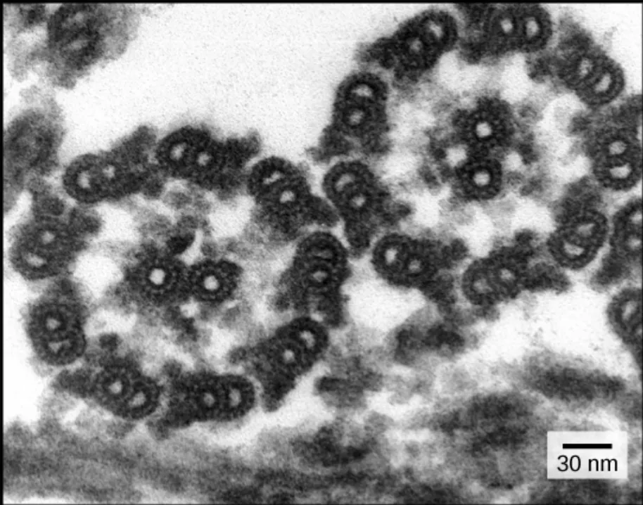
but they share the same internal arrangement of microtubules, called the 9 + 2 array.
1 / 2Next ▶
An electron micrograph in cross-section showing the 9 + 2 pattern of microtubules: nine doublets surround one central doublet in each flagellum. Image: OpenStax, Biology for AP Courses, CC BY 4.0.
Flagella and cilia
 ⤢ Enlarge
1
One flagellum or cilium is a ring of nine microtubule doublets surrounding a single central microtubule doublet.
2
The flagella and cilia of eukaryotes are structurally different from those of prokaryotes.
3
The electron micrograph below shows the 9 + 2 pattern in cross-section.
◀ Previous2 / 2
Science Practice Connection: common ancestry
Science Practice Connection: common ancestry
Think about it: Ribosomes in bacterial cells and in human cells are both made of proteins and ribosomal RNA
which suggests that the two cell types share a common ancestor. What other features of cells provide evidence for common ancestry?
Suggested answer: Nearly all organisms use DNA as their genetic material and read it with the same genetic code.
ATP is the universal energy carrier in cells.
Glucose breakdown through glycolysis is a core pathway shared by many organisms.
All cells are also bounded by a phospholipid bilayer plasma membrane.
Features this widely shared point to a common origin rather than to separate, independent origins.
Components of prokaryotic and eukaryotic cells
Components of prokaryotic and eukaryotic cells
This table summarizes the cell components covered in the chapter, with the cell types in which each one is found.
| Component | Function | Prokaryotes | Animal cells | Plant cells |
|---|---|---|---|---|
| Plasma membrane | Forms the cell boundary and controls the movement of molecules, ions, water, oxygen and wastes | Yes | Yes | Yes |
| Cytoplasm | Fluid that fills the cell, gives turgor pressure in plant cells, hosts many metabolic reactions and holds the organelles | Yes | Yes | Yes |
| Nucleolus | Dark region inside the nucleus where ribosomal subunits are made | No | Yes | Yes |
| Nucleus | Houses DNA and directs the synthesis of ribosomes and proteins | No | Yes | Yes |
| Ribosomes | Protein synthesis | Yes | Yes | Yes |
| Mitochondria | Produce ATP through cellular respiration | No | Yes | Yes |
| Peroxisomes | Break down fatty acids and amino acids and detoxify poisons | No | Yes | Yes |
| Vesicles and vacuoles | Store and transport materials; digest materials in plant cells | No | Yes | Yes |
| Centrosome | Source of microtubules in animal cells; its role in cell division is not fully defined | No | Yes | No |
| Lysosomes | Digest macromolecules and recycle worn-out organelles | No | Yes | No |
| Cell wall | Protects the cell, supports it and maintains its shape; made mainly of peptidoglycan in bacteria and mainly of cellulose in plants | Yes (peptidoglycan) | No | Yes (cellulose) |
| Chloroplasts | Carry out photosynthesis | No | No | Yes |
| Endoplasmic reticulum | Modifies proteins and synthesizes lipids | No | Yes | Yes |
| Golgi apparatus | Modifies, sorts, tags, packages and distributes lipids and proteins | No | Yes | Yes |
| Cytoskeleton | Maintains cell shape, holds organelles in position, moves vesicles within the cell and enables unicellular organisms to move | Yes | Yes | Yes |
| Flagella | Cell locomotion | Some | Some | No, except some plant sperm cells |
| Cilia | Cell locomotion, movement of particles along the outer surface of the plasma membrane, and filtration | Some | Some | No |
Key terms
| Term | Meaning |
|---|---|
| Cytoskeleton | A network of protein fibers in the cell that gives shape, holds organelles in place and enables movement. |
| Microfilament | The narrowest cytoskeleton fiber, about 7 nm wide, made of two intertwined strands of actin. |
| Actin | A globular protein that assembles, using ATP, into microfilaments that serve as tracks for myosin. |
| Myosin | A motor protein that moves along actin filaments to produce movement, including muscle contraction. |
| Cytoplasmic streaming | The circular flow of cytoplasm in plant cells, driven by actin and myosin. |
| Intermediate filament | A cytoskeleton fiber, 8 to 10 nm wide, made of wound fibrous proteins that provides structural support. |
| Keratin | A fibrous protein that forms intermediate filaments in hair, nails and the outer skin layer. |
| Microtubule | A hollow tube, about 25 nm wide, whose wall is made of alpha-tubulin and beta-tubulin dimers. |
| Centrosome | A region of animal cells that contains the centrioles and organizes microtubules. |
| Centriole | One of two perpendicular microtubule bodies that make up the centrosome. |
| Flagellum | A long, hair-like structure that moves an entire cell; a cell usually has one or a few. |
| Cilium | A short, hair-like structure; many extend over the cell surface to move cells or substances. |
| 9 + 2 array | The microtubule arrangement in flagella and cilia: nine doublets in a ring around one central doublet. |
| Depolymerize | To break a polymer, such as an actin or tubulin filament, into its smaller subunits. |
1Avoid these mistakes2Avoid these mistakes3Avoid these mistakes