Curriculum
Biology for AP Courses
Unit 1: The Study of Life
0/6
Unit 2: The Chemical Foundation of Life
0/8
Unit 3: Biological Macromolecules
0/12
-
Unit 3 overview: Biological Macromolecules
 Preview
-
3.1 Synthesis of Biological Macromolecules
Preview
-
3.1 Synthesis of Biological Macromolecules - Check yourself
 Preview
-
3.2 Carbohydrates
Preview
-
3.2 Carbohydrates - Check yourself
Preview
-
3.3 Lipids
Preview
-
3.3 Lipids - Check yourself
Preview
-
3.4 Proteins
Preview
-
3.4 Proteins - Check yourself
Preview
-
3.5 Nucleic Acids
Preview
-
3.5 Nucleic Acids - Check yourself
Preview
-
Unit 3 review quiz: Biological Macromolecules
Preview
Unit 4: Cell Structure
0/14
Unit 5: Structure and Function of Plasma Membranes
0/10
Unit 6: Metabolism
0/12
Unit 7: Cellular Respiration
0/16
Unit 8: Photosynthesis
0/8
Unit 9: Cell Communication
0/10
Unit 10: Cell Reproduction
0/12
Unit 11: Meiosis and Sexual Reproduction
0/6
Unit 12: Mendel's Experiments and Heredity
0/8
Unit 13: Modern Understandings of Inheritance
0/6
Unit 14: DNA Structure and Function
0/14
Unit 15: Genes and Proteins
0/13
Unit 16: Gene Regulation
0/16
Unit 17: Biotechnology and Genomics
0/12
Unit 18: Evolution and Origin of Species
0/8
Unit 19: The Evolution of Populations
0/8
Unit 20: Phylogenies and the History of Life
0/8
Unit 21: Viruses
0/10
Unit 22: Prokaryotes: Bacteria and Archaea
0/12
Unit 23: Plant Form and Physiology
0/14
Unit 24: The Animal Body: Basic Form and Function
0/8
Unit 25: Animal Nutrition and the Digestive System
0/10
Unit 26: The Nervous System
0/12
Unit 27: Sensory Systems
0/12
Unit 28: The Endocrine System
0/12
Unit 29: The Musculoskeletal System
0/10
Unit 30: The Respiratory System
0/10
Unit 31: The Circulatory System
0/10
Unit 32: Osmotic Regulation and Excretion
0/12
Unit 33: The Immune System
0/10
Unit 34: Animal Reproduction and Development
0/16
Unit 35: Ecology and the Biosphere
0/12
Unit 36: Population and Community Ecology
0/16
Unit 37: Ecosystems
0/8
Unit 38: Conservation Biology and Biodiversity
0/10
Text lesson
3.4 Proteins
3.4 Proteins — BIOLOGY FOR AP COURSES notes: You will be able to describe the functions of proteins, explain how amino acids link into polypeptides, and relate the four levels of protein structure to protein shape and function.
1Steroids and cholesterol2Steroids and cholesterol
Steroids and cholesterol
Steroids and cholesterol
Before proteins, it helps to know one other major group of lipids.
Steroids are lipids built from four fused hydrocarbon rings.
Cholesterol is the most common steroid in animal bodies.
It is made mainly in the liver and is the starting material (precursor) for steroid hormones such as testosterone and estradiol,
which are released by the gonads and other endocrine glands.
The body also uses cholesterol to make vitamin D and bile salts
which help break up fats so that cells can absorb them.
1 / 2Next ▶
The four fused rings that form the steroid skeleton of cholesterol and related steroids such as cortisol. Image: OpenStax, Biology for AP Courses, CC BY 4.0.
Steroids and cholesterol
 ⤢ Enlarge
1
Cholesterol is often described negatively
but the body needs it. It is a component of the plasma membrane of animal cellswhere it sits inside the phospholipid bilayer. The plasma membrane is the outer boundary of animal cells. It controls the transport of materials, lets cells recognize one another, and takes part in cell-to-cell communication.
2
Link to learning: an online video explains the different types of fat. The main idea is that fats and related lipids differ in structure, and that difference changes how they behave in the body.
3
Check your learning: What is cholesterol specifically classified as? a. a triglyceride, b. a phospholipid, c. a steroid, d. a wax. Answer: c, a steroid.
◀ Previous2 / 2
1Learning objectives2Learning objectives
1Connection for AP courses2Connection for AP courses
1Types and functions of proteins2Types and functions of proteins3Types and functions of proteins4Types and functions of proteins
1Amino acids2Amino acids3Amino acids4Amino acids
Amino acids
1
Amino acids are the monomers that make up proteins.
2
Every amino acid has the same basic structure: a central carbon atom, called the alpha carbon, bonded to an amino group, a carboxyl group and a hydrogen atom.
3
The central carbon is also bonded to a fourth group called the R group, or side chain.
4
The R group is the only part that differs from one amino acid to another.
1 / 4Next ▶
The general structure of an amino acid: a central carbon with an amino group, a carboxyl group, a hydrogen atom and a variable side chain. Image: OpenStax, Biology for AP Courses, CC BY 4.0.
Amino acids
 ⤢ Enlarge
1
The name amino acid comes from the fact that each molecule contains both an amino group and a carboxyl group.
2
Humans use 20 common amino acids to build proteins.
3
Nine of them are essential amino acids
which means the body cannot make them and they must come from the diet.
4
The nine essential amino acids for humans are histidine, isoleucine, leucine, lysine, methionine, phenylalanine, threonine, tryptophan and valine.
5
Cysteine is normally made by the body
so it is not on this list.
6
Which amino acids are essential depends on the organism.
◀ Previous2 / 4Next ▶
The 20 common amino acids, each drawn with its own R group that gives it its chemical character. Image: OpenStax, Biology for AP Courses, CC BY 4.0.
Amino acids
 ⤢ Enlarge
Amino acids
The chemical nature of the R group determines whether an amino acid is acidic, basic, polar or nonpolar.
Glycine has just a hydrogen atom as its R group.
Valine, methionine and alanine are nonpolar
which means they are hydrophobic.
Serine, threonine and cysteine are polar and have hydrophilic side chains.
Lysine and arginine have positively charged side chains
so they are called basic amino acids.
Proline is unusual because its R group bonds back to the amino group and forms a ring
so its amino group is not separate from the side chain.
◀ Previous3 / 4Next ▶
Amino acids
1
Each amino acid can be written with a single capital letter or a three-letter abbreviation. For example, valine is V or val.
2
Visual connection: Which categories of amino acid would you expect on the surface of a soluble protein, and which in the interior? a.
3
Polar and charged amino acids on the surface, nonpolar amino acids in the interior. b.
4
Polar and charged amino acids in the interior, nonpolar amino acids on the surface. c.
5
Nonpolar and uncharged amino acids both on the surface and in the interior. Answer: a.
A soluble protein sits in water,so its polar and charged side chains face outward toward the water,while its nonpolar side chains pack into the interior away from water.
◀ Previous4 / 4
1Peptide bonds and polypeptides2Peptide bonds and polypeptides3Peptide bonds and polypeptides
Peptide bond formation: the carboxyl group of one amino acid joins the amino group of the next, releasing a water molecule. Image: OpenStax, Biology for AP Courses, CC BY 4.0.
Peptide bonds and polypeptides
 ⤢ Enlarge
Peptide bonds and polypeptides
The sequence and number of amino acids determine a protein’s shape, size and function. Each amino acid is joined to the next by a covalent peptide bond. The carboxyl group of one amino acid combines with the amino group of the incoming amino acid, and a molecule of water is released. This is a dehydration synthesis reaction.
Short chains are called peptides.
As more amino acids join, the chain becomes a polypeptide.
A polypeptide has a free amino group at one end, the N-terminal or amino terminal,
and a free carboxyl group at the other, the C-terminal or carboxyl terminal.
The words polypeptide and protein are sometimes used interchangeably
but they are not the same.
A polypeptide is a polymer of amino acids.
1 / 3Next ▶
Peptide bonds and polypeptides
Peptide bonds and polypeptides
A protein is one or more polypeptides that have folded into a distinct shape, often with non-peptide groups attached,
and that perform a specific function.
After translation, most proteins undergo post-translational modifications, such as cleavage, phosphorylation or the addition of other chemical groups.
A protein is fully functional only after these modifications.
Link to learning: an interactive tutorial walks through the steps of protein synthesis. Protein synthesis matters because it produces the proteins that build tissues and form hormones and enzymes.
Check your learning: Why is the process of protein synthesis critical to life? a.
Protein is the body’s preferred source of energy for rapid energy production. b.
◀ Previous2 / 3Next ▶
Peptide bonds and polypeptides
1
Protein is stored in the liver and muscles as an energy reserve. c.
2
Protein is required for tissue formation and makes up hormones and enzymes. d.
3
Proteins are required to absorb all fat-soluble vitamins. Answer: c.
◀ Previous3 / 3
1Evolution connection: cytochrome c2Evolution connection: cytochrome c3Evolution connection: cytochrome c
1Primary structure2Primary structure3Primary structure4Primary structure
Primary structure
Primary structure
The unique sequence of amino acids in a polypeptide chain is its primary structure.
For example, the hormone insulin has two polypeptide chains, A and B, linked together by disulfide bonds.
The A chain begins with glycine at its N-terminal end and ends with asparagine at its C-terminal end.
The sequences of the A and B chains are unique to insulin.
Two cysteine side chains can react in the presence of oxygen to form a disulfide (S-S) bond.
Insulin has two disulfide bonds joining the A and B chains and a third that helps the A chain fold into the correct shape.
1 / 4Next ▶
The two chains of the insulin hormone, A and B, joined by disulfide bonds, with the amino acids of each chain listed in order. Image: OpenStax, Biology for AP Courses, CC BY 4.0.
Primary structure
 ⤢ Enlarge
Primary structure
The sequence of every protein is determined by the gene that codes for it.
A change in the nucleotide sequence of the coding region can insert a different amino acid into the chain,
which can change the protein’s structure and function.
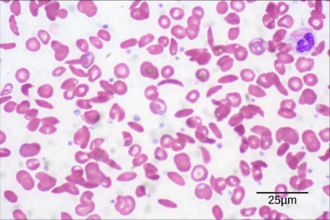
Sickle cell anemia is a clear example.
The hemoglobin beta chain has a single amino acid substitution in which glutamic acid is replaced by valine.
A hemoglobin molecule has two alpha chains and two beta chains, about 600 amino acids in total.
A single amino acid out of those 600 is enough to cause a serious disease.
◀ Previous2 / 4Next ▶
The sickle cell change in the hemoglobin beta chain: at position seven, glutamic acid in normal hemoglobin is replaced by valine in sickle cell hemoglobin. Image: OpenStax, Biology for AP Courses, CC BY 4.0.
Primary structure
 ⤢ Enlarge
1
The change comes from a single base change, a point mutation, in the gene.
2
Because of this single amino acid change, hemoglobin molecules stick together to form long fibers. These fibers distort the normally disc-shaped red blood cells into a crescent or sickle shape. The sickled cells clog blood vessels
which can cause breathlessness, dizziness, headaches and abdominal pain.
◀ Previous3 / 4Next ▶
A blood smear with crescent-shaped sickle cells mixed among normal disc-shaped red blood cells. Image: OpenStax, Biology for AP Courses, CC BY 4.0.
Primary structure
◀ Previous4 / 4
1Secondary structure2Secondary structure
Secondary structure
Secondary structure
Local folding of the polypeptide in some regions creates its secondary structure.
The two most common forms are the alpha helix and the beta-pleated sheet.
Both are held together by hydrogen bonds between the carbonyl (C=O) group of one amino acid and the amino (N-H) group of another.
In an alpha helix, the hydrogen bond forms between an amino acid and the one four positions further along the chain.
Each turn of the helix contains about 3.6 amino acids, and the R groups stick outward from the helix.
In a beta-pleated sheet, the pleats form from hydrogen bonds between backbone atoms of neighboring segments.
1 / 2Next ▶
The alpha helix and the beta-pleated sheet, the two common secondary structures stabilized by hydrogen bonds along the peptide backbone. Image: OpenStax, Biology for AP Courses, CC BY 4.0.
Secondary structure
 ⤢ Enlarge
1
The segments can run parallel or antiparallel, and the R groups stick out above and below the folds.
2
Both structures are common in globular and fibrous proteins
where they provide structural support.
3
Certain amino acids are more likely to form an alpha helix
while others favor a beta-pleated sheet.
◀ Previous2 / 2
1Tertiary structure2Tertiary structure
Tertiary structure
Tertiary structure
The unique three-dimensional structure of a single polypeptide is its tertiary structure.
It comes mainly from interactions among the R groups.
These interactions can work against the hydrogen bonds of the secondary structure.
R groups with like charges repel each other, and R groups with opposite charges attract each other through ionic bonds.
During folding, the hydrophobic R groups of nonpolar amino acids move into the interior of the protein,
while hydrophilic R groups stay on the outside.
This clustering is called the hydrophobic interaction.
1 / 2Next ▶
The chemical interactions that stabilize tertiary structure: hydrophobic interactions, ionic bonds, hydrogen bonds and disulfide linkages. Image: OpenStax, Biology for AP Courses, CC BY 4.0.
Tertiary structure
 ⤢ Enlarge
Tertiary structure
Disulfide linkages form between cysteine side chains in the presence of oxygen.
A disulfide linkage is the only covalent bond that forms during protein folding.
All of these interactions, weak and strong, together set the final three-dimensional shape of the protein. When a protein loses this shape, it may stop working.
◀ Previous2 / 2
1Quaternary structure2Quaternary structure
Quaternary structure
1
Some proteins are built from several polypeptides, called subunits.
2
The way these subunits fit together is the quaternary structure.
3
Weak interactions between subunits help stabilize the whole assembly.
4
Insulin is a globular protein.
5
Hydrogen bonds and disulfide bonds hold it together in a compact, ball-like shape.
6
Silk, by contrast, is a fibrous protein whose beta-pleated sheets form from hydrogen bonds between different chains.
1 / 2Next ▶
The four levels of protein structure, from the primary sequence of amino acids to the assembly of several subunits. Image: OpenStax, Biology for AP Courses, CC BY 4.0.
Quaternary structure
 ⤢ Enlarge
1
Hemoglobin is another example of a protein made of several subunits.
2
The four levels of protein structure, from primary to quaternary, are illustrated in the diagram below.
◀ Previous2 / 2
1Denaturation and protein folding2Denaturation and protein folding3Denaturation and protein folding4Denaturation and protein folding
1Science practice connection2Science practice connection3Science practice connection
1Extra resource and check your learning2Extra resource and check your learning
1Example 1: Counting amino acids in insulin2Example 2: Nucleotides in the hemoglobin coding region3Example 3: Fraction of conserved amino acid positions in cyt
Key terms
| Term | Meaning |
|---|---|
| Protein | A functional molecule made of one or more polypeptides that have folded into a specific shape. |
| Amino acid | A monomer with an amino group, a carboxyl group, a hydrogen and a variable R group, which joins others to form proteins. |
| Peptide bond | A covalent bond between the carboxyl group of one amino acid and the amino group of the next, formed with loss of water. |
| Polypeptide | A linear polymer of many amino acids joined by peptide bonds. |
| R group | The variable side group attached to the central carbon of an amino acid, which determines its chemical nature. |
| Essential amino acid | An amino acid that the body cannot make and that must be obtained from the diet. |
| Enzyme | A protein produced by living cells that acts as a catalyst for a biochemical reaction. |
| Substrate | The reactant that binds to an enzyme and is acted on in the reaction. |
| Active site | The region of an enzyme where its specific substrate binds. |
| Hormone | A chemical signal, usually a small protein or steroid, released by endocrine cells to regulate body processes. |
| Primary structure | The unique linear sequence of amino acids in a polypeptide chain. |
| Secondary structure | Local folding of a polypeptide into regular shapes, such as the alpha helix or beta-pleated sheet, stabilized by hydrogen bonds. |
| Alpha helix | A coiled secondary structure in which hydrogen bonds link each amino acid to one about four positions along the chain. |
| Beta-pleated sheet | A secondary structure formed by segments of the chain folded into pleats and held together by hydrogen bonds. |
| Tertiary structure | The overall three-dimensional shape of a single polypeptide, produced by interactions among R groups. |
| Quaternary structure | The arrangement of several polypeptide subunits into one protein. |
| Hydrophobic interaction | The tendency of nonpolar R groups to cluster in the interior of a folded protein away from water. |
| Disulfide bond | A covalent bond formed between two cysteine side chains in the presence of oxygen, which helps stabilize a protein. |
| Denaturation | Loss of a protein’s three-dimensional shape, and often its function, caused by changes in temperature, pH or chemicals. |
| Chaperone | A helper protein that binds to a target protein during folding to help it reach its correct shape. |
1Avoid these mistakes2Avoid these mistakes3Avoid these mistakes