Curriculum
Biology for AP Courses
Unit 1: The Study of Life
0/6
Unit 2: The Chemical Foundation of Life
0/8
Unit 3: Biological Macromolecules
0/12
Unit 4: Cell Structure
0/14
Unit 5: Structure and Function of Plasma Membranes
0/10
Unit 6: Metabolism
0/12
Unit 7: Cellular Respiration
0/16
Unit 8: Photosynthesis
0/8
Unit 9: Cell Communication
0/10
Unit 10: Cell Reproduction
0/12
Unit 11: Meiosis and Sexual Reproduction
0/6
Unit 12: Mendel's Experiments and Heredity
0/8
Unit 13: Modern Understandings of Inheritance
0/6
Unit 14: DNA Structure and Function
0/14
Unit 15: Genes and Proteins
0/13
Unit 16: Gene Regulation
0/16
Unit 17: Biotechnology and Genomics
0/12
Unit 18: Evolution and Origin of Species
0/8
Unit 19: The Evolution of Populations
0/8
Unit 20: Phylogenies and the History of Life
0/8
Unit 21: Viruses
0/10
Unit 22: Prokaryotes: Bacteria and Archaea
0/12
Unit 23: Plant Form and Physiology
0/14
Unit 24: The Animal Body: Basic Form and Function
0/8
Unit 25: Animal Nutrition and the Digestive System
0/10
Unit 26: The Nervous System
0/12
Unit 27: Sensory Systems
0/12
Unit 28: The Endocrine System
0/12
Unit 29: The Musculoskeletal System
0/10
Unit 30: The Respiratory System
0/10
Unit 31: The Circulatory System
0/10
Unit 32: Osmotic Regulation and Excretion
0/12
Unit 33: The Immune System
0/10
Unit 34: Animal Reproduction and Development
0/16
-
Unit 34 overview: Animal Reproduction and Development
 Preview
-
34.1 Reproduction Methods
Preview
-
34.1 Reproduction Methods - Check yourself
 Preview
-
34.2 Fertilization
Preview
-
34.2 Fertilization - Check yourself
Preview
-
34.3 Human Reproductive Anatomy and Gametogenesis
Preview
-
34.3 Human Reproductive Anatomy and Gametogenesis - Check yourself
Preview
-
34.4 Hormonal Control of Human Reproduction
Preview
-
34.4 Hormonal Control of Human Reproduction - Check yourself
Preview
-
34.5 Fertilization and Early Embryonic Development
Preview
-
34.5 Fertilization and Early Embryonic Development - Check yourself
Preview
-
34.6 Organogenesis and Vertebrate Axis Formation
Preview
-
34.6 Organogenesis and Vertebrate Axis Formation - Check yourself
Preview
-
34.7 Human Pregnancy and Birth
Preview
-
34.7 Human Pregnancy and Birth - Check yourself
Preview
-
Unit 34 review quiz: Animal Reproduction and Development
Preview
Unit 35: Ecology and the Biosphere
0/12
Unit 36: Population and Community Ecology
0/16
Unit 37: Ecosystems
0/8
Unit 38: Conservation Biology and Biodiversity
0/10
Text lesson
34.5 Fertilization and Early Embryonic Development
34.5 Fertilization and Early Embryonic Development — BIOLOGY FOR AP COURSES notes: By the end of this lesson you will be able to describe how a sperm and an egg fuse to form a zygote, explain how cleavage builds a blastula and then a blastocyst, and outline how gastrulation produces the three germ layers.
Learning objectives
Learning objectives
This section answers three questions. How does fertilization occur? How does an embryo form from the zygote? What roles do cleavage and fertilization play in animal development?
Connection for AP courses
1
The material in this section is outside the scope of the AP Biology exam
but it connects to the wider theme of development.
2
A single-celled zygote becomes a multicellular organism with specialized cells, tissues and organs through a process that is complex and tightly regulated.
3
The organism’s genes control this organization.
4
Cell differentiation results from the expression of specific genes,
and homeotic (HOX) genes and morphogens set the pattern and timing of each developmental stage.
5
The examples in this section do not align with the AP Curriculum Framework
so read them as background that shows how the later stages of development fit together.
1Fertilization2Fertilization
Fertilization
Fertilization
Fertilization is the process in which a gamete from each parent fuses to form a zygote. Each gamete, an egg or a sperm, carries one set of chromosomes
so each is haploid. Only one sperm should fuse with one eggbecause the offspring needs exactly one complete diploid set of chromosomes from its two parents.
In mammals the egg is protected by an extracellular layer of glycoproteins called the zona pellucida.
When a sperm binds to this layer, a series of biochemical events called the acrosomal reaction begins.
In placental mammals, the sperm’s acrosome contains digestive enzymes that start to break down the glycoprotein matrix around the egg.
This clears the way for the sperm’s plasma membrane to fuse with the egg’s plasma membrane.
1 / 2Next ▶
Panel a is a micrograph of a sperm binding to an egg. Panel b labels the sperm nucleus, acrosomal granule and zona pellucida, and shows how changes in the egg’s proteins block the entry of other sperm. Image: OpenStax, Biology for AP Courses, CC BY 4.0.
Fertilization
 ⤢ Enlarge
Fertilization
The fusion of the two membranes opens a channel through which the sperm nucleus enters the egg. The nuclear envelopes of the egg and the sperm then break down. The two haploid genomes condense and combine into a single diploid genome.
Fertilization must also prevent more than one sperm from entering.
Once the acrosomal reaction occurs at one point on the egg membrane, the egg releases proteins at other locations that stop additional sperm from fusing with it.
If this block fails, several sperm can fuse with the egg.
This condition is called polyspermy.
The resulting embryo has an abnormal number of chromosome sets, is not genetically viable and dies within a few days.
◀ Previous2 / 2
1Cleavage and blastula stage2Cleavage and blastula stage3Cleavage and blastula stage
Cleavage and blastula stage
1
Development of a multicellular organism begins when the single-celled zygote undergoes rapid cell division. These repeated rounds of division are called cleavage. During cleavage the zygote does not increase in size. One large cell becomes many smaller cells, and each cell within the embryo is called a blastomere.
2
When cleavage has produced more than 100 cells, the embryo is called a blastula.
3
The blastula is usually a hollow ball: a spherical layer of cells, known as the blastoderm, surrounds a cavity called the blastocoel.
4
The blastocoel is filled with fluid or yolk, depending on the species.
5
The diagram below shows the successive divisions of the zygote and the hollow blastula that results from them.
1 / 3Next ▶
Panel a shows the zygote dividing repeatedly inside the zona pellucida without growing larger. Panel b compares a solid early ball of cells with a hollow blastula that has a central blastocoel. Image: OpenStax, Biology for AP Courses, CC BY 4.0.
Cleavage and blastula stage
 ⤢ Enlarge
1
Cleavage can be holoblastic (total) or meroblastic (partial), and the type depends on how much yolk the egg contains.
2
Placental mammals,
including humans, receive nourishment from the mother’s body,so their eggs contain very little yolk and undergo holoblastic cleavage, in which the whole egg divides.
3
Birds have yolk-rich eggs that feed the developing embryo
so they undergo meroblastic cleavage, in which only part of the egg divides.
4
In mammals, the blastula develops into a blastocyst.
5
Its cells arrange themselves into two layers.
6
The inner cell mass, also called the embryoblast, sits inside and will form the embryo itself.
◀ Previous2 / 3Next ▶
A cross-section of a blastocyst showing the fluid-filled blastocoel, the outer ring of trophoblast cells and the inner cell mass on one side of the cavity. Image: OpenStax, Biology for AP Courses, CC BY 4.0.
Cleavage and blastula stage
 ⤢ Enlarge
Cleavage and blastula stage
Its cells are embryonic stem cells that can differentiate into all the cell types the organism needs.
The outer layer, called the trophoblast, will contribute to the placenta and nourish the embryo.
◀ Previous3 / 3
Link to Learning
Link to Learning
The Virtual Human Embryo project, hosted by the Endowment for Human Development, is an online interactive resource. It steps through the stages of human embryo development using micrographs and rotating three-dimensional images. It is a useful way to see how the stages described in this section look in real embryos.
Check your learning
Check your learning
Question: Which early embryonic stage is characterized by a fluid-filled cavity with an inner cell mass surrounded by a layer of cells? a. blastocyst b. blastula c. blastocoel d. gastrula
Answer: a (blastocyst). The blastocyst is the mammalian stage that has a fluid-filled blastocoel, an inner cell mass and an outer layer of trophoblast cells. The blastocoel is only the cavity, not the whole embryo, and the gastrula is a later stage with three germ layers.
1Gastrulation2Gastrulation
Gastrulation
Gastrulation
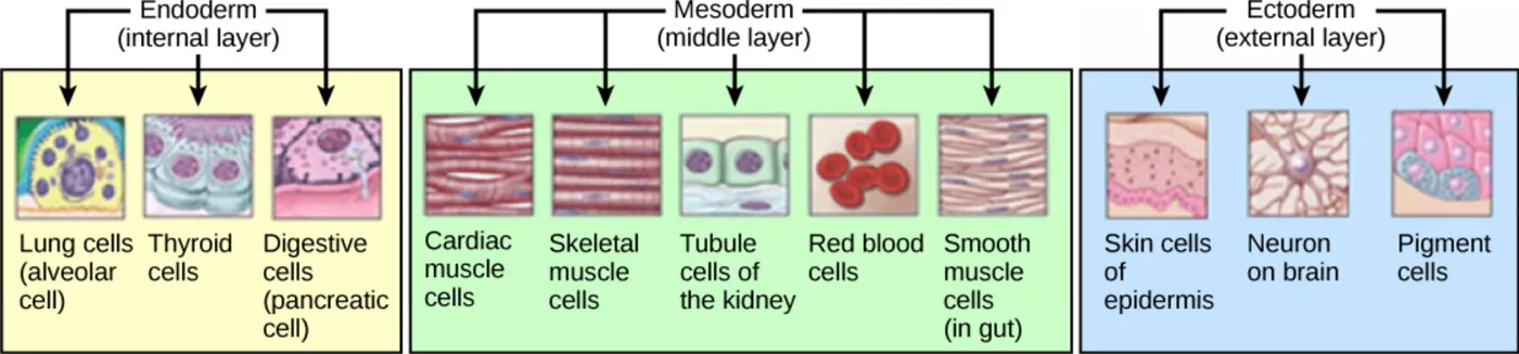
The next stage is the formation of the body plan. During gastrulation, the cells of the blastula rearrange themselves in space to form three layers of cells. The blastula folds upon itself to create these layers. Each layer is called a germ layer, and each germ layer differentiates into particular organ systems.
The ectoderm is the outer germ layer, and it gives rise to the nervous system and the epidermis of the skin.
The mesoderm is the middle germ layer, and it gives rise to muscle cells and connective tissue.
The endoderm is the inner germ layer, and it gives rise to the columnar cells that line the digestive system and many internal organs.
1 / 2Next ▶
Example cell types sorted by germ layer of origin. Endoderm gives lung, thyroid and pancreatic cells, mesoderm gives several muscle, kidney and blood cell types, and ectoderm gives skin, nerve and pigment cells. Image: OpenStax, Biology for AP Courses, CC BY 4.0.
Gastrulation
◀ Previous2 / 2
1Everyday Connection: Are Designer Babies in Our Future?2Everyday Connection: Are Designer Babies in Our Future?3Everyday Connection: Are Designer Babies in Our Future?4Everyday Connection: Are Designer Babies in Our Future?5Everyday Connection: Are Designer Babies in Our Future?
Everyday Connection: Are Designer Babies in Our Future?
1
If you could prevent your child from inheriting a devastating genetic disease, would you?
2
Would you choose a child’s sex, or select for appearance, strength or intelligence?
3
Genetic engineering of a human child to produce a “designer baby” with desirable traits was once only science fiction.
4
Many trait choices for offspring are already possible, and more may become possible.
5
Which traits should be selected, and how, are heavily debated, and the ethical line is not always clear.
6
Eugenics is the use of information and technology from many sources to improve the genetic makeup of the human race.
1 / 5Next ▶
Everyday Connection: Are Designer Babies in Our Future?
Everyday Connection: Are Designer Babies in Our Future?
The goal of creating genetically superior humans was widespread, though controversial, in several countries in the early twentieth century.
It fell out of favor after Nazi Germany ran a large eugenics program in the 1930s and 1940s.
That program forcibly sterilized hundreds of thousands of people labeled unfit and killed tens of thousands of disabled people in institutions.
Eugenic ideas have been much less openly expressed since then
but some people still promote them.
◀ Previous2 / 5Next ▶
A 1921 conference logo shows eugenics as a tree whose roots draw on many fields, such as genetics, anthropology and statistics, to build one harmonious discipline. Image: OpenStax, Biology for AP Courses, CC BY 4.0.
Everyday Connection: Are Designer Babies in Our Future?
 ⤢ Enlarge
1
Efforts to choose traits in children are not new. A sperm bank opened in 1980 that accepted donations only from men with high IQ scores
but it did not attract public interest and closed in 1999.
2
Prenatal genetic diagnosis (PGD) is a technique used with in vitro fertilization, in which embryos are created and grown outside the mother’s body for a period before implantation.
3
The term PGD usually covers the diagnosis, the selection and the implantation of the chosen embryos.
4
The least controversial use of PGD tests embryos for alleles that cause serious genetic diseases,
such as sickle cell disease, muscular dystrophy and hemophilia.
5
Embryos carrying the disease alleles are not implanted
so the disease is prevented.
6
The unused embryos are either donated to science or discarded.
◀ Previous3 / 5Next ▶
Everyday Connection: Are Designer Babies in Our Future?
Everyday Connection: Are Designer Babies in Our Future?
Even where the ethics seem clear, not everyone agrees.
People who believe human life begins at conception find the discarding of embryos unacceptable in any circumstance.
Selecting a child’s sex is murkier.
Great Britain bans sex selection unless it prevents sex-linked diseases, some countries allow “family balancing” so that parents can have at least one child of each sex,
and the United States leaves the decision largely to individual physicians.
Some people with deafness or dwarfism have used PGD to choose embryos that share their condition, citing its positive aspects and culture,
while others say this violates the principle of “first, do no harm.” Such procedures are legal in most countries but raise hard ethical questions.
The major limitation is cost.
◀ Previous4 / 5Next ▶
Everyday Connection: Are Designer Babies in Our Future?
Everyday Connection: Are Designer Babies in Our Future?
PGD is usually not covered by medical insurance
so only a very small percentage of live births use these methods.
Looking ahead, lawmakers may struggle to keep pace with technology that changes quickly and unpredictably.
Other bioethicists argue that only existing technologies should be debated,
because these procedures are expensive and rare,so fears of eugenics are overstated.
The debate continues.
Question: Prenatal genetic diagnosis is often sought by parents before an embryo is implanted. Which of the following CANNOT be diagnosed by PGD? a. inheritance of sickle cell anemia b. Down syndrome c. determination of sex d. inheritance of sexually transmitted diseases
Answer: d. PGD examines the embryo’s genetic alleles and chromosomes
so it can detect sickle cell anemia, Down syndrome and sex. It cannot diagnose sexually transmitted diseaseswhich are infections rather than inherited genetic conditions.
◀ Previous5 / 5
Key terms
| Term | Meaning |
|---|---|
| Fertilization | Fertilization is the fusion of a sperm and an egg to form a zygote. |
| Zygote | A zygote is the single diploid cell formed when a sperm and an egg fuse. |
| Zona pellucida | The zona pellucida is the glycoprotein layer that surrounds and protects a mammalian egg. |
| Acrosomal reaction | The acrosomal reaction is a series of biochemical events in a sperm, triggered by binding to the zona pellucida, that lets the sperm penetrate the egg. |
| Polyspermy | Polyspermy is the fusion of more than one sperm with an egg, which produces a nonviable embryo. |
| Cleavage | Cleavage is the rapid series of cell divisions after fertilization that produces many smaller cells without increasing the total size. |
| Blastomere | A blastomere is one of the cells produced by cleavage. |
| Blastoderm | The blastoderm is the single layer of cells that forms the wall of a blastula. |
| Blastocoel | The blastocoel is the fluid- or yolk-filled cavity inside a blastula. |
| Blastula | A blastula is the hollow ball of cells that forms once cleavage has produced more than 100 cells. |
| Blastocyst | A blastocyst is the mammalian blastula stage in which cells form an inner cell mass and an outer trophoblast. |
| Inner cell mass (embryoblast) | The inner cell mass is the group of embryonic stem cells inside the blastocyst that will form the embryo. |
| Trophoblast | The trophoblast is the outer layer of the blastocyst that nourishes the embryo and contributes to the placenta. |
| Holoblastic cleavage | Holoblastic cleavage is cleavage in which the whole egg divides, typical of eggs with little yolk. |
| Meroblastic cleavage | Meroblastic cleavage is cleavage in which only part of the egg divides, typical of yolk-rich eggs. |
| Gastrulation | Gastrulation is the process that rearranges the blastula into three germ layers. |
| Germ layer | A germ layer is one of the three layers of cells formed in gastrulation, each of which gives rise to particular tissues and organs. |
| Ectoderm | The ectoderm is the outer germ layer that forms the nervous system and the epidermis. |
| Mesoderm | The mesoderm is the middle germ layer that forms muscle cells and connective tissue. |
| Endoderm | The endoderm is the inner germ layer that forms the lining of the digestive system and many internal organs. |
| Eugenics | Eugenics is the attempt to improve the genetic makeup of the human race using information and technology. |
| Prenatal genetic diagnosis (PGD) | Prenatal genetic diagnosis is the screening of embryos created by in vitro fertilization for disease-causing alleles before implantation. |
1Avoid these mistakes2Avoid these mistakes3Avoid these mistakes